. 2018 Jan 10:15:3.
doi: 10.1186/s12950-018-0179-6. eCollection 2018.

Histone deacetylase 2 (HDAC2) attenuates lipopolysaccharide (LPS)-induced inflammation by regulating PAI-1 expression

Affiliations

Histone deacetylase 2 (HDAC2) attenuates lipopolysaccharide (LPS)-induced inflammation by regulating PAI-1 expression

Wen-Feng Fang et al. J Inflamm (Lond). .

Abstract

Background: Sepsis is a life-threatening organ dysfunction caused by dysregulated host response to infection, and is primarily characterized by an uncontrolled systemic inflammatory response. In the present study, we developed an effective adjunct therapy mediated by a novel mechanism, to attenuate overt inflammation. LPS-treated macrophages were adopted as an in vitro model of endotoxin-induced inflammation during sepsis. Experiments were carried out using primary mouse peritoneal macrophages and the murine macrophage cell line RAW264.7, to elucidate the mechanisms by which HDAC2 modulates endotoxin-induced inflammation.

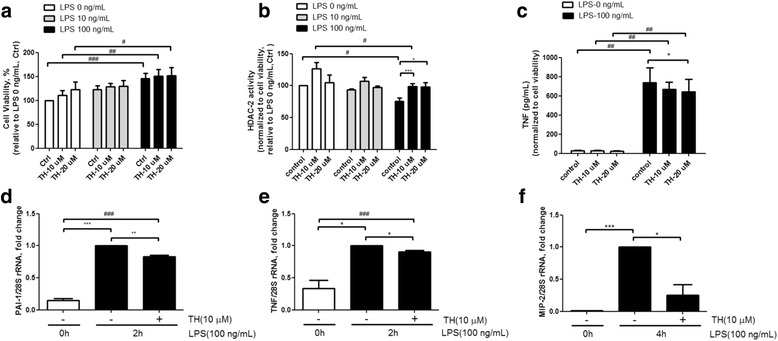
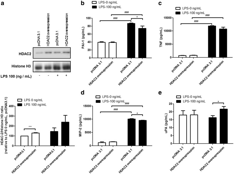
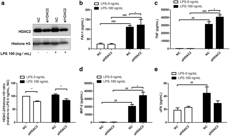
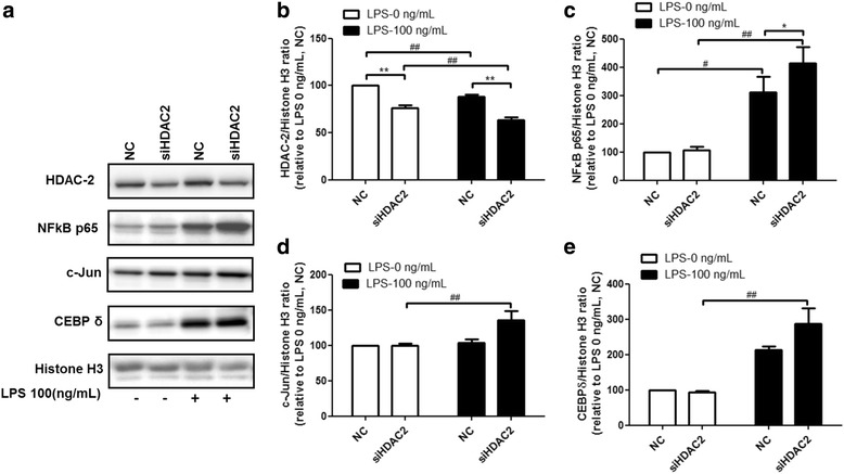
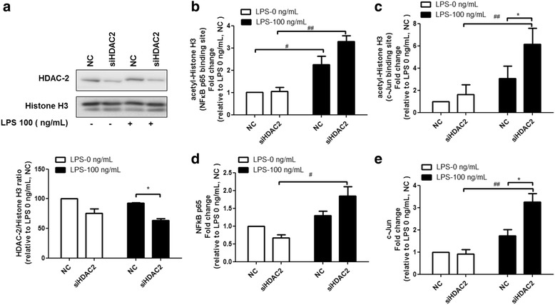
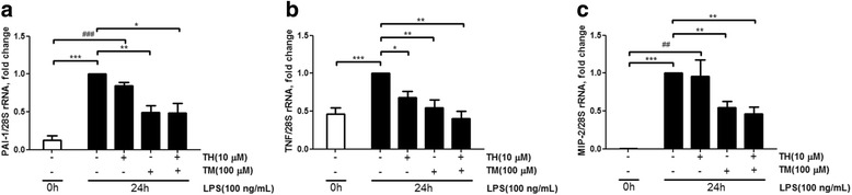
Results: Results revealed that PAI-1, TNF, and MIP-2 expression were inhibited by theophylline, an HDAC2 enhancer, in a RAW macrophage cell line, following LPS-induced inflammation. Thus, HDAC2 plays an important role in immune defense by regulating the expression of inflammatory genes via the c-Jun/PAI-1 pathway. During LPS-induced inflammation, overexpression of HDAC2 was found to inhibit PAI-1, TNF, and MIP-2 expression. Following LPS stimulation, HDAC2 knockdown increased nuclear translocation and DNA binding of c-Jun to the PAI-1 gene promoter, thereby activating PAI-1 gene transcription. Furthermore, inhibition of PAI-1 by TM5275 alone or in combination with theophylline notably suppressed TNF and MIP-2 expression.

Conclusion: HDAC2 can attenuate lipopolysaccharide-induced inflammation by regulating c-Jun and PAI-1 expression in macrophages.

Keywords: Histone deacetylase 2 (HDAC2); Lipopolysaccharide (LPS); Plasminogen activator inhibitor (PAI).


Conflict of interest statement

Procedures for the care of the animals and experiments were followed in accordance with the Guide of the Care and Use of Laboratory Animals from Chang Gung Memorial Hospital institutional animal care and use committee. The Institutional Biosafety Committee of Chang Gung University approved the experimental procedures.Not applicable.The authors declare that they have no competing interests.Springer Nature remains neutral with regard to jurisdictional claims in published maps and institutional affiliations.

Figures

Fig. 1
Fig. 1
Theophylline increases HDAC2 activity and attenuates LPS-induced expression of pro-inflammatory genes. Mouse primary peritoneal macrophages cells were pretreated with theophylline (TH) for 30 min and then stimulated with LPS for 1 h. Theophylline increased cell viability (a), and enhanced HDAC2 activity (b) of peritoneal macrophages in the control and LPS-treated groups. Theophylline inhibited TNF secretion in the control and LPS-treated groups (c), and significantly repressed LPS-induced mRNA expression of PAI-1 (d), TNF (e), and MIP-2 (f) in RAW264.7 macrophages. Data are expressed as the mean relative expression ± SEM and are representative of at least three independent experiments. For all figures, * indicates p < 0.05; **,p < 0.01; ***,p < 0.001 in paired t test. # indicates p < 0.05; ##, p < 0.01; ###, p < 0.00.1 in ANOVA test
Fig. 2
Fig. 2
Overexpression of HDAC2 attenuates LPS-induced PAI-1, TNF, and MIP-2 secretion, but enhances uPA secretion. RAW264.7 cells were transfected with HDAC2 expression vector for 48 h, and then stimulated with 100 ng/ml LPS for 2 h. HDAC2 overexpression resulted in a 1.5-fold increase in protein levels of HDAC2 (a). HDAC2 inhibited LPS-induced secretion of PAI-1 (b), TNF (c), and MIP-2 (d), but increased uPA secretion (e). Data are expressed as the mean relative expression ± SEM of at least three independent experiments
Fig. 3
Fig. 3
HDAC2 knockdown enhances PAI-1, TNF, and MIP-2 secretion, but represses uPA secretion during LPS-induced inflammation. RAW264.7 cells were transfected with siRNA targeting HDAC2 for 48 h, and then stimulated with 100 ng/ml LPS for 2 h. HDAC2 knockdown resulted in 20% reduction in protein levels of HDAC2 (a). HDAC2 significantly enhanced LPS-induced secretion of PAI-1 (b), TNF (c), and MIP-2 (d), but inhibited uPA secretion (e). Data are expressed as the mean relative expression ± SEM of at least three independent experiments
Fig. 4
Fig. 4
NF-κB p65, c-Jun and CEBPδ are involved in HDAC2 regulation of LPS-induced inflammation. RAW264.7 cells were transfected with siRNA targeting HDAC2 for 48 h, and then stimulated with 100 ng/ml LPS for 2 h. HDAC-2 knockdown (a, b) increased the translocation of the inflammation-related transcription factors NF-κB p65 (a, c), c-Jun (a, d), and CEBPδ (a, e) to the cell nucleus. Data are expressed as the mean relative expression ± SEM of at least three independent experiments
Fig. 5
Fig. 5
Epigenetic regulation of PAI-1 gene expression by HDAC2. RAW264.7 cells were transfected with siRNA targeting HDAC2 for 48 h, and then stimulated with 100 ng/ml LPS for 2 h. Cells were harvested and subjected to the ChIP assay to analyze the status of acetyl-Histone H3, NFκB p65, and c-Jun at the PAI-1 gene promoter. Immunoprecipitated chromatin was analyzed via qPCR using primers (shown with arrows) targeting the promoter regions of PAI-1. HDAC2 knockdown (a) increased the binding of acetyl-histone H3 to the NFκB p65 and c-Jun binding sites of the PAI-1 gene promoter (b, c) following LPS treatment. HDAC2 knockdown also increased the binding of NFκB p65 and c-Jun to the PAI-1 gene promoter (d, e) after LPS stimulation. Data are expressed as the mean relative expression ± SEM of at least three independent experiments
Fig. 6
Fig. 6
Effects of theophylline and TM5275 on RAW264.7 cells. RAW264.7 cells were pretreated with either 10 μM theophylline (TH) or 100 μM TM5275 (TM) for 30 min, and then stimulated with 100 ng/ml LPS for 24 h. Transcript levels of PAI-1, TNF, and MIP-2 were measured via qRT-PCR. Only the PAI-1 inhibitor TM5275 significantly downregulated mRNA levels of PAI-1, TNF, and MIP-2 following LPS induction. Combined treatment with theophylline and TM5275 caused a stronger inhibition of the expression of PAI-1 (a), TNF (b), and MIP-2 (c). Data are shown as the mean relative expression ± SEM of at least three independent experiments

References

    1. Singer M, Deutschman CS, Seymour CW, Shankar-Hari M, Annane D, Bauer M, Bellomo R, Bernard GR, Chiche JD, Coopersmith CM, et al. The third international consensus definitions for sepsis and septic shock (Sepsis-3) JAMA. 2016;315:801–810. doi: 10.1001/jama.2016.0287. - DOI - PMC - PubMed
    1. Angus DC, van der Poll T. Severe sepsis and septic shock. N Engl J Med. 2013;369:2063. doi: 10.1056/NEJMra1208623. - DOI - PubMed
    1. Dellinger RP, Levy MM, Rhodes A, Annane D, Gerlach H, Opal SM, Sevransky JE, Sprung CL, Douglas IS, Jaeschke R, et al. Surviving sepsis campaign: international guidelines for management of severe sepsis and septic shock, 2012. Intensive Care Med. 2013;39:165–228. doi: 10.1007/s00134-012-2769-8. - DOI - PMC - PubMed
    1. Phua J, Koh Y, Du B, Tang YQ, Divatia JV, Tan CC, Gomersall CD, Faruq MO, Shrestha BR, Gia Binh N, et al. Management of severe sepsis in patients admitted to Asian intensive care units: prospective cohort study. BMJ. 2011;342:d3245. doi: 10.1136/bmj.d3245. - DOI - PMC - PubMed
    1. Fang WF, Douglas IS, Wang CC, Kao HC, Chang YT, Tseng CC, Huang KT, Chang HC, Lin MC. 5-lipoxygenase activating protein (FLAP) dependent leukotriene biosynthesis inhibition (MK591) attenuates lipid a endotoxin-induced inflammation. PLoS One. 2014;9:e102622. doi: 10.1371/journal.pone.0102622. - DOI - PMC - PubMed

LinkOut - more resources