. 2017 Dec;4(1):48-51.
doi: 10.1159/000478102. Epub 2017 Jul 19.

Unilateral Multifocal Choroidal Melanoma

Affiliations

Unilateral Multifocal Choroidal Melanoma

Debarshi Mustafi et al. Ocul Oncol Pathol. 2017 Dec.

Abstract

We report a case of multifocal choroidal melanoma in the same eye, separated in presentation by 20 years. A 57-year-old Caucasian male initially presented with a choroidal melanoma of the right eye that was treated with transpupillary thermotherapy. Due to recurrence, the patient underwent proton beam therapy with subsequent tumor regression. A second small choroidal lesion was noted in the right eye during his surveillance examinations that was closely monitored and demonstrated stable dimensions and features suggestive of a choroidal nevus. Twenty years after his first presentation, the second lesion exhibited accelerated growth with imaging studies indicative of transformation to a distinct choroidal melanoma. The patient underwent a second globe salvage treatment of proton beam therapy. We describe the clinical course, radiographic, and imaging findings of this rare choroidal melanoma.

Keywords: Choroidal melanoma; Melanoma; Multifocal melanoma; Optical coherence tomography angiography; Proton beam radiotherapy; Tumor recurrence.


Figures

Fig. 1
Fig. 1
Serial fundus photographs documenting the transformation of the second choroidal lesion from nevus to melanoma. a Fundus photograph of the right eye showing the original choroidal melanoma previously treated by transpupillary thermotherapy in 1996 and proton beam radiotherapy in 2008. The second choroidal lesion superonasal to the optic disc is flat and without any high-risk features. b Small raised choroidal lesion superonasal to the optic disc noted on presentation in 2016. c Lesion superonasal to the optic disc 1 month later without discernable change in appearance or size. d Interval increases in size and coloration of the choroidal lesion after 12 months. e Fundus photograph of the left eye with no abnormal features.
Fig. 2
Fig. 2
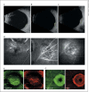
Ultrasonography, fluorescein angiography (FA), and optical coherence tomography angiography (OCTA) in documenting features of progression to choroidal melanoma. a Ultrasonography at presentation in 2016 showing the superonasal choroidal lesion measuring 3.6 mm in basal dimension and 1.1 mm in thickness. b Ultrasonography at 1 month with similar dimensions. c Ultrasonography at 12 months showing interval increase in dimensions to 5.2 in basal dimension and 2.0 mm in thickness. d FA of the right eye at 12 months revealing hypofluorescence of the treated lesion with small punctate areas of leakage into the macula along with double circulation and late dye leakage emanating from the second choroidal lesion. e OCTA of the treated lesion at 12 months illustrating sclerosis of the underlying retinal vasculature. f OCTA of the new untreated choroidal tumor at 12 months illustrating disruption of both the superficial and deep retinal vascular layers.

References

    1. Singh AD, Turell ME, Topham AK. Uveal melanoma: trends in incidence, treatment, and survival. Ophthalmology. 2011;118:1881–1885. - PubMed
    1. Singh AD, Shields CL, Shields JA, De Potter P. Bilateral primary uveal melanoma. Bad luck or bad genes? Ophthalmology. 1996;103:256–262. - PubMed
    1. Shammas HF, Watzke RC. Bilateral choroidal melanomas. Case report and incidence. Arch Ophthalmol. 1977;95:617–623. - PubMed
    1. Honavar SG, Shields CL, Singh AD, et al. Two discrete choroidal melanomas in an eye with ocular melanocytosis. Surv Ophthalmol. 2002;47:36–41. - PubMed
    1. Harbour JW, Onken MD, Roberson ED, et al. Frequent mutation of BAP1 in metastasizing uveal melanomas. Science. 2010;330:1410–1413. - PMC - PubMed

LinkOut - more resources