Review
. 2018 Jan 17;113(2):10.
doi: 10.1007/s00395-018-0668-z.

Molecular imaging of cardiac remodelling after myocardial infarction

Affiliations
Review

Molecular imaging of cardiac remodelling after myocardial infarction

Daniel Curley et al. Basic Res Cardiol. .

Abstract

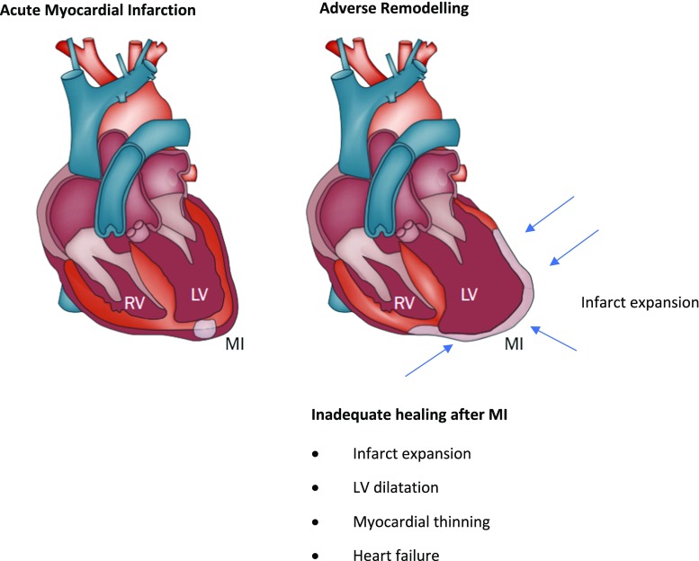
Myocardial infarction and subsequent heart failure is a major health burden associated with significant mortality and morbidity in western societies. The ability of cardiac tissue to recover after myocardial infarction is affected by numerous complex cellular and molecular pathways. Unbalance or failure of these pathways can lead to adverse remodelling of the heart and poor prognosis. Current clinical cardiac imaging modalities assess anatomy, perfusion, function, and viability of the myocardium, yet do not offer any insight into the specific molecular pathways involved in the repair process. Novel imaging techniques allow visualisation of these molecular processes and may have significant diagnostic and prognostic values, which could aid clinical management. Single photon-emission tomography, positron-emission tomography, and magnetic resonance imaging are used to visualise various aspects of these molecular processes. Imaging probes are usually attached to radioisotopes or paramagnetic nanoparticles to specifically target biological processes such as: apoptosis, necrosis, inflammation, angiogenesis, and scar formation. Although the results from preclinical studies are promising, translating this work to a clinical environment in a valuable and cost-effective way is extremely challenging. Extensive evaluation evidence of diagnostic and prognostic values in multi-centre clinical trials is still required.

Keywords: Cardiac remodelling; Cardiovascular imaging; MRI; Myocardial infarction.


Figures

Fig. 1
Fig. 1
Schematic showing the gross changes in adverse cardiac remodelling post-MI (figure adapted from [143])
Fig. 2
Fig. 2
Thalium-201 perfusion imaging, Tc-99m-labelled MMP imaging, and fused images before (SHAM), 1 week and 3 week post-MI using SPECT/CT. Arrows show infarct zone, which is poorly perfused, where MMPs are detected [132]
Fig. 3
Fig. 3
T2-weighted CMR image before injection of EP-3533 (a) and b an inversion recovery CMR image 40 min after the injection of EP-3533. The arrow highlights the hyper-intense region indicating high levels of collagen, therefore, scar [11]
Fig. 4
Fig. 4
Myocardial short-axis images from base to apex with 201Tl perfusion SPECT, 99mTc-RAFT-RGD SPECT scan and fused images. The arrows highlight areas of infarct (figure adapted from [21])
Fig. 5
Fig. 5
Post-MI state in mouse models injected with AnxCLIO-Cy5.5 (a) and a control probe inact_CLIO-Cy5.5 (b). Significant hypo-intensity can be seen with AnxCLIO-Cy5.5 (a), depicted by yellow arrows. There are no areas of significant uptake seen using the control probe (b). Regions of hypo-intensity represent the visualisation of active apoptosis [128]
Fig. 6
Fig. 6
Uptake of 68 Ga pentixafor in patients after acute ST-segment elevation myocardial infarction indicating various levels of CXCR4 expression in myocardial segments with different patterns of myocardial injury as defined by the presence (+) or absence (−) of gadolinium-diethylenetriamine pentaacetic acid (DTPA) late enhancement (LE) or edema on T2-sequences (T2) at cardiac magnetic resonance imaging. a Representative short-axis slices characterizing four different types of segments. b Results of segmental pentixafor uptake score in respective segment types. *p < 0.05 versus remote; **p < 0.05 versus all others. HLA horizontal long axis, SA short axis [139]
Fig. 7
Fig. 7
Typical 31P-magnetic resonance spectroscopy spectrum showing 2,3-diphosphoglycerate (2,3-DGP), phosphodiester (PDE), phosphocreatine (PCr), and the three phosphorus peaks of ATP (γ, α, and β). The x-axis is expressed in parts per million (ppm) [108]
Fig. 8
Fig. 8
Multimodal characterization of the myocardial tissue after AMI using PET/MRI. Short-axis images of a patient who was imaged shortly after acute MI using simultaneous 18F-FDG and 13N-NH3 PET/MRI. Myocardial scarring can be imaged using LGE MRI (left column, top; white arrows pointing at subendocardial non-transmural infarction). The area of myocardial infarction is exceeded by the myocardial oedema imaged using T2-weighted sequences (right column, top; red arrows). Using fasting-heparin 18F-FDG-PET/MRI, the area of post-ischemic inflammation or ischemic memory can be assessed. After revascularization by percutaneous coronary intervention (PCI), only a slightly reduced perfusion of the inferior wall was observed in this patient [113]
Fig. 9
Fig. 9
Diffusion tractography in a healthy rat heart (a, b). Two infarcted rat hearts show severe distortion of myofibre architecture (c, d) [125, 129]

References

    1. Abubakar I, Tillmann T, Banerjee A. Global, regional, and national age-sex specific all-cause and cause-specific mortality for 240 causes of death, 1990–2013: a systematic analysis for the Global Burden of Disease Study 2013. Lancet. 2015;385:117–171. doi: 10.1016/S0140-6736(14)61638-X. - DOI - PMC - PubMed
    1. Ahmadi A, Ohira H, Mielniczuk LM. FDG PET imaging for identifying pulmonary hypertension and right heart failure. Curr Cardiol Rep. 2015;17:555. doi: 10.1007/s11886-014-0555-7. - DOI - PubMed
    1. Akki A, Gupta A, Weiss RG. Magnetic resonance imaging and spectroscopy of the murine cardiovascular system. Am J Physiol Heart Circ Physiol. 2013;304:H633–H648. doi: 10.1152/ajpheart.00771.2011. - DOI - PMC - PubMed
    1. Asano Y, Ihn H, Yamane K, Jinnin M, Mimura Y, Tamaki K. Increased expression of integrin alpha(v)beta3 contributes to the establishment of autocrine TGF-beta signaling in scleroderma fibroblasts. J Immunol. 2005;175:7708–7718. doi: 10.4049/jimmunol.175.11.7708. - DOI - PubMed
    1. Atluri P, Woo YJ. Pro-angiogenic cytokines as cardiovascular therapeutics. BioDrugs. 2008;22:209–222. doi: 10.2165/00063030-200822040-00001. - DOI - PubMed

Publication types

MeSH terms