. 2018 Mar;103(3):395-407.
doi: 10.1002/JLB.4HI0517-210R. Epub 2018 Jan 3.

Frontline Science: High fat diet and leptin promote tumor progression by inducing myeloid-derived suppressor cells

Affiliations

Frontline Science: High fat diet and leptin promote tumor progression by inducing myeloid-derived suppressor cells

Virginia K Clements et al. J Leukoc Biol. 2018 Mar.

Abstract

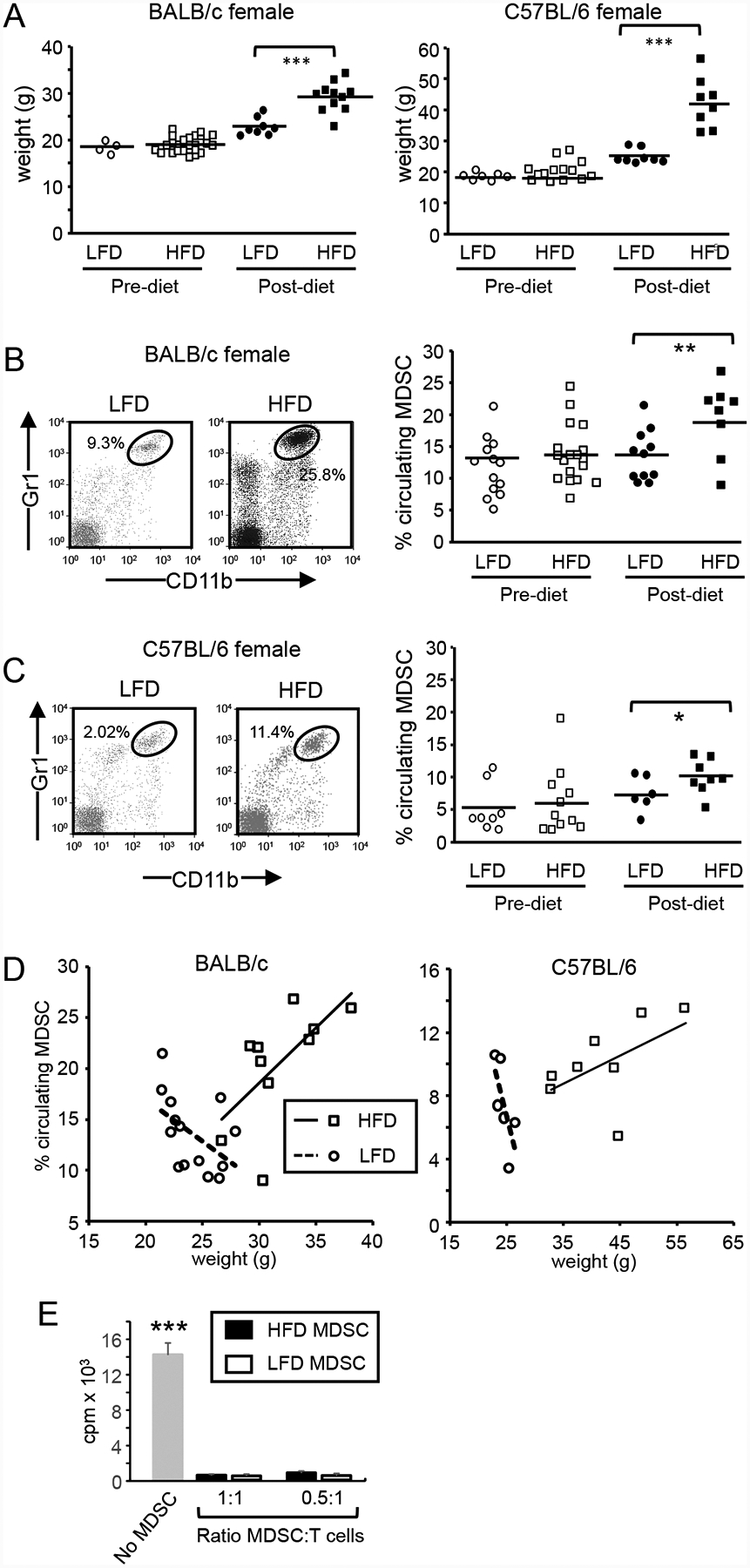
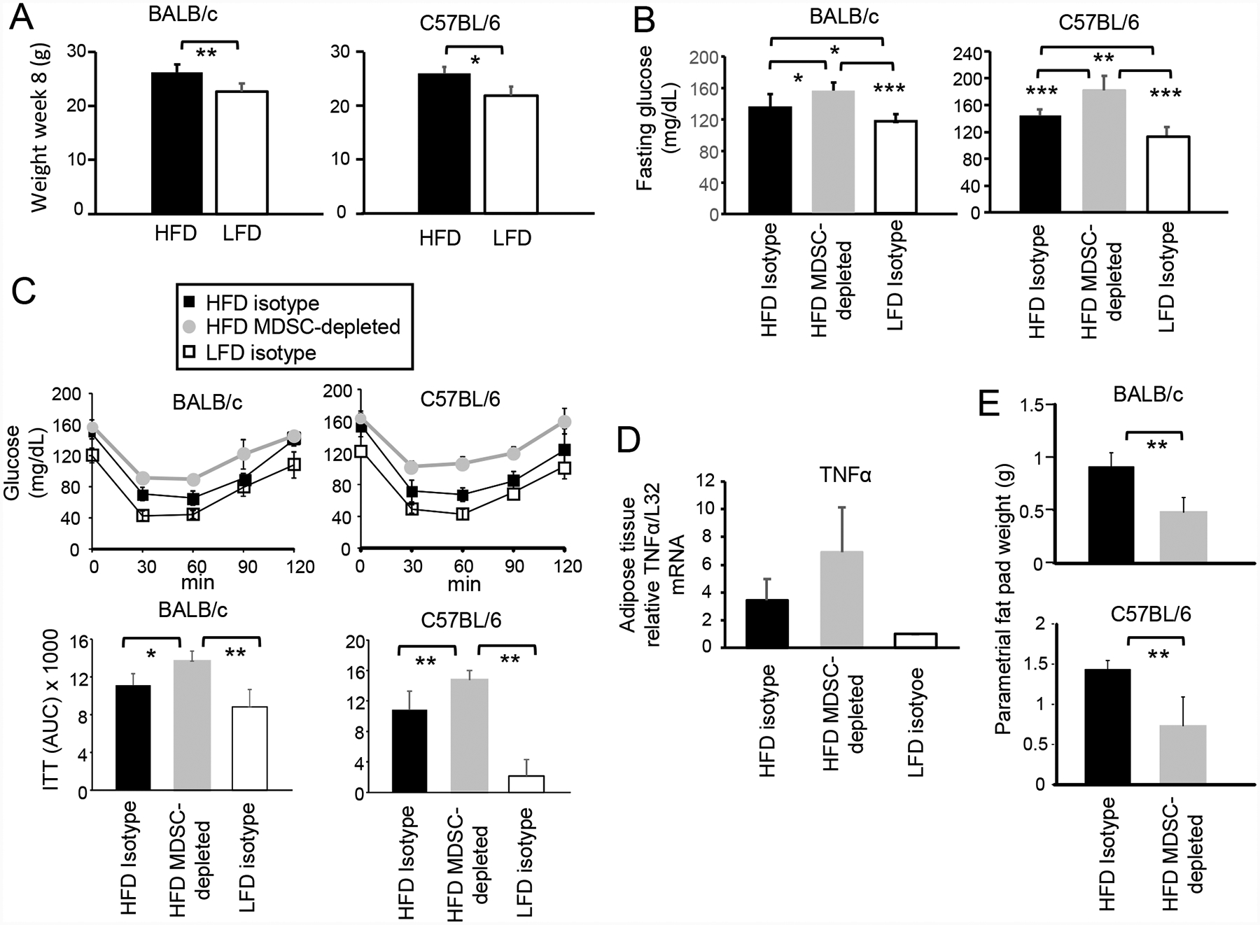
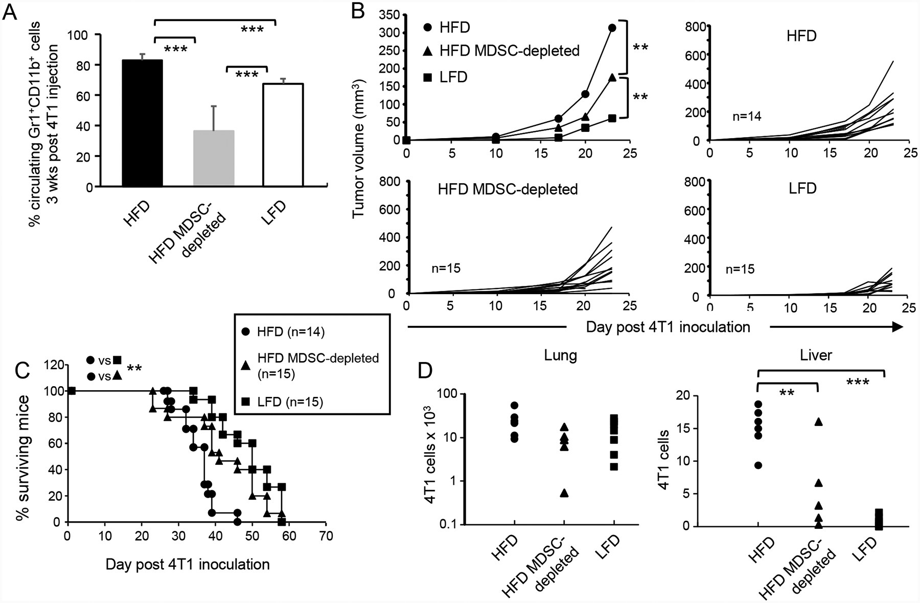
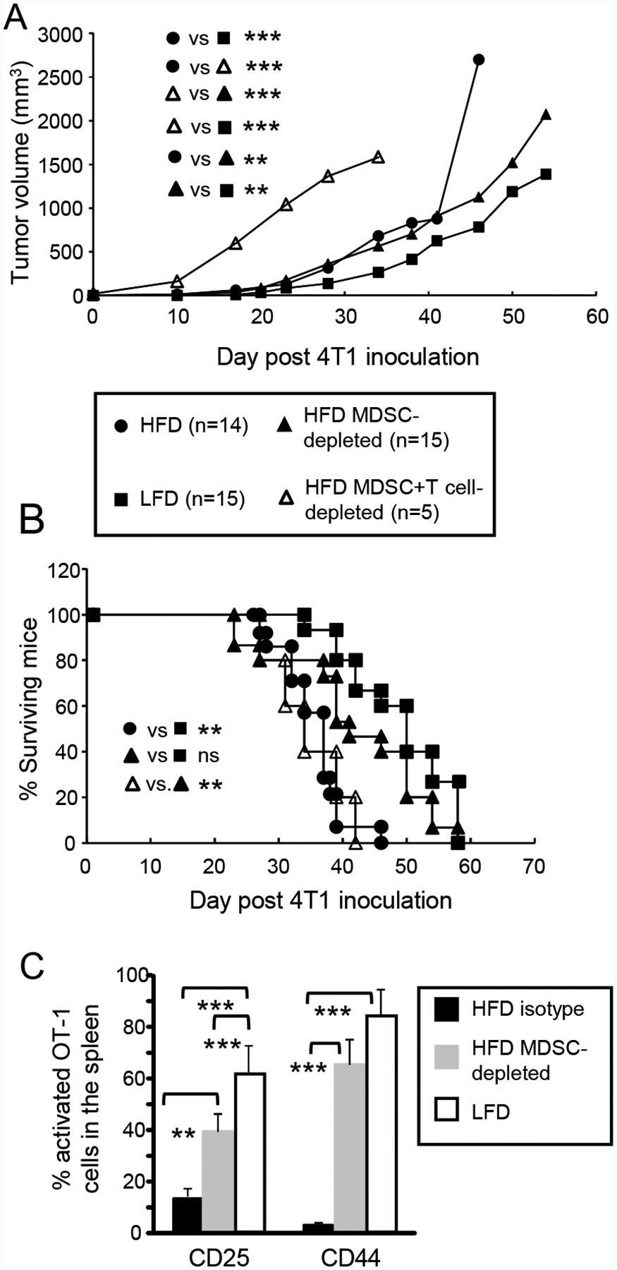
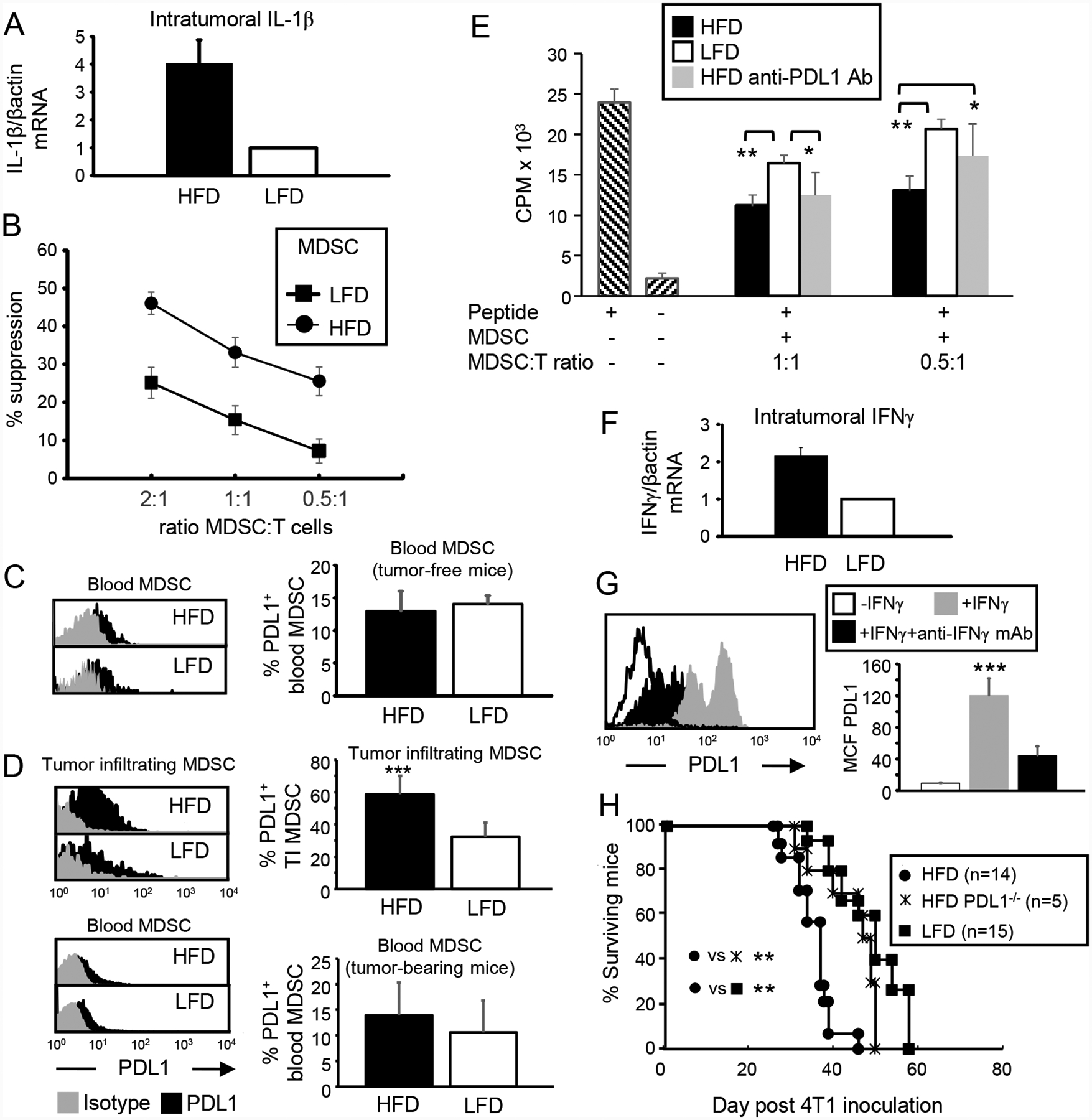
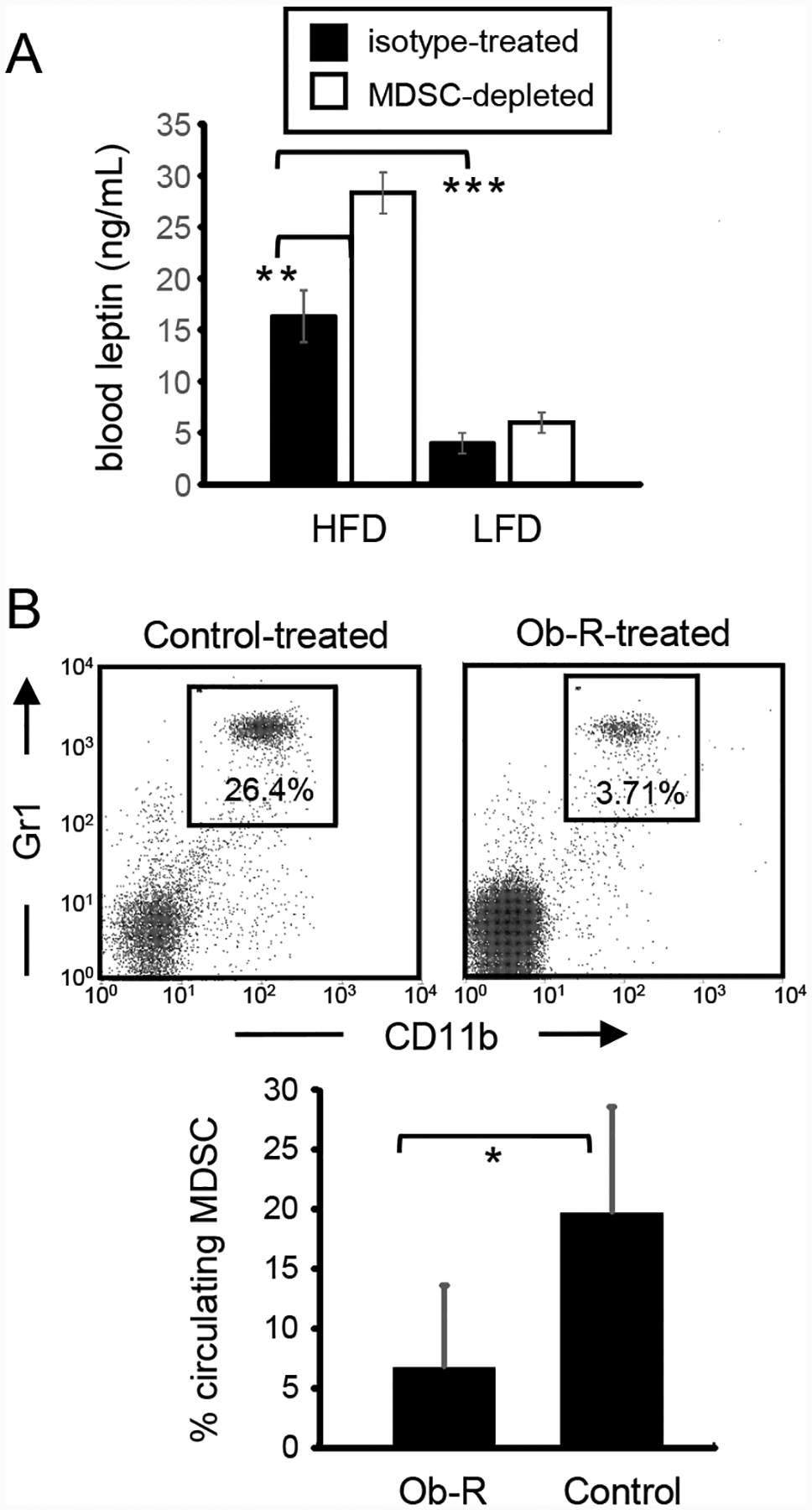
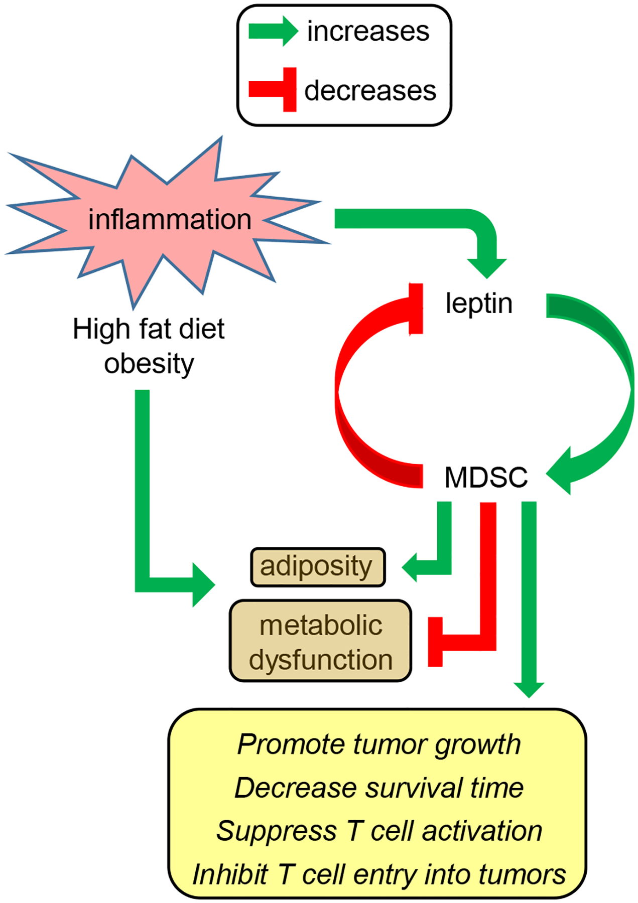
Obesity is a risk factor for cancer incidence and cancer mortality. The association of obesity and cancer is attributed to multiple factors, but the tightest linkage is with the chronic, low-grade inflammation that accompanies obesity. Myeloid-derived suppressor cells (MDSC) are known facilitators of cancer progression that act by suppressing the activation and function of tumor-reactive T cells. Because MDSC quantity and function are driven by chronic inflammation, we hypothesized that MDSC may accumulate in obese individuals and facilitate tumor growth by suppressing antitumor immunity. To test this hypothesis, tumor-bearing mice on a high fat or low fat diet (HFD or LFD) were assessed for tumor progression and the metabolic dysfunction associated with obesity. HFD enhanced the accumulation of MDSC, and the resulting MDSC had both beneficial and detrimental effects. HFD-induced MDSC protected mice against diet-induced metabolic dysfunction and reduced HFD-associated inflammation, but also increased the accumulation of fat, enhanced tumor progression, and spontaneous metastasis and reduced survival time. HFD-induced MDSC facilitated tumor growth by limiting the activation of tumor-reactive CD8+ T cells. Leptin, an adipokine that regulates appetite satiety and is overexpressed in obesity, undergoes crosstalk with MDSC in which leptin drives the accumulation of MDSC while MDSC down-regulate the production of leptin. Collectively, these studies demonstrate that although MDSC protect against some metabolic dysfunction associated with HFD they enhance tumor growth in HFD mice and that leptin is a key regulator linking HFD, chronic inflammation, immune suppression, and tumor progression.

Keywords: immune suppression; inflammation; obesity; programmed death ligand 1.


Conflict of interest statement

Conflict of Interest Disclosure

The authors declare no conflicts of interest

Figures

Figure 1.
Figure 1.. HFD increases body weight and the quantity of immune suppressive MDSC in blood.
Female 6–8 week old BALB/c and C57BL/6 mice were maintained on a HFD or LFD for 12 weeks and (A) weighed. Six to 8 week old female BALB/c (B) and C57BL/6 (C) mice were bled and then placed on a HFD or LFD for 12 weeks and the percent of circulating total, PMN-MDSC and M-MDSC was determined by flow cytometry pre and post-diet. (D) HFD increases and LFD decreases the number of circulating MDSC in proportion to weight. The % of MDSC in the blood of female BALB/c and C57BL/6 female mice on a HFD or LFD was plotted relative to weight. (E) Gr1+CD11b+ cells from HFD and LFD mice are immune suppressive. Gr1+CD11b+ cells from the spleens of 8 HFD mice were purified by magnetic bead sorting, and cultured with splenocytes from Clone4 TcR transgenic mice plus cognate peptide at ratios of 1:1 and 0.5:1 MDSC:T cells. T cell activation was assessed by incorporation of tritiated thymidine. All values with MDSC are statistically significantly different from no MDSC at p<0.001.
Figure 2.
Figure 2.. HFD-induced MDSC protect against metabolic dysfunction and reduce inflammation, but promote adiposity.
BALB/c and C57BL/6 female mice were maintained on a HFD or LFD diet for eight weeks. Some groups were concurrently depleted for MDSC or treated with isotype control antibodies. (A) HFD increases body weight. Mice on a HFD or LFD for 8 weeks were weighed. BALB/c: HFD n=20, LFD n=10; C57BL/6: HFD n=5, LFD n=5. (B) MDSC protect against HFD-induced elevated serum glucose. BALB/c and C57BL/6 mice on a HFD or LFD for 8 weeks were fasted for 6 hr and then tested for glucose levels in the blood. BALB/c: HFD and LFD n=10 mice/group; C57BL/6: HFD n= 5 mice/group, LFD n = 4 mice/group. (C) MDSC protect against insulin tolerance. Mice were injected with insulin, bled at 30 minute intervals, and the blood tested for glucose (insulin tolerance test). Area under the curve (AUC) values are pooled for 5 mice/group for each strain. (D) MDSC reduce inflammation in adipose tissue. RNA was isolated from the parametrial fat pads of BALB/c mice and assayed by qRT-PCR for expression of TNFα. Data are fold-change in HFD tissue relative to the same tissue from LFD mice. n=5 mice/group for HFD and LFD mice, and n=4 mice/group for HFD MDSC-depleted group. Data are from one of two independent experiments. (E) MDSC promote the accumulation of visceral adipose tissue. BALB/c and C57BL/6 HFD mice were MDSC-depleted or treated with control isotype mAb and their parametrial fat pads were dissected and weighed. n=4–5 mice/group.
Figure 3.
Figure 3.. HFD-induced MDSC facilitate primary tumor growth and metastatic disease, and reduce survival time.
Female BALB/c mice were maintained on a HFD or LFD for eight weeks and then inoculated with 7000 4T1 mammary carcinoma cells. Mice were then either depleted for MDSC or treated with PBS. (A) MDSC levels are elevated in HFD tumor-bearing mice. Three weeks after tumor inoculation, the level of MDSC in the blood was determined. n=9, 6, and 8 mice/group for HFD, HFD-MDSC-depleted, and LFD, respectively. (B) HFD-induced MDSC facilitate primary tumor growth and (C) HFD-induced MDSC decrease survival time. Mice were followed for primary tumor growth and survival time. (D) HFD-induced MDSC enhance spontaneous metastatic disease. Twenty-eight days post tumor inoculation, mice were sacrificed and their lungs and livers assayed by clonogenic assay for metastatic 4T1 cells. Each marker represents an individual mouse. Data for A-C are pooled from two independent experiments.
Figure 4.
Figure 4.. HFD-induced MDSC facilitate tumor progression by suppressing T cell activation.
Female BALB/c mice were maintained on a HFD or LFD for nine weeks and then inoculated with 4T1 mammary carcinoma cells. HFD mice were then either depleted for MDSC, depleted for MDSC and CD4+ and CD8+ T cells, or treated with an isotype-matched control mAb. (A, B) MDSC enhance tumor progression and survival by acting on T cells. Mice were monitored for (A) primary tumor progression and (B) survival time. Legend applies to both (A) and (B). (C) HFD-induced MDSC inhibit T cell activation in vivo. Tumor-bearing BALB/c HFD, HFD MDSC-depleted, and LFD mice were adoptively transferred with violet-labeled CD8+ OT1 cells and one day later were injected with cognate peptide. Two days after peptide inoculation, mice were sacrificed and their spleens analyzed for activated CD3+violet+ OT1 T cells as assessed by the activation markers CD25 and CD44. n=3–4 mice/group.
Figure 5.
Figure 5.. The tumor microenvironment induces MDSC expression of PDL1 and enhances tumor progression. BALB/c HFD and LFD mice were inoculated with 4T1 tumor cells.
(A) Tumor microenvironment in HFD mice has elevated levels of IL-1β. RNA from 4T1 tumors was assayed by qRT-PCR for IL-1β. Data are the pooled average ± SEM of 3 independent experiments with a total of 9 mice/group. (B) Tumor-infiltrating HFD-induced MDSC have enhanced suppressive activity. Splenocytes from Clone 4 TCR transgenic mice were incubated with cognate peptide in the presence of varying amounts of 4T1-induced HFD or LFD MDSC. Data are pooled from 3 independent experiments with 2 mice/group/experiment. (C) Blood MDSC from non-tumor-bearing mice express similar levels of PDL1. Mice were bled and their leukocytes labeled for Gr1, CD11b, and PDL1. Gated Gr1+CD11b+ cells were analyzed for PDL1. n=4 mice/group. (D) Tumor-infiltrating HFD-induced MDSC express more PDL1. Tumor-bearing mice were sacrificed on days 30–40 after 4T1 inoculation and their blood cells and tumor-infiltrating MDSC analyzed for PDL1. Blood: HFD n= 9, LFD n=8; TIMDSC: HFD n=8, LFD n=3. (E) Antibody blocking of PDL1 reduces the suppressive potency of HFD-induced MDSC. Splenocytes from Clone 4 transgenic mice were incubated with cognate peptide ± varying amounts of 4T1-induced MDSC ± antibody to PDL1. n=3,5,3 mice/group for HFD, HFD PDL1 antibody-treated, and LFD MDSC, respectively. (F) IFNγ is elevated in the tumor microenvironment of HFD mice. RNA from 4T1 tumors was assayed by qRT-PCR for IFNγ. Data are the pooled average ± SEM of 4 independent experiments with a total of 9 mice/group. (G) MDSC expression of PDL1 is up-regulated by IFNγ. Blood MDSC were cultured for 24 hrs ± 100 units/ml IFNγ ± antibody to IFNγ, then stained for Gr1, CD11b, and PDL1. Gated Gr1+CD11b+ cells were analyzed for PDL1. Representative histogram; data are pooled from 3 mice and 3 independent experiments. (H) BALB/c HFD, BALB/c LFD, and BALB/c PDL1−/− mice were inoculated with 4T1 cells and followed for survival time.
Figure 6.
Figure 6.. Leptin drives the expansion of MDSC.
(A) HFD mice have elevated levels of leptin. Leptin levels in the plasma of HFD, LFD, and HFD-MDSC-depleted mice were measured by ELISA (n=3 mice/group). (B) In vivo blocking of the leptin receptor in HFD mice decreases the level of circulating MDSC. Female BALB/c mice on a HFD were administered soluble leptin receptor (Ob-R-Fc) or an irrelevant soluble recombinant protein or mouse IgG1 (control). MDSC levels in the blood were determined three days later. Data are the average of 3 Ob-R-treated and 14 control-treated mice pooled from two independent experiments.
Figure 7.
Figure 7.. MDSC-leptin crosstalk enhances immune suppression and promotes tumor progression, while increasing adiposity but limiting metabolic dysfunction in mice on a high fat diet.
The chronic low grade inflammation that is induced by and accompanies obesity results in the upregulation of the adipokine leptin, the accumulation of adipose tissue, and metabolic dysfunction. Leptin and MDSC undergo cross-talk in which leptin induces the accumulation of MDSC while MDSC down-regulate the production of leptin. The elevated levels of MDSC that exist in mice on a high fat diet promote adiposity and enhance tumor progression while diminishing some of the metabolic dysfunction that accompanies high fat diet and obesity.

Comment in

References

    1. Calle EE, Rodriguez C, Walker-Thurmond K, Thun MJ (2003) Overweight, obesity, and mortality from cancer in a prospectively studied cohort of U.S. adults. N. Engl. J. Med 348, 1625–38. - PubMed
    1. Renehan AG, Tyson M, Egger M, Heller RF, Zwahlen M (2008) Body-mass index and incidence of cancer: a systematic review and meta-analysis of prospective observational studies. Lancet 371, 569–78. - PubMed
    1. Font-Burgada J, Sun B, Karin M (2016) Obesity and Cancer: The Oil that Feeds the Flame. Cell. Metab 23, 48–62. - PubMed
    1. van Kruijsdijk RC, van der Wall E, Visseren FL (2009) Obesity and cancer: the role of dysfunctional adipose tissue. Cancer Epidemiol. Biomarkers Prev 18, 2569–78. - PubMed
    1. Feola A, Ricci S, Kouidhi S, Rizzo A, Penon A, Formisano P, Giordano A, Di Carlo A, Di Domenico M (2017) Multifaceted Breast Cancer: The Molecular Connection With Obesity. J. Cell. Physiol 232, 69–77. - PubMed

Publication types

MeSH terms