. 2018 May 1;314(5):G583-G596.
doi: 10.1152/ajpgi.00351.2017. Epub 2018 Jan 18.

Mature gastric chief cells are not required for the development of metaplasia

Affiliations

Mature gastric chief cells are not required for the development of metaplasia

Hiroto Kinoshita et al. Am J Physiol Gastrointest Liver Physiol. .

Abstract

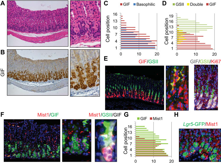
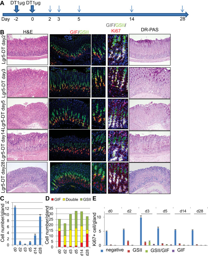
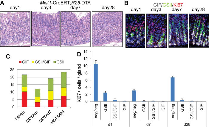
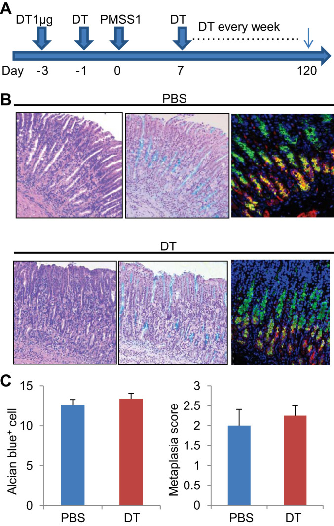
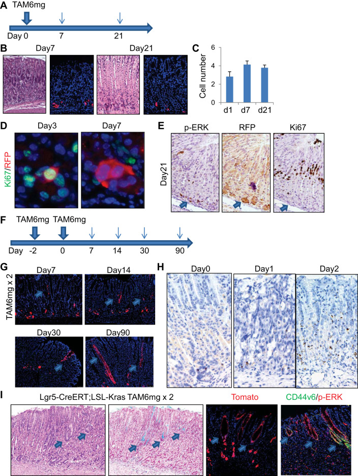
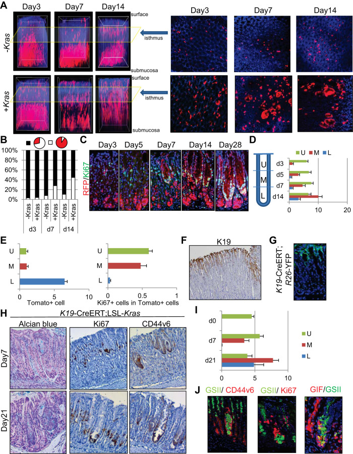
During human gastric carcinogenesis, intestinal metaplasia is frequently seen in the atrophic stomach. In mice, a distinct type of metaplasia known as spasmolytic polypeptide-expressing metaplasia (SPEM) is found in several inflammatory and genetically engineered models. Given the diversity of long- and short-term models of mouse SPEM, it remains unclear whether all models have a shared or distinct molecular mechanism. The origin of SPEM in mice is presently under debate. It is postulated that stem or progenitor cells acquire genetic alterations that then supply metaplastic cell clones, whereas the possibility of transdifferentiation or dedifferentiation from mature gastric chief cells has also been suggested. In this study, we report that loss of chief cells was sufficient to induce short-term regenerative SPEM-like lesions that originated from chief cell precursors in the gastric neck region. Furthermore, Lgr5+ mature chief cells failed to contribute to both short- and long-term metaplasia, whereas isthmus stem and progenitor cells efficiently contributed to long-term metaplasia. Interestingly, multiple administrations of high-dose pulsed tamoxifen induced expansion of Lgr5 expression and Lgr5-CreERT recombination within the isthmus progenitors apart from basal chief cells. Thus we conclude that short-term SPEM represents a regenerative process arising from neck progenitors following chief cell loss, whereas true long-term SPEM originates from isthmus progenitors. Mature gastric chief cells may be dispensable for SPEM development. NEW & NOTEWORTHY Recently, dedifferentiation ability in gastric chief cells during metaplasia development has been proposed. Our findings reveal that lesions that were thought to be acute metaplasia in fact represent normal regeneration supplied from neck lineage and that isthmus stem/progenitors are more responsible for sustained metaplastic changes. Cellular plasticity in gastric chief cells may be more limited than recently highlighted.

Keywords: Lgr5; gastric chief cell; metaplasia; stem cell.


Conflict of interest statement

No conflicts of interest, financial or otherwise, are declared by the authors.

Figures

Fig. 1.
Fig. 1.
Defining mature chief cells in the mouse stomach. AC: hematoxylin and eosin (H and E) (A) and gastric intrinsic factor (GIF) (B) staining of wild-type mouse gastric corpus. Sequential sections were stained with the same region. Basophilic cell position on H and E-stained and GIF+ cells are quantified in C. D and E: simultaneous staining for GIF (red) and Griffonia simplicifolia lectin II (GSII) (green) of wild-type mouse corpus (E, left and middle) and cell position quantification of GSII+, GIF+GSII+, and GIF+ cells (D). E, right is triple immunofluorescence for GIF (gray), GSII (green), and Ki67 (red). F, left and middle: immunofluorescence for Mist1 (red) and GIF (green) of wild-type mouse corpus. F, right: immunofluorescence of Mist1 (red), GSII (green), and GIF (gray). G: cell position quantification of GIF+ and Mist1+ cells. H: green fluorescent protein (GFP) (green) and Mist1 (red) staining of Lgr5-DTR-EGFP mice.
Fig. 2.
Fig. 2.
Loss of chief cells induces regenerative metaplasia. A: protocol of diphtheria toxin (DT) administration. B: hematoxylin and eosin (H and E), gastric intrinsic factor (GIF) (red), and Griffonia simplicifolia lectin II (GSII) (green), GIF (gray)/GSII (green)/Ki67 (red), and diastase-resistant Periodic acid Schiff (DR-PAS) staining of Lgr5-DTR mouse corpus after DT treatment at the indicated time points. C: average numbers of basophilic chief cells on H and E staining per gland at the indicated time points after Lgr5 ablation. D: numbers of GSII+, GIF+GSII+, and GIF+ cells per gland at the indicated time points after Lgr5 ablation. E: average numbers of Ki67+ cells in GSII+, GIF+GSII+, GIF+, and double-negative cells per gland at the indicated time points after Lgr5 ablation.
Fig. 3.
Fig. 3.
Regenerative metaplasia in Mist1-CreERT; R26-DTA mice. A and B: hematoxylin and eosin (H and E) (A) and gastric intrinsic factor (GIF) (gray)/Griffonia simplicifolia lectin II (GSII) (green)/Ki67 (red; B) staining of Mist1-CreERT; R26-DTA mouse corpus at the indicated time points after the administration of 2 mg tamoxifen. C: numbers of GSII+, GIF+GSII+, and GIF+ cells per gland in Mist1-CreERT; R26-DTA mice at the indicated time points after the administration of tamoxifen. D: average numbers of Ki67+ cells in GSII+, GIF+GSII+, GIF+, and double-negative cells per gland in Mist1-CreERT; R26-DTA mice.
Fig. 4.
Fig. 4.
Lgr5+ chief cells are dispensable for the development of metaplasia. A: high-dose tamoxifen (HDT) administration protocol. B and C: hematoxylin and eosin (H and E) (B) and gastric intrinsic factor (GIF) (gray)/Griffonia simplicifolia lectin II (GSII) (green)/Ki67 (red; C) staining of mouse corpus at the indicated time points after HDT administration. Arrows indicate Ki67+ cells at the base. D: TdTomato expression (left) and numbers of TdTomato+ cells (right) in Lgr5-EGFP-IRES-CreERT; R26-TdTomato mouse corpus. E and F: GSII (E, green) and Ki67 (F, green) staining of Lgr5-EGFP-IRES-CreERT; R26-TdTomato mouse corpus after HDT treatment. G: HDT + diphtheria toxin (DT) administration protocol. H: H and E (left), GIF (red)/GSII (green; middle, left), GIF (gray)/GSII (green)/Ki67 (red; middle, right), and diastase-resistant Periodic acid Schiff (DR-PAS) (right) staining of HDT + DT-treated Lgr5-DTR mouse corpus at the indicated time points. I: average numbers of GSII+, GIF+GSII+, and GIF+ cells per gland at the indicated time points after HDT treatment with or without Lgr5 ablation. J: average numbers of Ki67+ cells in GSII+, GIF+GSII+, GIF+, and double-negative cells per gland at the indicated time points after HDT treatment with or without Lgr5 ablation.
Fig. 5.
Fig. 5.
Lgr5+ chief cells are dispensable for Helicobacter-induced metaplasia. A: protocol for PMSS1 infection and diphtheria toxin (DT) ablation. B: Helicobacter pylori infection-induced metaplastic glands shown by hematoxylin and eosin (H and E), Alcian blue, and gastric intrinsic factor (GIF) (red)/Griffonia simplicifolia lectin II (GSII) (green) staining with (bottom) or without (top) Lgr5 ablation. C: average numbers of Alcian blue-stained metaplastic cells per gland and metaplastic score in Helicobacter pylori-infected Lgr5-DTR mouse corpus with or without Lgr5-DT ablation. Mice were analyzed 4 mo after Helicobacter pylori infection.
Fig. 6.
Fig. 6.
Chief cells are not responsible for Kras-induced metaplasia. A: protocol. B: hematoxylin and eosin (H and E) staining and TdTomato expression in high-dose tamoxifen (HDT)-treated Lgr5-EGFP-IRES-CreERT; R26-TdTomato mouse corpus. C: average numbers of TdTomato+ cells per gland at the indicated time points. D: Ki67 (green)/red fluorescent protein (RFP) (red) staining of HDT-treated Lgr5-EGFP-IRES-CreERT; R26-TdTomato mouse corpus. E: phosphorylated ERK (p-ERK, left), RFP (middle), and Ki67 staining on Lgr5-EGFP-IRES-CreERT; R26-TdTomato mouse corpus 28 days after HDT treatment. Sequential sections were used for staining, with the same region shown each time. F: protocol for tamoxifen pulse treatment. G: Lgr5-EGFP-IRES-CreERT; R26-TdTomato lineage tracing after tamoxifen pulse treatment. Arrows indicate isthmus-derived clones. H: in situ hybridization for Lgr5 using RNA scope with or without HDT treatment. I: H and E, Alcian blue, TdTomato, and CD44v6 (green)/p-ERK (red) staining on Lgr5-EGFP-IRES-CreERT; LSL-KrasG12D; R26-TdTomato mouse corpus 28 days after the tamoxifen pulse protocol. Arrows indicate isthmus-derived metaplastic clusters.
Fig. 7.
Fig. 7.
Isthmus progenitors are responsible for Kras-induced metaplasia. A: 3D reconstituted (left) and cross-sectional isthmal (right) images of Mist1-CreERT; R26-TdTomato (top) and Mist1-CreERT; LSL-KrasG12D; R26-TdTomato mice at the indicated time points after treatment with 3 mg tamoxifen. B: proportion of glands in which TdTomato+ cells are >50% (white) or <50% (black) at the isthmus level in cross-sectional analysis. C: red fluorescent protein (RFP) (red)/Ki67 (green) staining on Mist1-CreERT; LSL-KrasG12D; R26-TdTomato mouse corpus after treatment with 3 mg tamoxifen. D: average numbers of Ki67+ cells per gland in the upper (U), middle (M), and lower (L) portion of Mist1-CreERT; LSL-KrasG12D; R26-TdTomato mouse corpus glands at the indicated time points. E, left: average numbers of TdTomato+ cells per gland at each portion of Mist1-CreERT; LSL-KrasG12D; R26-TdTomato corpus glands 3 days after treatment with 3 mg tamoxifen. E, right: average numbers of Ki67+ cells in TdTomato+ cells at each portion of Mist1-CreERT; LSL-KrasG12D; R26-TdTomato corpus glands 7 days after tamoxifen treatment. F: K19 immunostaining of mouse corpus. G: green fluorescent protein (GFP) staining (green) on K19-CreERT; R26-YFP mouse corpus 1 day after the administration of 3 mg tamoxifen. H: Alcian blue, Ki67, and CD44v6 staining of K19-CreERT; R26-YFP mouse corpus 7 and 21 days after treatment with 3 mg tamoxifen. I: average numbers of Ki67+ cells per gland in each portion of K19-CreERT; LSL-KrasG12D mouse corpus glands. J, left: GSII (green)/CD44v6 (red). J, middle: GSII (green)/Ki67 (red). J, right: gastric intrinsic factor (GIF) (red)/GSII (green) staining of K19-CreERT; LSL-KrasG12D mouse corpus 21 days after tamoxifen treatment.
Fig. 8.
Fig. 8.
Distinct marker expression in acute and chronic metaplasia. A: Griffonia simplicifolia lectin II (GSII) (green)/CD44v6 (red; top) and Alcian blue (bottom) staining of Lgr5-DTR mice 14 days after diphtheria toxin (DT) treatment, wild-type mice 7 days after high-dose tamoxifen (HDT) treatment, Mist1-CreERT; LSL-KrasG12D mice 14 days after tamoxifen treatment, and wild-type mice 4 mo after PMSS1 Helicobacter pylori strain infection. B: pepsinogen C (PGC)/GSII staining in normal human corpus. C: in situ hybridization for Pgc and dapB (negative control) in mouse corpus. D: Ki67 (green)/PGC (red) staining in mouse corpus. E: schematic model of metaplasia development in the mouse corpus gland. SPEM, spasmolytic polypeptide-expressing metaplasia.

Similar articles

Cited by

References

    1. Arnold IC, Lee JY, Amieva MR, Roers A, Flavell RA, Sparwasser T, Müller A. Tolerance rather than immunity protects from Helicobacter pylori-induced gastric preneoplasia. Gastroenterology 140: 199–209, 2011. doi:10.1053/j.gastro.2010.06.047. - DOI - PMC - PubMed
    1. Barker N, Huch M, Kujala P, van de Wetering M, Snippert HJ, van Es JH, Sato T, Stange DE, Begthel H, van den Born M, Danenberg E, van den Brink S, Korving J, Abo A, Peters PJ, Wright N, Poulsom R, Clevers H. Lgr5(+ve) stem cells drive self-renewal in the stomach and build long-lived gastric units in vitro. Cell Stem Cell 6: 25–36, 2010. doi:10.1016/j.stem.2009.11.013. - DOI - PubMed
    1. Barker N, Ridgway RA, van Es JH, van de Wetering M, Begthel H, van den Born M, Danenberg E, Clarke AR, Sansom OJ, Clevers H. Crypt stem cells as the cells-of-origin of intestinal cancer. Nature 457: 608–611, 2009. doi:10.1038/nature07602. - DOI - PubMed
    1. Bredemeyer AJ, Geahlen JH, Weis VG, Huh WJ, Zinselmeyer BH, Srivatsan S, Miller MJ, Shaw AS, Mills JC. The gastric epithelial progenitor cell niche and differentiation of the zymogenic (chief) cell lineage. Dev Biol 325: 211–224, 2009. doi:10.1016/j.ydbio.2008.10.025. - DOI - PMC - PubMed
    1. Burclaff J, Osaki LH, Liu D, Goldenring JR, Mills JC. Targeted apoptosis of parietal cells is insufficient to induce metaplasia in stomach. Gastroenterology 152: 762-766, 2016. doi:10.1053/j.gastro.2016.12.001. - DOI - PMC - PubMed

Publication types

Substances

LinkOut - more resources