. 2018 Apr 2;128(4):1326-1337.
doi: 10.1172/JCI97325. Epub 2018 Feb 26.

The deubiquitinase USP9X regulates FBW7 stability and suppresses colorectal cancer

Affiliations

The deubiquitinase USP9X regulates FBW7 stability and suppresses colorectal cancer

Omar M Khan et al. J Clin Invest. .

Abstract

The tumor suppressor FBW7 targets oncoproteins such as c-MYC for ubiquitylation and is mutated in several human cancers. We noted that in a substantial percentage of colon cancers, FBW7 protein is undetectable despite the presence of FBW7 mRNA. To understand the molecular mechanism of FBW7 regulation in these cancers, we employed proteomics and identified the deubiquitinase (DUB) USP9X as an FBW7 interactor. USP9X antagonized FBW7 ubiquitylation, and Usp9x deletion caused Fbw7 destabilization. Mice lacking Usp9x in the gut showed reduced secretory cell differentiation and increased progenitor proliferation, phenocopying Fbw7 loss. In addition, Usp9x inactivation impaired intestinal regeneration and increased tumor burden in colitis-associated intestinal cancer. c-Myc heterozygosity abrogated increased progenitor proliferation and tumor burden in Usp9x-deficient mice, suggesting that Usp9x suppresses tumor formation by regulating Fbw7 protein stability and thereby reducing c-Myc. Thus, we identify a tumor suppressor mechanism in the mammalian intestine that arises from the posttranslational regulation of FBW7 by USP9X independent of somatic FBW7 mutations.

Keywords: Cell Biology; Colorectal cancer; Gastroenterology; Ubiquitin-proteosome system.


Conflict of interest statement

Conflict of interest: The authors have declared that no conflict of interest exists.

Figures

Figure 1
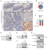
Figure 1. FBW7 is downregulated in human CRC.
(A) Representative images of RNAscope for PPIB and FBW7, and FBW7 IHC on serial sections from human TMAs. u.d., undetected. Scale bars: 10 μm. (B) Quantification of FBW7 mRNA on CRC TMAs in 28 tissue cores positive for a control PPIB mRNA. (C) Quantification of FBW7 IHC in tissue cores from A. (DF) Endogenous FBW7 interacts with endogenous USP9X and USP9X interacts with endogenous FBW7 in HEK293 cells. Black line in D indicates noncontiguous lanes from the same gel. siFBW7 control in F confirms antibody specificity of FBW7 and represents a negative control for FBW7-USP9X interaction. (G) Endogenous USP9X interacts with epitope-tagged FBW7 isoforms α and β.
Figure 2
Figure 2. FBW7 is a USP9X substrate.
(A and B) USP9X knockdown using multiple shRNAs reduced FBW7 protein levels, with no effect on its mRNA. shScr, scrambled control; shNT, nontargeting control. (C) MG132 rescues reduced FBW7 protein levels in USP9X-silenced cells. (D) USP9X knockdown reduced the half-life of FBW7 protein. CHX, cycloheximide. (E) Quantification of 3 independent experiments performed as in D. (F) Increased ubiquitylation (Ubi) of FBW7 in USP9X-silenced cells. UM, unmodified. (G) In vitro deubiquitylation of FBW7 by recombinant GST-USP9X. (H and I) Western blots for Flag-FBW7α in HEK293 cells co-overexpressing WT (V5-Usp9x) or catalytically dead (V5-C1566S) mouse Usp9x. (J) Western blots on Ni-NTA pulldown samples from cells cotransfected with Flag-FBW7α and the indicated constructs. All experiments were done in HEK293 cells.
Figure 3
Figure 3. USP9X negatively regulates SCF(FBW7) substrates.
(A) Accumulation of SCF(FBW7) substrates in USP9X-silenced HEK293 cells. (B) Accumulation of SCF(Fbw7) substrates in Usp9x-knockout murine adult fibroblasts. (C) Western blots showing levels of SCF(FBW7) substrates in USP9X- and ITCH-silenced HEK293 cells. (D) Accumulation of SCF(FBW7) substrates with USP9X silencing was abolished in the HCT116-FBW7Δ/Δ CRC cell line. (E and F) Western blots for ubiquitylated c-Myc on Ni-NTA pulldown from HEK293 cells overexpressing Flag–c-Myc and 6x-His–tagged ubiquitin and cotransfected with the indicated shRNAs. c-MYC mRNA levels in the same cells are shown in F. (G) c-MYC luciferase activity in HCT116 cells with the indicated genotypes and siRNA treatments. Mean of 2 independent experiments is shown.
Figure 4
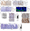
Figure 4. Usp9x controls intestinal tissue homeostasis.
(AD) Representative IHC sections for Usp9x (A), transit-amplifying cells (BrdU, B), goblet cells (AB/PAS, C), and Paneth cells (lysozyme, D) in WT (Usp9x+/+) and Usp9x-knockout mouse gut. Scale bars: 50 μm. Right panels, quantification from BD; n = 5–8 mice/group. Dashed lines outline crypts. (E) Western blots for the indicated proteins in freshly isolated crypts from WT and Usp9x-knockout mouse gut. Black line indicates noncontiguous lanes from the same gel. (F) BrdU staining on gut cross sections from Usp9xΔG mice. Scale bars: 50 μm. Right: quantification from F; n = 3–4 mice/group. (G) IHC for NICD1 in mouse gut. Scale bars: 50 μm. Right panels are ×2 magnification of the dashed areas on the left. (H) AB/PAS staining on guts from the indicated mice treated with a vehicle or a Notch inhibitor (DBZ). Scale bars: 50 μm. Right: quantification from H; n = 3–4 mice/group. Data are shown as mean + SD, and statistical significance was calculated by Student’s t test.
Figure 5
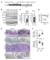
Figure 5. Usp9x is required for tissue regeneration during acute colitis.
(A) Schematic of acute colitis protocol. (B) Western blots for the indicated proteins in 6 independent WT mice fed with (day 7, “peak” and day 21, “recovery”) or without (day 0, “normal”) 2.5% DSS in water. (C) qRT-PCR analysis for Usp9x and Fbw7 mRNA in different phases of colitis from the experiment in B. Statistical significance calculated by 1-way ANOVA. (D) Weight curves from DSS-induced colitis experiment in the indicated mice; n = 7–8/group. *P < 0.05. (E) Representative IHC sections for H&E, BrdU, AB/PAS, and NICD1 from Usp9x+/+ and Usp9xΔG mice. Scale bars: 100 μm. (F) Quantification of BrdU+ and AB/PAS+ cells from the experiment in D. Data are presented as mean; statistical significance was calculated by Student’s t test in D and F.
Figure 6
Figure 6. Usp9x suppresses CRC.
(A) Schematic of AOM/DSS-induced CRC model. (B) H&E on sections from transformed colons of the indicated genotypes. Arrowheads indicate tumors. Scale bars: 100 mm. Right panels: ×4 magnification of the dashed areas on the left; dashed lines outline tumors. (C) Number of tumors in each mouse from experiment in B; n = 5–13/group. (D) Tumor area represented as mm2 in mice from B; n = 5–13/group. (E) Western blots showing levels of indicated proteins in 4 independent tumors from B. (F) H&E and c-Myc staining on sections from transformed colons of the indicated genotypes. Scale bars: 50 mm (H&E); 100 μm (c-Myc). Arrowheads indicate tumors. (G) Average number of tumors in indicated mice from experiment in F; n = 3–5/group. Data are presented as mean; statistical significance calculated by 1-way ANOVA in C and D, and by Student’s t test in G.
Figure 7
Figure 7. Reduced USP9X is associated with poor prognosis in human CRC.
(A) IHC for the indicated proteins on human CRC TMAs, including associated normal tissue. Scale bars: 50 μm. Insets show x2 magnifications of areas in the main image. (B) Quantification of staining intensities from sections in A, and their Spearman’s rank correlation. (C) Western blots showing positive correlation of USP9X and FBW7 protein levels in 8 different CRC cell lines. Red: USP9X mutant, pink: FBW7 truncation mutant (homozygous) cell lines. Black line indicates noncontiguous lanes from the same gel. (D) Kaplan-Meier plot showing comparison of survival between USP9X-low and -high expression groups of CRC patients. Data are from TCGA. (E) Mutational status of USP9X and FBW7 in CRC patients. Data are from the TCGA dataset of 212 patients and the DFCI Colorectal Adenocarcinoma dataset of 619 patients (published in refs. , [Nature 2012 and Cell Reports 2016, respectively]). Data viewed with cBioPortal. Only samples with alterations in either USP9X or FBW7 are shown. Fisher’s exact test P = 0.01 for females and P = 1 for males. WCL, whole cell lysate.

References

    1. Scoville DH, Sato T, He XC, Li L. Current view: intestinal stem cells and signaling. Gastroenterology. 2008;134(3):849–864. doi: 10.1053/j.gastro.2008.01.079. - DOI - PubMed
    1. de Lau W, Barker N, Clevers H. WNT signaling in the normal intestine and colorectal cancer. Front Biosci. 2007;12:471–491. doi: 10.2741/2076. - DOI - PubMed
    1. Clevers H. Wnt/beta-catenin signaling in development and disease. Cell. 2006;127(3):469–480. doi: 10.1016/j.cell.2006.10.018. - DOI - PubMed
    1. Fre S, Huyghe M, Mourikis P, Robine S, Louvard D, Artavanis-Tsakonas S. Notch signals control the fate of immature progenitor cells in the intestine. Nature. 2005;435(7044):964–968. doi: 10.1038/nature03589. - DOI - PubMed
    1. Kinzler KW, Vogelstein B. Lessons from hereditary colorectal cancer. Cell. 1996;87(2):159–170. doi: 10.1016/S0092-8674(00)81333-1. - DOI - PubMed

Publication types

MeSH terms