. 2018 Jan 18;10(1):21.
doi: 10.3390/cancers10010021.

IRF4 Mediates the Oncogenic Effects of STAT3 in Anaplastic Large Cell Lymphomas

Affiliations

IRF4 Mediates the Oncogenic Effects of STAT3 in Anaplastic Large Cell Lymphomas

Cecilia Bandini et al. Cancers (Basel). .

Abstract

Systemic anaplastic large cell lymphomas (ALCL) are a category of T-cell non-Hodgkin's lymphomas which can be divided into anaplastic lymphoma kinase (ALK) positive and ALK negative subgroups, based on ALK gene rearrangements. Among several pathways aberrantly activated in ALCL, the constitutive activation of signal transducer and activator of transcription 3 (STAT3) is shared by all ALK positive ALCL and has been detected in a subgroup of ALK negative ALCL. To discover essential mediators of STAT3 oncogenic activity that may represent feasible targets for ALCL therapies, we combined gene expression profiling analysis and RNA interference functional approaches. A shRNA screening of STAT3-modulated genes identified interferon regulatory factor 4 (IRF4) as a key driver of ALCL cell survival. Accordingly, ectopic IRF4 expression partially rescued STAT3 knock-down effects. Treatment with immunomodulatory drugs (IMiDs) induced IRF4 down regulation and resulted in cell death, a phenotype rescued by IRF4 overexpression. However, the majority of ALCL cell lines were poorly responsive to IMiDs treatment. Combination with JQ1, a bromodomain and extra-terminal (BET) family antagonist known to inhibit MYC and IRF4, increased sensitivity to IMiDs. Overall, these results show that IRF4 is involved in STAT3-oncogenic signaling and its inhibition provides alternative avenues for the design of novel/combination therapies of ALCL.

Keywords: ALK; IRF4; JQ1; STAT3; anaplastic large cell lymphomas; immunomodulatory drugs.


Conflict of interest statement

The authors have no conflicts of interest.

Figures

Figure 1
Figure 1
Kinetics of signal transducer and activator of transcription 3 (STAT3)-regulated genes in anaplastic large cell lymphomas (ALCL). (A) RT-qPCR analysis shows progressive decrease of STAT3 mRNA levels in the anaplastic lymphoma kinase (ALK) positive cell line TS-SUP-M2 S3S after doxycycline treatment (1 µg/mL). Pellet were collected at 72, 96, 120, 144 h. Error bars represent the standard deviation (s.d.) of triplicate measurements. (B) Heatmap representation of gene expression profile analysis after STAT3 inducible knockdown in the ALK positive cell line TS-SUP-M2 S3S. Biological triplicate were used for each experimental condition. Hybridization was carried out on HumanHT-12 v4.0 Expression BeadChip (Illumina Inc., San Diego, CA, USA). STAT3 modulated genes were grouped in 12 clusters. Upregulated RNAs are shown in red, downregulated RNA are shown in green. The colour bar represents relative gene expression changes. In brackets are shown genes selected for functional validation.
Figure 2
Figure 2
Interferon regulatory factor 4 (IRF4) is required for proliferation and survival of ALCL cells. (A) TS-SUP-M2 S3S cells were transduced with lentiviral particles expressing three shRNA (45A, 45B, 45E) targeting IRF4. IRF4 silencing was monitored by RT-qPCR 96 h post transduction (upper panel) and by immunoblotting at the indicated time points (bottom panel). (B) Viability of TS-SUP-M2 S3S cells transduced with the indicated shRNAs was monitored by tetrametylrodamine methyl ester (TMRM) staining-flow cytometry at different time points. (C) TS-SUP-M2 S3S cells were transduced with human IRF4 open reading frame (ORF) and empty vector as a control, selected by blasticidin (5 µg/mL), infected with a shRNA (45A) targeting IRF4 5′UTR or a control shRNA (85E), then selected with puromycin (1 mg/mL). Endogenous and ectopic IRF4 levels were detected by RT-qPCR 96 h post-infection. (D) Viability of TS-SUP-M2 S3S cells transduced with the indicated constructs was monitored by TMRM staining-flow cytometry at different time points. Error bars represent the s.d. of triplicate measurements (*** p < 0.001).
Figure 3
Figure 3
STAT3 binds to IRF4 regulatory regions in ALK-positive ALCL cells. STAT3 (blue), H3K4me (green), H3K27Ac (purple) and H3K27me3 (orange) bindings to IRF4 in TS-SUP-M2 cells. Loss of STAT3 binding (light blue) was achieved after crizotinib treatment (6 h, 200 nM). y-axis values represent read densities normalized to total number of reads.
Figure 4
Figure 4
IRF4 partially mediates STAT3 oncogenic properties in ALCL cells. (A) TS-SUP-M2 S3S cells were transduced with lentiviral particles expressing human IRF4 open reading frame (ORF), an empty vector (EV), or left untransduced (UTR) as negative controls. Cells were cultured in the presence of 1 µg/mL doxycycline to induce STAT3 KD. Kinetics of cell death induced by conditional STAT3 KD revealed that cells expressing IRF4 displayed lower apoptotic rates compared to controls. Apoptosis analysis was performed by TMRM staining-flow cytometry at the indicated time points after doxycycline treatment. Error bars represent the s.d. of triplicate measurements (*** p < 0.001). (B) Western blot analysis of the experiment described above revealing that IRF4 over-expressing cells display lower levels of processed PARP, and invariant levels of cyclin A, B1 and D3 following STAT3 KD as compared to control cells. (C) Propidium iodide staining analysis of the experiment described above indicating that IRF4 over-expressing cells display reduced sub-G0/G1 fraction, indicative of decreased apoptosis. These findings are representative of three independent experiments.
Figure 5
Figure 5
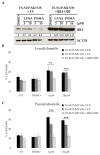
Immunomodulatory drugs downregulate IRF4 expression and increase cell death in TS-SUP-M2 cells. (A) TS-SUP-M2 S3S cells were transduced with human IRF4 ORF or with an empty vector (EV) and treated with the indicated concentrations of lenalidomide or pomalidomide. Western blot analysis revealed IRF4 downregulation after pomalidomide treatment both at 3 µM and 10 µM. Pellet for western blot were collected 4 days after treatment. Quantitative densiometric analysis were performed with ImageJ software. (B,C) Viability of TS-SUP-M2 S3S cells transduced with IRF4 ORF, empty vector (EV) or untransduced (UTR) as negative controls. Cells were treated with the indicated concentrations of lenalidomide, pomalidomide, diluent (DMSO), or left untreated (NT). Analysis of cell death revealed that ectopic expression of IRF4 completely rescued apoptosis induced by lenalidomide and pomalidomide. Apoptosis analysis was performed by TMRM staining-flow cytometry 6 days post treatments. Error bars represent the s.d. of triplicate measurements (** p < 0.01; *** p < 0.001).
Figure 6
Figure 6
The bromodomain and extra-terminal (BET)-inhibitor JQ1 sensitizes ALCL cells to Pomalidomide treatment. Apoptosis and western blot analysis of KARPAS 299 (A), L82 (B), SU-DHL-1 (C) and FePd (D) cells treated with DMSO, pomalidomide, the BET inhibitor JQ1, or the combination of the two drugs. Cell death analysis revealed increased sensitivity to pomalidomide in combination with JQ1 (left panels) which correlates with a stronger down-regulation of IRF4 and c-MYC protein levels (right panels). Error bars represent the s.d. of triplicate measurements (** p < 0.01; *** p < 0.001). Pellet for western blot were collected 48 h after treatments.

Similar articles

Cited by

References

    1. Stein H., Mason D.Y., Gerdes J., O’Connor N., Wainscoat J., Pallesen G., Gatter K., Falini B., Delsol G., Lemke H., et al. The expression of the hodgkin’s disease associated antigen Ki-1 in reactive and neoplastic lymphoid tissue: Evidence that reed-sternberg cells and histiocytic malignancies are derived from activated lymphoid cells. Blood. 1985;66:848–858. - PubMed
    1. Turner S.D., Lamant L., Kenner L., Brugieres L. Anaplastic large cell lymphoma in paediatric and young adult patients. Br. J. Haematol. 2016;173:560–572. doi: 10.1111/bjh.13958. - DOI - PubMed
    1. Swerdlow S.H., Campo E., Pileri S.A., Harris N.L., Stein H., Siebert R., Advani R., Ghielmini M., Salles G.A., Zelenetz A.D., et al. The 2016 revision of the world health organization classification of lymphoid neoplasms. Blood. 2016;127:2375–2390. doi: 10.1182/blood-2016-01-643569. - DOI - PMC - PubMed
    1. Iwahara T., Fujimoto J., Wen D., Cupples R., Bucay N., Arakawa T., Mori S., Ratzkin B., Yamamoto T. Molecular characterization of ALK, a receptor tyrosine kinase expressed specifically in the nervous system. Oncogene. 1997;14:439–449. doi: 10.1038/sj.onc.1200849. - DOI - PubMed
    1. Morris S.W., Naeve C., Mathew P., James P.L., Kirstein M.N., Cui X., Witte D.P. ALK, the chromosome 2 gene locus altered by the t(2;5) in non-Hodgkin’s lymphoma, encodes a novel neural receptor tyrosine kinase that is highly related to leukocyte tyrosine kinase (LTK) Oncogene. 1997;14:2175–2188. doi: 10.1038/sj.onc.1201062. - DOI - PubMed