. 2018 Jan 18;14(1):e1007106.
doi: 10.1371/journal.pgen.1007106. eCollection 2018 Jan.

Mutations in Caenorhabditis elegans neuroligin-like glit-1, the apoptosis pathway and the calcium chaperone crt-1 increase dopaminergic neurodegeneration after 6-OHDA treatment

Affiliations

Mutations in Caenorhabditis elegans neuroligin-like glit-1, the apoptosis pathway and the calcium chaperone crt-1 increase dopaminergic neurodegeneration after 6-OHDA treatment

Sarah-Lena Offenburger et al. PLoS Genet. .

Abstract

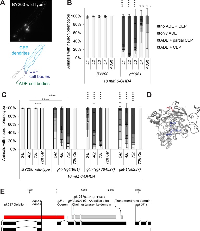
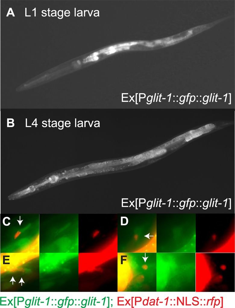
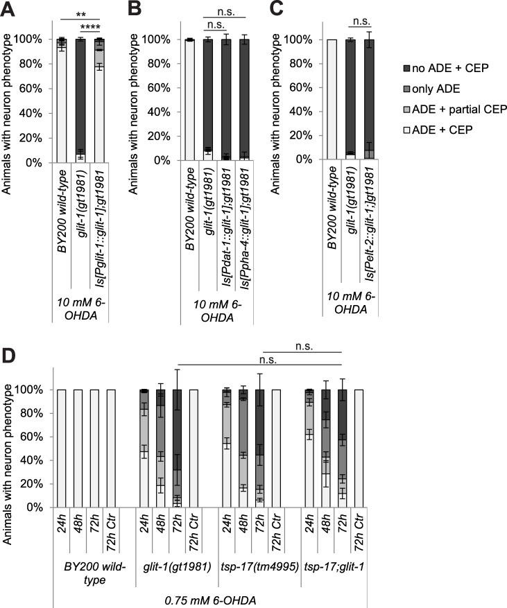
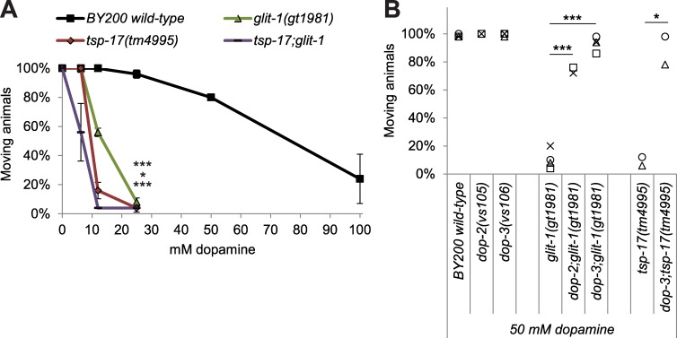
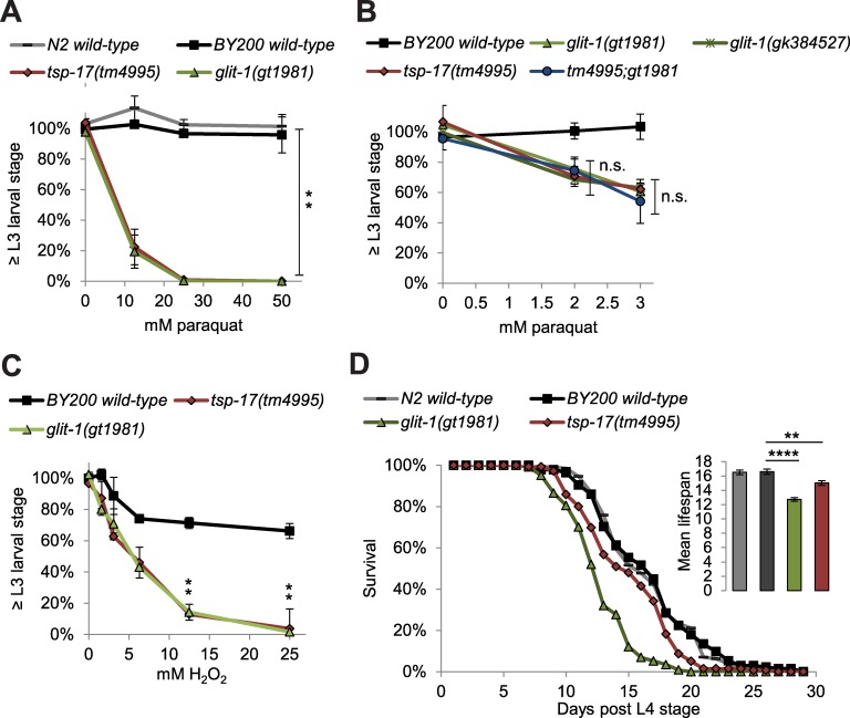
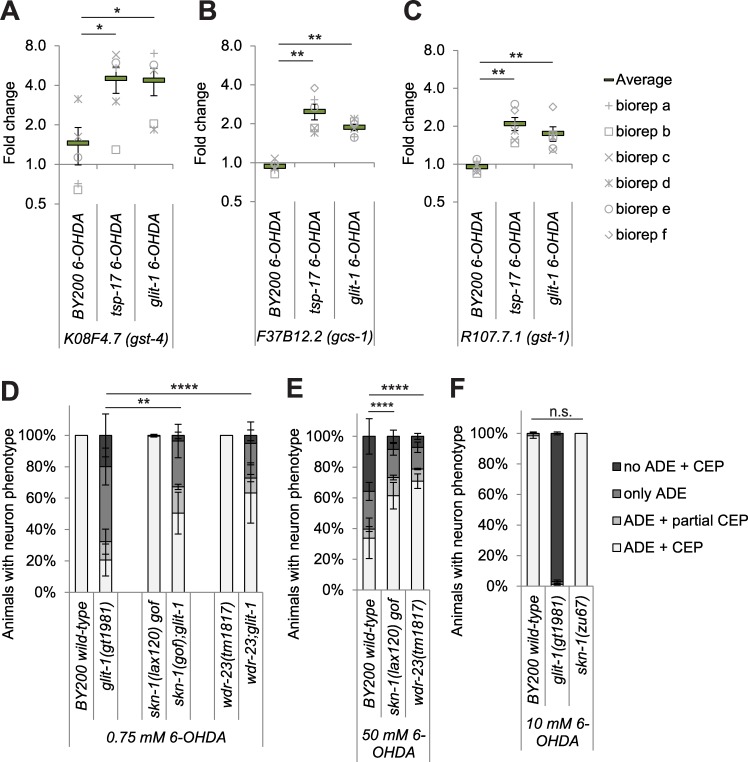
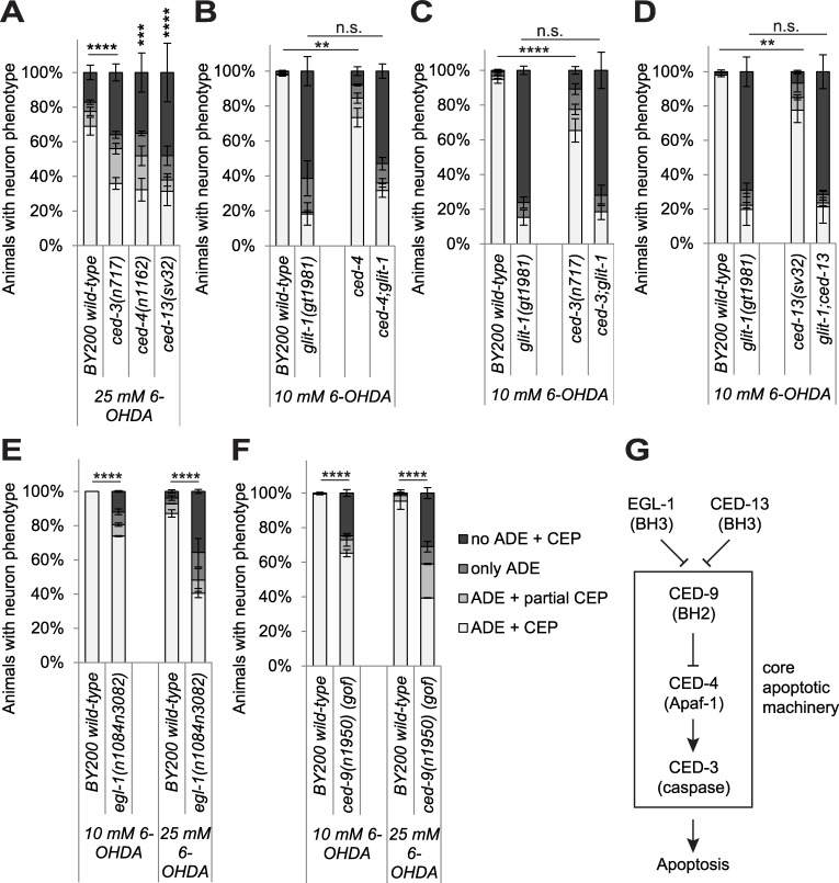
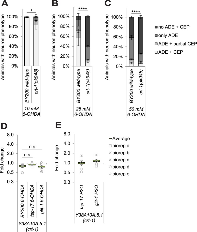
The loss of dopaminergic neurons is a hallmark of Parkinson's disease, the aetiology of which is associated with increased levels of oxidative stress. We used C. elegans to screen for genes that protect dopaminergic neurons against oxidative stress and isolated glit-1 (gliotactin (Drosophila neuroligin-like) homologue). Loss of the C. elegans neuroligin-like glit-1 causes increased dopaminergic neurodegeneration after treatment with 6-hydroxydopamine (6-OHDA), an oxidative-stress inducing drug that is specifically taken up into dopaminergic neurons. Furthermore, glit-1 mutants exhibit increased sensitivity to oxidative stress induced by H2O2 and paraquat. We provide evidence that GLIT-1 acts in the same genetic pathway as the previously identified tetraspanin TSP-17. After exposure to 6-OHDA and paraquat, glit-1 and tsp-17 mutants show almost identical, non-additive hypersensitivity phenotypes and exhibit highly increased induction of oxidative stress reporters. TSP-17 and GLIT-1 are both expressed in dopaminergic neurons. In addition, the neuroligin-like GLIT-1 is expressed in pharynx, intestine and several unidentified cells in the head. GLIT-1 is homologous, but not orthologous to neuroligins, transmembrane proteins required for the function of synapses. The Drosophila GLIT-1 homologue Gliotactin in contrast is required for epithelial junction formation. We report that GLIT-1 likely acts in multiple tissues to protect against 6-OHDA, and that the epithelial barrier of C. elegans glit-1 mutants does not appear to be compromised. We further describe that hyperactivation of the SKN-1 oxidative stress response pathway alleviates 6-OHDA-induced neurodegeneration. In addition, we find that mutations in the canonical apoptosis pathway and the calcium chaperone crt-1 cause increased 6-OHDA-induced dopaminergic neuron loss. In summary, we report that the neuroligin-like GLIT-1, the canonical apoptosis pathway and the calreticulin CRT-1 are required to prevent 6-OHDA-induced dopaminergic neurodegeneration.


Conflict of interest statement

The authors have declared that no competing interests exist.

Figures

Fig 1
Fig 1. glit-1(gt1981) exhibits increased 6-OHDA-induced dopaminergic neurodegeneration.
(A) GFP-labelled C. elegans dopaminergic head neurons– 4 CEP neurons and 2 ADE neurons–in BY200 wild-type animals. (B) Remaining dopaminergic head neurons in BY200 wild-type animals and gt1981 mutants 48 hours after treating L1-L4 larval stages or adult animals with 10 mM 6-OHDA. Animals possessing all neurons were scored as ‘ADE + CEP’ (white bar), those with partial loss of CEP but intact ADE neurons as ‘ADE + partial CEP’ (light grey bar), those with complete loss of CEP but intact ADE neurons as ‘only ADE’ (dark grey bar) and those with complete loss of dopaminergic head neurons as ‘no ADE + CEP’ (black bar). Error bars = SEM of 2 biological replicates, each with 25–40 animals per stage and strain. Total number of animals per condition n = 50–80 (****p<0.0001, n.s. p>0.05; G-Test comparing BY200 wild-type and mutant data of the same lifecycle stages). (C) Dopaminergic head neurons 24, 48 and 72 hours after treatment with 10 mM 6-OHDA and 72 hours after treatment with ascorbic acid only (‘72h Ctr’) for BY200 wild-type or glit-1 mutant animals. Error bars = S.E.M. of 2 biological replicates for glit-1(ok237) and 3 biological replicates for all the other strains, each with 60–115 animals per strain and concentration. Total number of animals for the ‘72h Ctr’ experiment n = 30–100 and for all other conditions n = 130–340 (****p<0.0001; G-Test comparing BY200 wild-type and mutant data of the same time point). (D) GLIT-1 protein structure prediction based on homology modelling with acetylcholinesterase (PDB ID: 2W6C). The gt1981 point mutation leads to a proline to glycine conversion (P113G) and is indicated in red. The amino acids replacing the acetylcholinesterase catalytic triad are indicated in blue. (E) glit-1 gene structure with positions of the glit-1(gt1981) point mutation, the glit-1(gk384527) splice site mutation and the glit-1(ok237) deletion. For the point mutations the nucleotide and amino acid changes are indicated in brackets. The ok237 deletion spans 5’UTR and first exon of glit-1 and 5’UTR and first exons of dnj-14 (DNaJ domain (prokaryotic heat shock protein)) and is indicated with a red bar. Also, glit-1 is located in an operon (grey bar) with the ribosomal protein rpl-25.1 (Ribosomal Protein, Large subunit).
Fig 2
Fig 2. glit-1 is expressed in the pharynx, the intestine and in several cells in the head including dopaminergic neurons.
(A) L1 stage larva expressing Ex[Pglit-1::gfp::glit-1]. (B) L4 stage larva expressing Ex[Pglit-1::gfp::glit-1]. This image was generated from three individual images in ImageJ using the Stitching plugin [60]. (C)–(F) Close up of dopaminergic neurons of the same L4 stage larva. The green channel shows expression of Ex[Pglit-1::gfp::glit-1]. The red channel shows expression of Is[Pdat-1::NLS::rfp;Pttx-3::mCherry] for labelling of dopaminergic neuron nuclei, as well as the pharynx muscle marker Ex[Pmyo-2::mCherry] and the body muscle marker Ex[Pmyo-3::mCherry] that were used for injections.
Fig 3
Fig 3. Single-copy expression of glit-1 alleviates neurodegeneration in glit-1 mutants and glit-1 likely functions in a pathway with the tetraspanin tsp-17.
(A) Effect of a single-copy Is[Pglit-1::glit-1] construct on dopaminergic neurodegeneration after treatment with 10 mM 6-OHDA. Error bars = SEM of 2 biological replicates, each with 100–105 animals per strain. Total number of animals per condition n = 200–205 (****p<0.0001, **p<0.01; G-Test). (B) Effect of single-copy Is[Pdat-1::glit-1] and Is[Ppha-4::glit-1] constructs on dopaminergic neurodegeneration after treatment with 10 mM 6-OHDA. Error bars = SEM of 2 biological replicates, each with 100–105 animals per strain. Total number of animals per condition n = 200–205 (n.s. p>0.05; G-Test). (C) Effect of a single-copy Is[Pelt-2::glit-1] construct on dopaminergic neurodegeneration after treatment with 10 mM 6-OHDA. Error bars = SEM of 2 biological replicates, each with 95–100 animals per strain. Total number of animals per condition n = 195–200 (n.s. p>0.05,; G-Test). (D) Dopaminergic head neurons in wild-type and glit-1 and tsp-17 single and double mutants 24, 48 and 72 hours after treatment with 0.75 mM 6-OHDA and 72 hours after control treatment with ascorbic acid only (‘72 h Ctr’). Error bars = SEM of 2–3 biological replicates, each with 60–115 animals per strain. Total number of animals per condition n = 180–350 (*p<0.05, n.s. p>0.05; G-Test). The ‘72 h Ctr’ was conducted twice with 30 animals per strain, resulting in a total n = 60.
Fig 4
Fig 4. glit-1 and tsp-17 exhibit increased dopamine-induced paralysis.
(A) Percentage of moving young adults on plates with indicated concentrations of dopamine. Error bars = StDev of 2 technical replicates, each with 25 animals per strain and condition. Total number of animals per condition n = 50 (***p<0.001, *p<0.05, n.s. p>0.05; two-tailed t-test comparing wild-type and mutant animal data at 25 mM dopamine). (B) Percentage of moving young adults on plates containing 50 mM dopamine. Indicated with grey symbols are the 2–4 biological replicates, each performed with 50 animals per strain. Total number of animals per condition n = 100–200 (***p<0.001, *p<0.05; two-tailed t-test).
Fig 5
Fig 5. glit-1 and tsp-17 mutants are hypersensitive to oxidative stress at the organismal level.
(A) Percentage of animals that developed to L3 stage 24 hours after 1 hour incubation with indicated concentration of paraquat. Error bars = SEM of 3 biological replicates, each with 85–365 animals per strain and concentration. Total number of animals per condition n = 350–650 (*p<0.05; two-tailed t-test comparing data of BY200 wild-type and mutant animal data at 50 mM paraquat). (B) Percentage of animals that developed to L3 stage 24 hours after 1 hour incubation with 50 mM paraquat. Error bars = SEM of 3 biological replicates, each with 85–380 animals per strain and concentration. Total number of animals per condition n = 340–830 (n.s. p>0.05; two-tailed t-test comparing the tsp-17(tm4995) and glit-1(gt1981) single mutants to the tsp-17(tm4995);glit-1(gt1981) double mutant at 2 and 3 mM paraquat). (C) Percentage of animals that developed to L3 stage or beyond 48 hours after 1 hour incubation with indicated concentration of H2O2. Error bars = SEM of 2 biological replicates, each with 165–547 animals per strain and concentration. Total number of animals per condition n = 311–942 (*p<0.05; two-tailed t-test comparing data of BY200 wild-type and mutant animal data at 12.5 and 25 mM H2O2). (D) Lifespan data for first biological replicate including 97–130 animals per strain. The inset shows the mean lifespan with the error bars depicting the standard error (****p<0.0001, **p<0.01; Bonferroni-corrected; Log-Rank Test).
Fig 6
Fig 6
Expression of oxidative stress reporters is increased in glit-1 and tsp-17 mutants and hyperactivation of skn-1 alleviates 6-OHDA-induced neurodegeneration (A) gst-4 (B) gcs-1 and (C) gst-1 mRNA levels in wild-type and mutant L1 stage larvae after 1 hour treatment with 10 mM 6-OHDA. (A)-(C) The data are normalised to the control gene Y45F10D.4. The average and the respective values for 5–6 biological replicates (biorep a-f) are indicated. Error bars = SEM of 5–6 biological replicates (**p<0.01, *p<0.05; two-tailed t-test). (D) Effect of skn-1 gain-of-function (gof) mutations skn-1(lax120) and wdr-23(tm1817) on dopaminergic neurodegeneration after treatment with 0.75 mM 6-OHDA. Error bars = SEM of 3–4 biological replicates, each with 40–120 animals per strain and concentration. Total number of animals per strain n = 180–430 (****p<0.0001, **p<0.01; G-Test). (E) Effect of skn-1 gain-of-function (gof) mutations skn-1(lax120) and wdr-23(tm1817) on dopaminergic neurodegeneration after treatment with 50 mM 6-OHDA. Error bars = SEM of 3–6 biological replicates, each with 70–115 animals per strain and concentration. Total number of animals per condition = 290–430 (****p<0.0001; G-Test). (F) Effect of skn-1 loss-of-function (lof) allele zu67 on dopaminergic neurodegeneration after treatment with 10 mM 6-OHDA. Error bars = SEM of 3 biological replicates, each with 30–105 animals per strain. Total number of animals per strain n = 170–270 (n.s. p>0.05; G-Test). Experiment was conducted with unstarved, filtered L1 larvae since an insufficient number of skn-1(zu67) mutants could be gained in liquid culture. Unstarved animals show a higher degree of degeneration.
Fig 7
Fig 7. Mutations in the canonical apoptosis pathway sensitise to 6-OHDA-induced dopaminergic neurodegeneration.
(A) Effect of apoptosis/mtROS pathway mutations on dopaminergic neurodegeneration after treatment with 25 mM 6-OHDA. Error bars = SEM of 3 biological replicates, each with 70–120 animals per strain and concentration. Total number of animals per condition n = 280–320 (****p<0.0001, ***p<0.001, n.s. p>0.05; G-Test). (B) Effect of ced-4, (C) ced-3 (D) ced-13, (E) egl-1 loss-of-function mutations, as well as the (F) ced-9 gain-of-function (gof) mutation on dopaminergic neurodegeneration after treatment with 10 mM 6-OHDA. Error bars = SEM of 2–5 biological replicates, each with 40–115 animals per strain and concentration. Total number of animals per condition n = 150–490 (****p<0.0001, **p<0.01, n.s. p>0.05; G-Test). (G) Simplified scheme of the apoptosis pathway (adapted from [61]).
Fig 8
Fig 8. Mutation of the calcium chaperone crt-1 sensitises to 6-OHDA-induced dopaminergic neurodegeneration.
(A) Effect of mutations in crt-1 mutation on dopaminergic neurodegeneration after treatment with 10 mM, (B) 25 mM and (C) 50 mM 6-OHDA. Error bars = SEM of 2–3 biological replicates with 90–130 animals per strain and concentration. Total number of animals per strain = 200–360 (****p<0.0001, *p<0.05; G-Test). (D) crt-1 mRNA level analysis after 1h of 6-OHDA treatment in wild-type and mutant L1 stage larvae. (E) crt-1 mRNA level analysis in mutant L1 stage larvae as compared to wild-type animals under control conditions (treatment with H2O instead of 6-OHDA). (D) and (E) The data are normalised to the control gene Y45F10D.4. The average and the respective values for 5 biological replicates (biorep a-e) are indicated. Error bars = SEM of 5 biological replicates (n.s. p>0.05; two-tailed t-test).

Similar articles

Cited by

References

    1. Schultz W. Multiple dopamine functions at different time courses. Annu Rev Neurosci. 2007;30: 259–88. doi: 10.1146/annurev.neuro.28.061604.135722 - DOI - PubMed
    1. Delamarre A, Meissner WG. Epidemiology, environmental risk factors and genetics of Parkinson’s disease. Presse Med. Elsevier Masson SAS; 2017;46: 175–181. doi: 10.1016/j.lpm.2017.01.001 - DOI - PubMed
    1. Thomas B, Beal MF. Parkinson’s disease. Hum Mol Genet. 2007;16: 183–194. doi: 10.1093/hmg/ddm159 - DOI - PubMed
    1. Klein C, Westenberger A. Genetics of Parkinson’s disease. Cold Spring Harb Perspect Med. 2012;2: a008888 doi: 10.1101/cshperspect.a008888 - DOI - PMC - PubMed
    1. Sies H, Berndt C, Jones DP. Oxidative stress. Annu Rev Biochem. 2017; doi: 10.1016/j.bpobgyn.2010.10.016 - DOI - PubMed

Publication types

MeSH terms