. 2018 Jan 18;14(1):e1007125.
doi: 10.1371/journal.pgen.1007125. eCollection 2018 Jan.

6-OHDA-induced dopaminergic neurodegeneration in Caenorhabditis elegans is promoted by the engulfment pathway and inhibited by the transthyretin-related protein TTR-33

Affiliations

6-OHDA-induced dopaminergic neurodegeneration in Caenorhabditis elegans is promoted by the engulfment pathway and inhibited by the transthyretin-related protein TTR-33

Sarah-Lena Offenburger et al. PLoS Genet. .

Abstract

Oxidative stress is linked to many pathological conditions including the loss of dopaminergic neurons in Parkinson's disease. The vast majority of disease cases appear to be caused by a combination of genetic mutations and environmental factors. We screened for genes protecting Caenorhabditis elegans dopaminergic neurons from oxidative stress induced by the neurotoxin 6-hydroxydopamine (6-OHDA) and identified the transthyretin-related gene ttr-33. The only described C. elegans transthyretin-related protein to date, TTR-52, has been shown to mediate corpse engulfment as well as axon repair. We demonstrate that TTR-52 and TTR-33 have distinct roles. TTR-33 is likely produced in the posterior arcade cells in the head of C. elegans larvae and is predicted to be a secreted protein. TTR-33 protects C. elegans from oxidative stress induced by paraquat or H2O2 at an organismal level. The increased oxidative stress sensitivity of ttr-33 mutants is alleviated by mutations affecting the KGB-1 MAPK kinase pathway, whereas it is enhanced by mutation of the JNK-1 MAPK kinase. Finally, we provide genetic evidence that the C. elegans cell corpse engulfment pathway is required for the degeneration of dopaminergic neurons after exposure to 6-OHDA. In summary, we describe a new neuroprotective mechanism and demonstrate that TTR-33 normally functions to protect dopaminergic neurons from oxidative stress-induced degeneration, potentially by acting as a secreted sensor or scavenger of oxidative stress.


Conflict of interest statement

The authors have declared that no competing interests exist.

Figures

Fig 1
Fig 1. ttr-33 mutations cause increased dopaminergic neurodegeneration after treatment with 6-OHDA.
(A) GFP-labelled C. elegans dopaminergic head neurons– 4 cephalic sensilla (CEP) neurons and 2 anterior deirid (ADE) neurons–in animals carrying the vtIs1 transgene and neurodegeneration after addition of 6-OHDA. (B) Remaining dopaminergic head neurons 24, 48 and 72 hours after treatment with 10 mM 6-OHDA and 72 hours after control treatment with ascorbic acid only (‘72h Ctr’) for BY200 wild-type and gt1983 mutant animals. Animals possessing all neurons were scored as ‘ADE + CEP’ (white bar), those with partial loss of CEP but intact ADE neurons as ‘ADE + partial CEP’ (light grey bar), those with complete loss of CEP but intact ADE neurons as ‘only ADE’ (dark grey bar) and those with complete loss of dopaminergic head neurons as ‘no ADE + CEP’ (black bar). Error bars = S.E.M. of 3 biological replicates for all the other strains, each with 60–120 animals per strain. Total number of animals per condition n = 30 for the ‘72h Ctr’ and n = 220–340 for all the other conditions (****p<0.0001; G-Test comparing BY200 wild-type and mutant data of the same time point). (C) ttr-33 gene structure with positions of ttr-33(gt1983) and ttr-33(gk567379) point mutations. Nucleotide and amino acid changes are indicated in parentheses. (D) Dopaminergic head neurons after treatment with 10 mM 6-OHDA in BY200 wild-type animals and ttr-33(gt1983) homozygous and ttr-33(gt1983)/+ heterozygous mutants. Error bars = SEM (Standard Error of the Mean) of 2 experiments, each with 50–105 animals per strain. Total number of animals per strain n = 120–205 (****p<0.0001, n.s. p>0.05; G-Test). (E) Dopaminergic head neurons after treatment with 10 mM 6-OHDA in BY200 wild-type animals, gt1983 and ttr-33(gk567379) homozygous mutants and gt1983/ttr-33(gk567379) trans heterozygous mutant animals. Error bars = SEM of 2–3 experiments, each with 100–175 animals per strain. Total number of animals per strain n = 210–340 (****p<0.0001; G-Test). (F) Effect of Pttr-33::ttr-33 and Pbath-15::ttr-33 extrachromosomal arrays on dopaminergic neurodegeneration of ttr-33 mutant animals after treatment with 10 mM 6-OHDA. Error bars = SEM of 2 biological replicates, each with 90–130 animals per strain and concentration. Total number of animals per condition n = 200–245 (****p<0.0001, ***p<0.001, **p<0.01, n.s. p>0.05; G-Test). At present, we speculate that the ttr-33 mutant defect is not be fully rescued by our ttr-33 wild-type plasmids because of a 3 amino acid linker sequence in the constructs. This linker sequence was introduced as part of the cloning strategy between the translational start site and the N-terminal secretion signal of TTR-33 and may compromise protein function. It is also possible that the construct do not contain all promoter elements. As extrachromosomal arrays lead to variable expression between tissues, it is possible that not all tested animals expressed TTR-33 in the arcade cells. Finally, overexpression of TTR-33 might interfere with its function.
Fig 2
Fig 2. ttr-33 mutants develop at a normal speed and 6-OHDA must enter dopaminergic neurons to cause dopaminergic neurodegeneration.
(A) TTR-33 protein structure prediction based on homology modelling using the structure of TTR-52 (PDB ID: 3UAF). The overlay of the TTR-33 and the TTR-52 structures is shown in S2B Fig. The ttr-33(gt1983) point mutation leading to a glycine to glutamate (G27E) conversion and the ttr-33(gk567379) mutation leading to a leucine to phenylalanine (L72F) conversion are both indicated in red. (B) Dopaminergic head neurons in wild-type animals and ttr-33 mutants 48 hours after treatment of L1-L4 larval stages or adult animals with 10 mM 6-OHDA. Error bars = SEM of 2 biological replicates, each with 25–45 animals per stage and strain. Total number of animals per condition n = 50–80 (****p<0.0001, **p<0.01, n.s. p>0.05; G-Test comparing BY200 wild-type and mutant animals data of the same lifecycle stages). (C) Developmental stages of wild-type and ttr-33 mutant L1 stage larvae 48 hours after treatment with and without 10 mM 6-OHDA. C. elegans L1 stage larvae develop via the L2 (in red), L3 (in orange), L4 (green) and young adult stage (in light blue) into adults (in dark blue). Error bars = SEM of 3 biological replicates, each with 60–175 animals per treatment and strain. Total number of animals per condition n = 205–350 (n.s. p>0.05; G-Test). (D) Effect of dat-1 mutation on dopaminergic neurodegeneration after treatment with 10 mM 6-OHDA. Error bars = SEM of 2 experiments, each with 90–115 animals per strain. Total number of animals per strain n = 200–535 (****p<0.0001; G-Test).
Fig 3
Fig 3. ttr-33 and ttr-52 have independent functions.
(A) Effect of ttr-52 mutation on dopaminergic neurodegeneration after treatment with 2.5 mM 6-OHDA. Error bars = SEM of 2 biological replicates, each with 95–115 animals per strain. Total number of animals per condition n = 200–225 (n.s. p>0.05; G-Test). (B) Effect of ttr-52 mutation on dopaminergic neurodegeneration after treatment with 25 mM 6-OHDA. Error bars = SEM of 4 biological replicates, each with 100–130 animals per strain. Total number of animals per condition n = 425–460 (n.s. p>0.05; G-Test). (C) Quantification of axonal regrowth, regeneration and fusion of the PLM axons 24 hours after UV-laser axotomy in ttr-33(gt1983) mutants and wild-type animals carrying the zdIs5 transgene to visualise PLM. The percentage of successful reconnection is a proportion of the axons that showed regrowth. The percentage of successful axonal fusion is a proportion of the axons that successfully reconnected. Error bars = standard error of proportion. The number of animals tested is indicated at the bottom of each bar (*p<0.05, n.s. p>0.05; t-test). (D) Length of axonal regrowth in ttr-33(gt1983) mutants and wild-type animals (n.s. p>0.05; t-test).
Fig 4
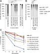
Fig 4. Mutations in the cell corpse engulfment pathway alleviate 6-OHDA-induced dopaminergic neurodegeneration.
(A) Cartoon of the engulfment pathway (adapted from [27]). The recognition of the conserved ‘eat-me’ signal phosphatidylserine (PS) on the surface of apoptotic and necrotic cells triggers two partially redundant pathways in the engulfing cell [73]. The genes tested in this study are highlighted in yellow and the part of the pathway that was shown to be required for axon regeneration [27] is boxed in blue. psr = phosphatidylserine receptor family, ced = cell death abnormality, nrf = nose resistant to fluoxetine (lipid-binding protein). (B) Effect of mutations in engulfment pathway genes on dopaminergic neurodegeneration after treatment with 10 mM 6-OHDA. Error bars = SEM of 2 biological replicates, each with 70–120 animals per strain. Total number of animals per condition n = 175–230 (****p<0.0001, ***p<0.002, **p<0.01, n.s. p>0.05; G-Test comparing ttr-33 mutant data to double mutant data). (C) Effect of mutations in engulfment pathway genes on dopaminergic neurodegeneration after treatment with 50 mM 6-OHDA. Error bars = SEM of 2 biological replicates, each with 85–140 animals per strain. Total number of animals per condition n = 225–235 (****p<0.0001, *p<0.05, n.s. p>0.05; G-Test).
Fig 5
Fig 5. Evidence that ttr-33 is expressed in the posterior arcade cells and in the pharynx.
(A) Head region of L1 stage larvae expressing the extrachromosomal transcriptional reporter Ex[Pttr-33::gfp]. Arrows indicate strongly expressing cells. (B) L1 stage larva expressing the transcriptional reporter Ex[Pttr-33::gfp]. (C) Pretzel stage embryo expression the transcriptional reporter Ex[Pttr-33::gfp]. The speckled signal is caused by intestinal autofluorescence.
Fig 6
Fig 6. ttr-33 mutants are sensitive to oxidative stress.
(A) Percentage of larvae reaching the L3 larval stage 24 hours after an 1 hour incubation with indicated concentration of paraquat. Error bars = SEM of 3 biological replicates for 25 and 50 mM paraquat and 2 biological replicates for 0 mM paraquat, each with 60–245 animals per strain and concentration. Total number of animals per condition n = 275–550 (****p<0.0001, *p<0.05; two-tailed t-test comparing BY200 wild-type and mutant animal data). (B) Percentage of animals developed to L3 stage 48 hours after an 1 hour incubation with indicated concentration of H2O2. Error bars = SEM of 2 biological replicates, each with 165–547 animals per strain and concentration. Total number of animals per condition n = 311–942 (*p<0.05; two-tailed t-test comparing data of BY200 wild-type and mutant animal data at 12.5 and 25 mM H2O2). (C) Lifespan data for first biological replicate including 110–125 animals per strain. The inset shows the mean lifespan with the error bars depicting the standard error. Total number of animals for N2 wild-type (which was tested once) n = 110, and for all other strains n = 200–235 (****Bonferroni p<0.0001; Log-Rank Test).
Fig 7
Fig 7. Interference with the jnk-1 MAPK pathway increases 6-OHDA sensitivity in ttr-33 mutants.
(A) Effect of p38 and JNK stress response pathway mutations on dopaminergic neurodegeneration after treatment with 2.5 mM 6-OHDA. Error bars = SEM of 2–4 biological replicates, each with 100–120 animals per strain and concentration. Total number of animals per condition n = 200–425 (*p<0.05, n.s. p>0.05; G-Test). (B) Effect of p38 and JNK stress response pathway mutations on dopaminergic neurodegeneration after treatment with 25 mM 6-OHDA. Error bars = SEM of 3 biological replicates, each with 100–110 animals per strain. Total number of animals per strain n = 310–330 (n.s. p>0.05; G-Test). (C) Effect of pmk-1 mutation on dopaminergic neurodegeneration after treatment with 25 mM 6-OHDA. Error bars = SEM of 3 biological replicates, each with 100–135 animals per strain. Total number of animals per strain n = 350–360 (n.s. p>0.05; G-Test).
Fig 8
Fig 8. Interference with the kgb-1 MAPK pathway decreases oxidative stress sensitivity in ttr-33 mutants.
(A) Effect of kgb-1 MAP kinase pathway mutations on dopaminergic neurodegeneration after treatment with 10 mM 6-OHDA. Error bars = SEM of 3 biological replicates, each with 90–115 animals per strain. Total number of animals per strain n = 300–340 (****p<0.0001, ***p<0.001; G-Test). (B) Effect of kgb-1 and mek-1 MAP kinase pathway mutation on dopaminergic neurodegeneration after treatment with 25 mM 6-OHDA. Error bars = SEM of 3 biological replicates, each with 50–130 animals per strain. Total number of animals per strain n = 255–330 (n.s. p>0.05; G-Test). (C) Percentage of larvae reaching the L3 larval stage 24 hours after an 1 hour incubation with indicated concentration of paraquat. Error bars = SEM of 3–4 biological replicates for 25 and 50 mM paraquat and 2 biological replicates for 0 mM paraquat, each with 70–270 animals per strain and concentration. Total number of animals per condition n = 220–650 (**p<0.01, *p<0.05; two-tailed t-test comparing ttr-33(gt1983) double mutants to ttr-33(gt1983) single mutant data).

Similar articles

Cited by

References

    1. Sies H, Berndt C, Jones DP. Oxidative stress. Annu Rev Biochem. 2017; doi: 10.1016/j.bpobgyn.2010.10.016 - DOI - PubMed
    1. Delamarre A, Meissner WG. Epidemiology, environmental risk factors and genetics of Parkinson’s disease. Presse Med. Elsevier Masson SAS; 2017;46: 175–181. doi: 10.1016/j.lpm.2017.01.001 - DOI - PubMed
    1. Klein C, Westenberger A. Genetics of Parkinson’s disease. Cold Spring Harb Perspect Med. 2012;2: a008888 doi: 10.1101/cshperspect.a008888 - DOI - PMC - PubMed
    1. Thomas B, Beal MF. Parkinson’s disease. Hum Mol Genet. 2007;16: 183–194. doi: 10.1093/hmg/ddm159 - DOI - PubMed
    1. Nass R, Miller DM, Blakely RD. C. elegans: A novel pharmacogenetic model to study Parkinson’s disease. Park Relat Disord. 2001;7: 185–191. doi: 10.1016/S1353-8020(00)00056-0 - DOI - PubMed

Publication types

MeSH terms