. 2018 Jan 18;13(1):e0191160.
doi: 10.1371/journal.pone.0191160. eCollection 2018.

Oxytocin alters cell fate selection of rat neural progenitor cells in vitro

Affiliations

Oxytocin alters cell fate selection of rat neural progenitor cells in vitro

Arvind Palanisamy et al. PLoS One. .

Abstract

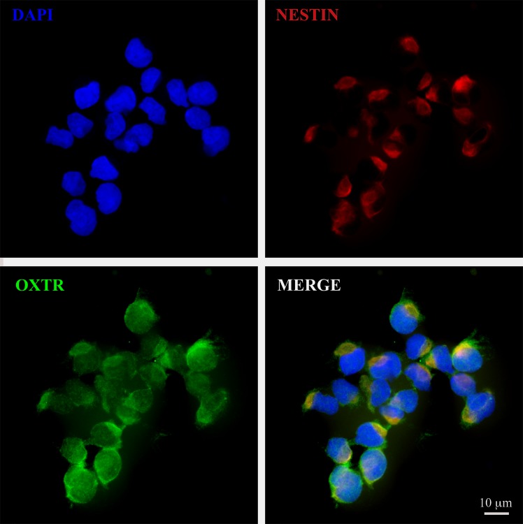
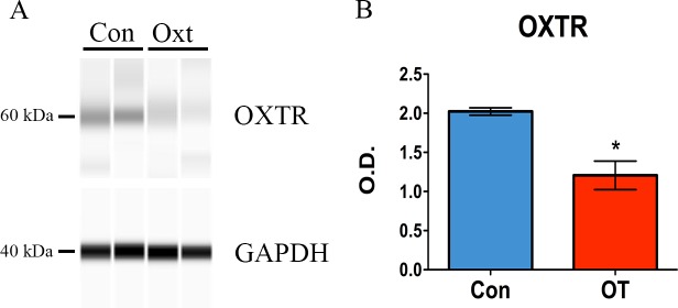
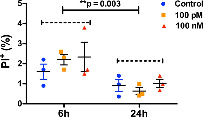
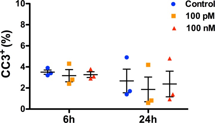
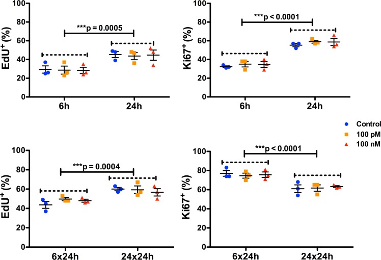
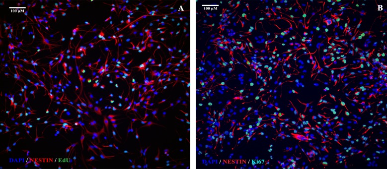
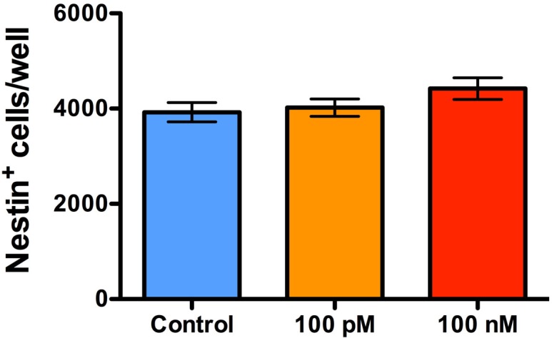
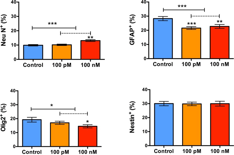
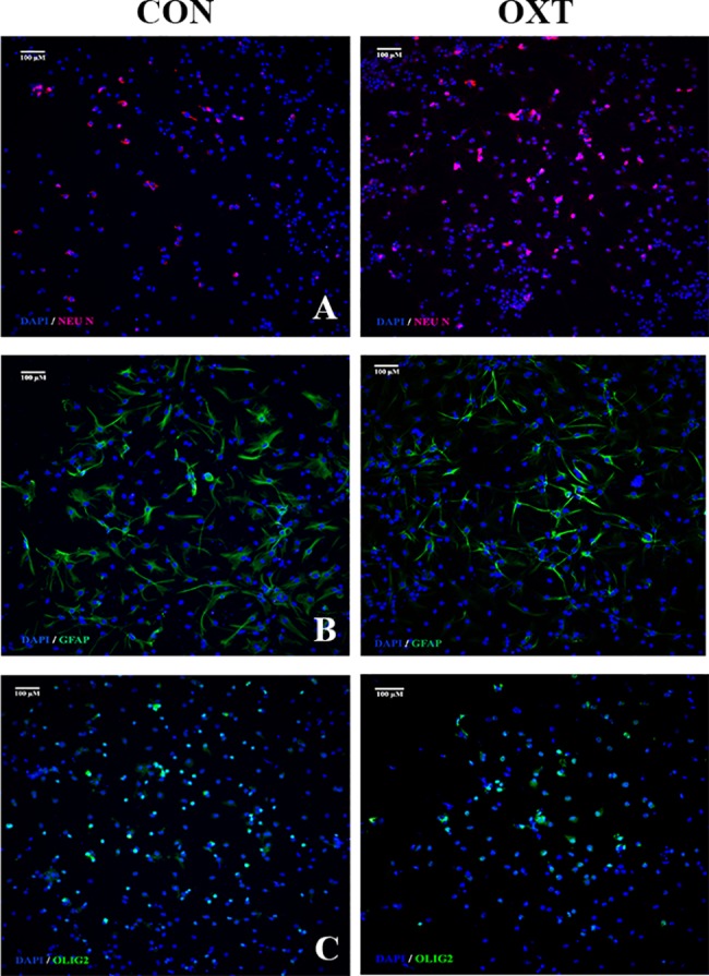
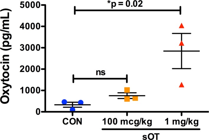
Synthetic oxytocin (sOT) is widely used during labor, yet little is known about its effects on fetal brain development despite evidence that it reaches the fetal circulation. Here, we tested the hypothesis that sOT would affect early neurodevelopment by investigating its effects on neural progenitor cells (NPC) from embryonic day 14 rat pups. NPCs expressed the oxytocin receptor (OXTR), which was downregulated by 45% upon prolonged treatment with sOT. Next, we examined the effects of sOT on NPC death, apoptosis, proliferation, and differentiation using antibodies to NeuN (neurons), Olig2 (oligodendrocytes), and GFAP (astrocytes). Treated NPCs were analysed with unbiased high-throughput immunocytochemistry. Neither 6 nor 24 h exposure to 100 pM or 100 nM sOT had an effect on viability as assessed by PI or CC-3 immunocytochemistry. Similarly, sOT had negligible effect on NPC proliferation, except that the overall rate of NPC proliferation was higher in the 24 h compared to the 6 h group regardless of sOT exposure. The most significant finding was that sOT exposure caused NPCs to select a predominantly neuronal lineage, along with a concomitant decrease in glial cells. Collectively, our data suggest that perinatal exposure to sOT can have neurodevelopmental consequences for the fetus, and support the need for in vivo anatomical and behavioral studies in offspring exposed to sOT in utero.


Conflict of interest statement

Competing Interests: The authors have declared that no competing interests exist.

Figures

Fig 1
Fig 1. NPCs express OXTR.
A 60x photomicrograph showing co-expression of OXTR (green) both in the cytoplasm as well as the plasma membrane of nestin+ NPCs (red). Imaging was performed in Olympus confocal FV1000 microscope and processed with Adobe Photoshop. Scale bar as noted.
Fig 2
Fig 2. Oxytocin downregulates OXTR in NPCS.
1.5 μg of total protein was analyzed with Protein Simple Wes™ automated Western blotting system. Goat anti-OXTR antibody was used at a dilution of 1:10. Human uterus lysate was used as positive control. Cropped representative pseudo-blots show the expression of OXTR (60 kDa), with GAPDH as the loading control (Fig 2A). Full-length pseudo-blots are presented in Supplementary S1 Fig. Compared to control treatment, 24 h of 100 nM oxytocin significantly downregulated OXTR protein by approximately 45% (*P = 0.01 by Student’s t test) (Fig 2B). Data expressed as mean ± S.E.M from three biologically independent NPC cultures.
Fig 3
Fig 3. No difference in NPC death at the end of oxytocin treatment.
Scatter plots showing the proportion of dead NPCs after treament with either 0, 100 pM, or 100 nM oxytocin for either 6 or 24 h, as noted. NPC death was quantified with PI staining. 2-way ANOVA analysis did not show a difference either with oxytocin treatment (F (2,12) = 0.59, p = 0.58, η2 = 0.04) or a treatment*time interaction (F (2, 12) = 0.66, p = 0.53, η2 = 0.04). However, there was a significant effect of time (F (1, 12) = 14, *p = 0.003, η2 = 0.49) with a lower rate of NPC death at 24h compared to 6h. Data are expressed as mean ± S.E.M from three biologically independent NPC cultures.
Fig 4
Fig 4. No difference in NPC apoptosis at the end of oxytocin treatment.
Scatter plots showing the proportion of apoptotic NPCs after treament with either 0, 100 pM, or 100 nM oxytocin for either 6 or 24 h, as noted. Apoptosis of NPCs was quantified with cleaved caspase-3 immunocytochemistry. 2-way ANOVA analysis did not show a significant difference either with oxytocin treatment (F (2,12) = 0.21, p = 0.81, η2 = 0.03), time (F (1, 12) = 2, p = 0.18, η2 = 0.14) or a treatment*time interaction (F (2, 12) = 0.043, p = 0.96, η2 = 0.006). Data are expressed as mean ± S.E.M from three biologically independent NPC cultures.
Fig 5
Fig 5. Oxytocin treatment has minimal effect on NPC proliferation.
Scatter plots showing the proportion of proliferating NPCs treated with either 100 pM or 100 nM of oxytocin for 6 or 24 h, and after 24 h following removal of oxytocin from the medium. Proliferation was quantified with EdU incorporation and Ki67 immunocytochemistry. There were no differences in NPC proliferation with oxytocin treatment either at 6 or 24 h, though the overall rate of proliferation was significantly higher in the 24 vs the 6 h group for both EdU incorporation and Ki-67 immunoreactivity. There were no significant treatment*time interactions for either EdU incorporation or Ki-67 immunoreactivity. The results were very similar in the oxytocin withdrawal experiments, except that Ki-67 immunoreactivity was significantly lower in the 24 compared to the 6 h group. Data are expressed as mean ± S.E.M from three biologically independent NPC cultures.
Fig 6
Fig 6. Representative photomicrographs showing EdU and Ki67 immunocytochemistry.
20x images of EdU and Ki67 immunoreactivity from control wells are shown in A and B, respectively. Approximately 35% of NPCs were positive for EdU and approximately 60% were positive for Ki67 immunoreactivity. Scale bar as noted.
Fig 7
Fig 7. Prolonged treatment with oxytocin does not decrease the neural stem cell pool.
A bar graph showing the proportion of NPCs 24 h after treatment with either 100 pM or 100 nM of oxytocin for 24 h. NPCs were phenotyped with nestin immunocytochemistry. There were no differences in the overall proportion of nestin-positive NPCs/well at 24 h after treatment with both concentrations, compared to control treatment. Data are expressed as mean ± S.E.M from three biologically independent NPC cultures.
Fig 8
Fig 8. Prolonged exposure to oxytocin enhances neuronal but impairs astrocytic and oligodendrocytic differentiation.
Bar graphs showing the proportion of neurons (Neu N), astrocytes (GFAP), oligodendrocytes (Olig2), and nestin+ NPCs, two weeks after treatment with 100 nM oxytocin for 24 h followed by mitogen withdrawal. Treatment with oxytocin increased the number of NeuN+ neurons (***P = 0.0005 by Kruskal-Wallis test), but decreased the number of both GFAP+ astrocytes (***P = 0.0009 by 1-way ANOVA) and Olig2+ oligodendrocytes (*P = 0.04 by 1-way ANOVA). These changes were not accompanied by a change in the overall proportion of nestin+ NPCs (approximately 30%) (P = 0.97 by Kruskal-Wallis test).
Fig 9
Fig 9. Representative photomicrographs of spontaneously differentiating NPCs after oxytocin exposure (100 nM).
A panel showing 10x photomicrographs of neuronal (A), astrocytic (B), and oligodendrocytic (C) differentiation as labeled by Neu N, GFAP, and Olig2, respectively, in control (left) and oxytocin (right) treated NPCs. Nuclei counterstained with DAPI. Scale bar as noted.
Fig 10
Fig 10. Dose-dependent transfer of sOT across the placenta.
Fetal plasma oxytocin level was quantified in pooled fetal cardiac blood samples from dams treated with either saline, 100 mcg/kg, or 1 mg/kg of sOT. Fetal plasma oxytocin increased in the 1 mg/kg dose group (*p = 0.02 by one-way ANOVA; mean ± S.E.M), suggesting that sOT can cross the placenta.

References

    1. Obstetrics ACoPB—. ACOG Practice Bulletin No. 107: Induction of labor. Obstetrics and gynecology. 2009;114(2 Pt 1):386–97. doi: 10.1097/AOG.0b013e3181b48ef5 . - DOI - PubMed
    1. Fuchs AR, Romero R, Keefe D, Parra M, Oyarzun E, Behnke E. Oxytocin secretion and human parturition: pulse frequency and duration increase during spontaneous labor in women. American journal of obstetrics and gynecology. 1991;165(5 Pt 1):1515–23. . - PubMed
    1. Malek A, Blann E, Mattison DR. Human placental transport of oxytocin. The Journal of maternal-fetal medicine. 1996;5(5):245–55. doi: 10.1002/(SICI)1520-6661(199609/10)5:5<245::AID-MFM3>3.0.CO;2-H . - DOI - PubMed
    1. Yoshimura R, Kimura T, Watanabe D, Kiyama H. Differential expression of oxytocin receptor mRNA in the developing rat brain. Neuroscience research. 1996;24(3):291–304. . - PubMed
    1. Hammock EA, Levitt P. Oxytocin receptor ligand binding in embryonic tissue and postnatal brain development of the C57BL/6J mouse. Frontiers in behavioral neuroscience. 2013;7:195 doi: 10.3389/fnbeh.2013.00195 ; PubMed Central PMCID: PMC3858721. - DOI - PMC - PubMed

Publication types

MeSH terms