. 2018 Jun 20;218(2):197-207.
doi: 10.1093/infdis/jiy018.

Receptor Usage of a Novel Bat Lineage C Betacoronavirus Reveals Evolution of Middle East Respiratory Syndrome-Related Coronavirus Spike Proteins for Human Dipeptidyl Peptidase 4 Binding

Affiliations

Receptor Usage of a Novel Bat Lineage C Betacoronavirus Reveals Evolution of Middle East Respiratory Syndrome-Related Coronavirus Spike Proteins for Human Dipeptidyl Peptidase 4 Binding

Susanna K P Lau et al. J Infect Dis. .

Abstract

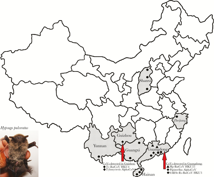
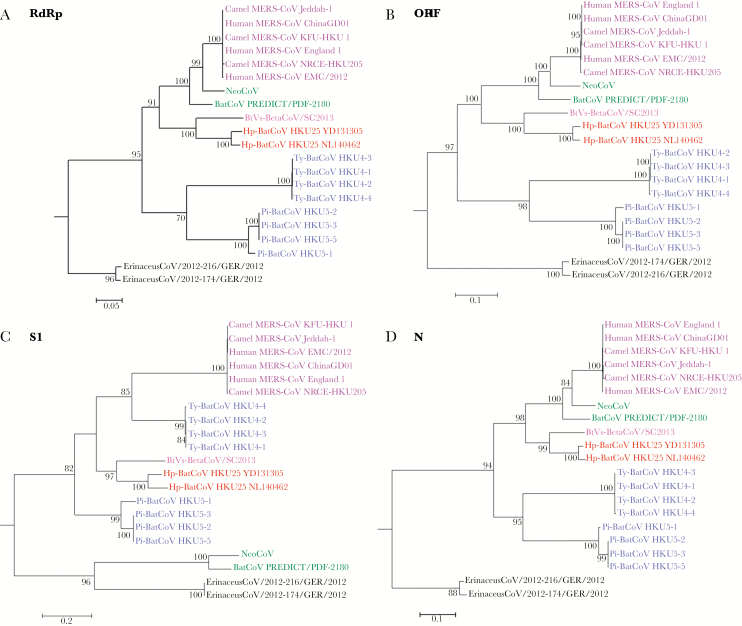
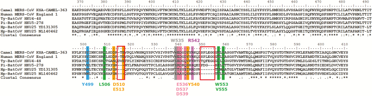
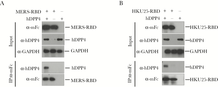
Although bats are known to harbor Middle East Respiratory Syndrome coronavirus (MERS-CoV)-related viruses, the role of bats in the evolutionary origin and pathway remains obscure. We identified a novel MERS-CoV-related betacoronavirus, Hp-BatCoV HKU25, from Chinese pipistrelle bats. Although it is closely related to MERS-CoV in most genome regions, its spike protein occupies a phylogenetic position between that of Ty-BatCoV HKU4 and Pi-BatCoV HKU5. Because Ty-BatCoV HKU4 but not Pi-BatCoV HKU5 can use the MERS-CoV receptor human dipeptidyl peptidase 4 (hDPP4) for cell entry, we tested the ability of Hp-BatCoV HKU25 to bind and use hDPP4. The HKU25-receptor binding domain (RBD) can bind to hDPP4 protein and hDPP4-expressing cells, but it does so with lower efficiency than that of MERS-RBD. Pseudovirus assays showed that HKU25-spike can use hDPP4 for entry to hDPP4-expressing cells, although with lower efficiency than that of MERS-spike and HKU4-spike. Our findings support a bat origin of MERS-CoV and suggest that bat CoV spike proteins may have evolved in a stepwise manner for binding to hDPP4.


Figures

Figure 1.
Figure 1.
Map showing various sampling locations in seven provinces of China (Guangxi, Guangdong, Shanxi, Zhejiang, Yunnan, Hainan and Guizhou). Sampling locations with Hp-BatCoV HKU25 and other CoVs detected are indicated with triangle and diamond respectively.
Figure 2.
Figure 2.
Phylogenetic analyses of RdRp, ORF1, S1 and N nucleotide sequences of Hp-BatCoV HKU25 and other lineage C betacoronaviruses (B). The trees were constructed by maximum likelihood method using GTR+G substitution models respectively and bootstrap values calculated from 1000 trees. Trees were rooted using corresponding sequences of HCoV HKU1 (GenBank accession number NC_006577). Only bootstrap values >70% are shown. (A) 2775 nt (B) 20694 nt (C) 3740 nt (D) 1167 nt positions respectively were included in the analyses. The scale bars represent (A) 20 (B) 20 (C) 10 (D) 10 substitutions per site respectively. The two Hp-BatCoV HKU25 strains, YD131305 and NL140462, detected in this study are bolded and underlined.
Figure 3.
Figure 3.
Multiple alignment of the amino acid sequences of the receptor-binding domain (RBD) of the spike protein of MERS-CoV and corresponding sequences in Hp-BatCoV HKU25 and other lineage C betacoronaviruses. Asterisks indicate positions with fully conserved residues. The two amino acid deletions in Hp-BatCoV HKU25 compared to MERS-CoV and Ty-BatCoV HKU4 are indicated with boxes. The 12 critical residues for receptor binding in MERS-CoV are highlighted. The 10 residues marked below the alignment are based on (Wang, 2013) [37] and the other two residues marked above the alignment are based on (Wang, 2014) [23]. Y499 formed hydrogen bond with DPP4 residue. L506, W553 and V555 formed a hydrophobic core surrounded by hydrophilic residues D510, E513 and Y540. D510 and E513 also contributed to salt bridge interaction and hydrogen bonding with DPP4 residues. E536, D537 and D539 formed negative-charged surface. W535 and R542 are residues that have strong polar contact with DPP4 residues.
Figure 4.
Figure 4.
Binding of HKU25-RBD with human cells was mediated by interacting with human dipeptidyl peptidase 4 (hDPP4) receptor. (A) Fluorescence-activated cell sorter (FACS) analysis of Middle East Respiratory Syndrome receptor-binding domain (MERS-RBD)-mFc (10 µg/mL) and HKU25-RBD-mFc (40 µg/mL) binding to Huh7 cells and hDPP4-knockdown Huh7 cells. (B) FACS analysis of MERS-RBD-mFc and HKU25-RBD-mFc binding to 293T cells and 293T cells transfected with hDPP4-expressing plasmid. The shaded area represents the secondary antibody control. (C) Determination of small interfering ribonucleic acid (siRNA) efficiency by quantitative reverse transcription-polymerase chain reaction (qRT-PCR) and Western blot analysis using primers and antibody specific for hDPP4. (D) MERS-RBD-mFc and HKU25-RBD-mFc binding to a molecule(s) located on the Huh7 cell surface. MERS-RBD-mFc and HKU25-RBD-mFc were detected by an Alexa Fluor 488-conjugated goat anti-mFc antibody. Empty expressing plasmid was used as a negative control.
Figure 5.
Figure 5.
Middle East Respiratory Syndrome receptor-binding domain (MERS-RBD)-mFc (A) and HKU25-RBD-mFc (B) proteins directly bind with human dipeptidyl peptidase 4 (hDPP4). HEK293 T cells were transfected with hDPP4-expressing plasmids, and MERS-RBD-mFc (A) and HKU25-RBD-mFc (B) proteins were used for immunoprecipitation of lysates of HEK293 T cells transfected with hDPP4-expressing or empty plasmids. Empty plasmid was mock-transfected as negative control. The hDPP4 was coprecipitated from the lysates, as detected by antibody specific for hDPP4. Glyceraldehyde 3-phosphate dehydrogenase (GAPDH) was used as a loading control.
Figure 6.
Figure 6.
HEK293 T cells transfected with empty plasmid or human dipeptidyl peptidase 4 (hDPP4) were infected by retroviruses pseudotyped with Middle East Respiratory Syndrome coronavirus (MERS-CoV), Ty-BatCoV HKU4, Pi-BatCoV HKU5, and Hp-BatCoV HKU25 S proteins with mock pseudovirus (∆env) as control. The cells were also preincubated with anti-hDPP4 antibodies (Ab) to test for cell entry inhibition. Cell entry efficiencies were assayed by luciferase activity measurement after 72 hours.

References

    1. Zaki AM, van Boheemen S, Bestebroer TM, Osterhaus AD, Fouchier RA. Isolation of a novel coronavirus from a man with pneumonia in Saudi Arabia. N Engl J Med 2012; 367:1814–20. - PubMed
    1. de Groot RJ, Baker SC, Baric RS et al. . Middle East respiratory syndrome coronavirus (MERS-CoV): announcement of the Coronavirus Study Group. J Virol 2013; 87:7790–2. - PMC - PubMed
    1. Raj VS, Mou H, Smits SL et al. . Dipeptidyl peptidase 4 is a functional receptor for the emerging human coronavirus-EMC. Nature 2013; 495:251–4. - PMC - PubMed
    1. Reusken CB, Haagmans BL, Müller MA et al. . Middle East respiratory syndrome coronavirus neutralising serum antibodies in dromedary camels: a comparative serological study. Lancet Infect Dis 2013; 13:859–66. - PMC - PubMed
    1. Haagmans BL, Al Dhahiry SH, Reusken CB et al. . Middle East respiratory syndrome coronavirus in dromedary camels: an outbreak investigation. Lancet Infect Dis 2014; 14:140–5. - PMC - PubMed

Publication types