

Review
doi: 10.1021/acs.jpcb.7b11370.
Epub 2018 Feb 1.
Two-Dimensional Spectroscopy Is Being Used to Address Core Scientific Questions in Biology and Materials Science
Affiliations
- PMID: 29346730
- PMCID: PMC6400462
- DOI: 10.1021/acs.jpcb.7b11370
Item in Clipboard
Review
Two-Dimensional Spectroscopy Is Being Used to Address Core Scientific Questions in Biology and Materials Science
J Phys Chem B.
.
Abstract
Two-dimensional spectroscopy is a powerful tool for extracting structural and dynamic information from a wide range of chemical systems. We provide a brief overview of the ways in which two-dimensional visible and infrared spectroscopies are being applied to elucidate fundamental details of important processes in biological and materials science. The topics covered include amyloid proteins, photosynthetic complexes, ion channels, photovoltaics, batteries, as well as a variety of promising new methods in two-dimensional spectroscopy.
Conflict of interest statement
The authors declare the following competing financial interest(s): Martin Zanni is co-owner of PhaseTech Spectroscopy, Inc., which sells mid-IR and visible pulse shapers and 2D spectrometers.
Figures
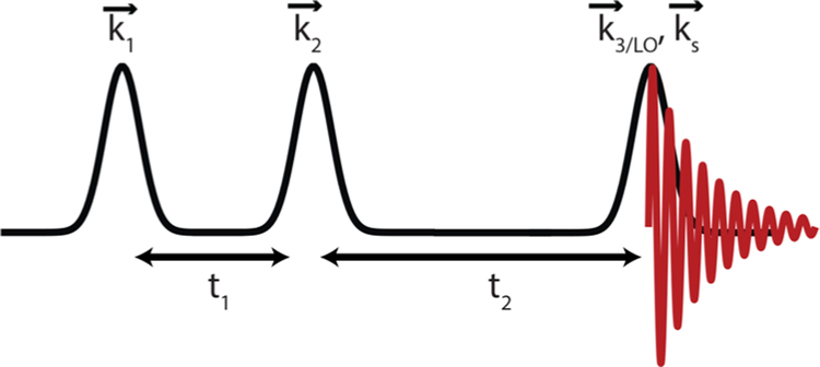
Pulse sequence used in a four-wave mixing experiment to obtain a two-dimensional infrared or electronic spectrum. Three field interactions with the sample produce an emitted signal that is heterodyned with either a local oscillator or the third laser pulse, depending on the experimental geometry.
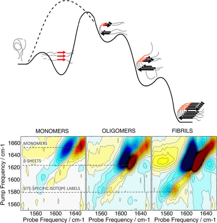
Top: Schematic free energy diagram of the multistep hIAPP aggregation process. The FGAIL region that participates in the formation of a transient β-sheet intermediate is highlighted in red. Introduction of a proline mutation into the FGAIL sequence inhibits aggregation by destabilizing the intermediate (dashed line). Bottom: 2D IR spectra of isotopically labeled (V17) hIAPP measured at different aggregation times are presented below. Dashed arrows show pump frequencies of 2D IR peaks originating from monomers, β-sheet aggregates, and site-specific isotope labels. Adapted with permission from ref .
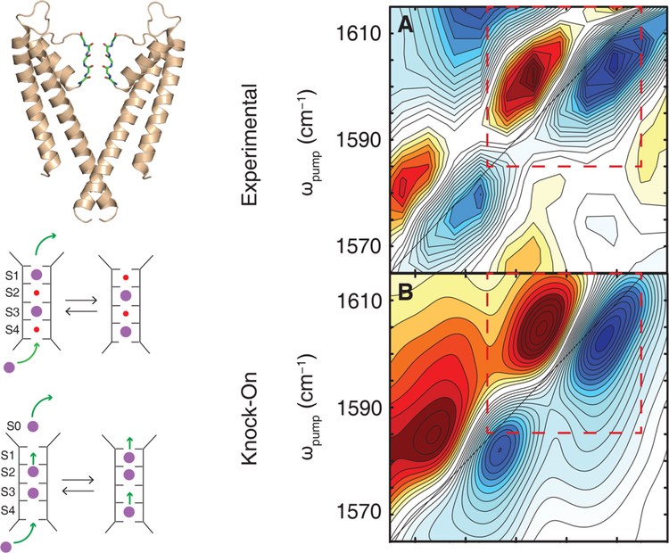
Left: The KcsA potassium ion channel and a schematic of the knock-on (top) and hard-knock (bottom) ion transport mechanisms. Right:(a) The experimental IR spectrum in the isotope labeled region and (b) the simulated 2D IR spectrum of the knock-on model. Reproduced with permission from refs and . Copyright 2017 American Chemical Society and 2016 American Association for the Advancement of Science, respectively.
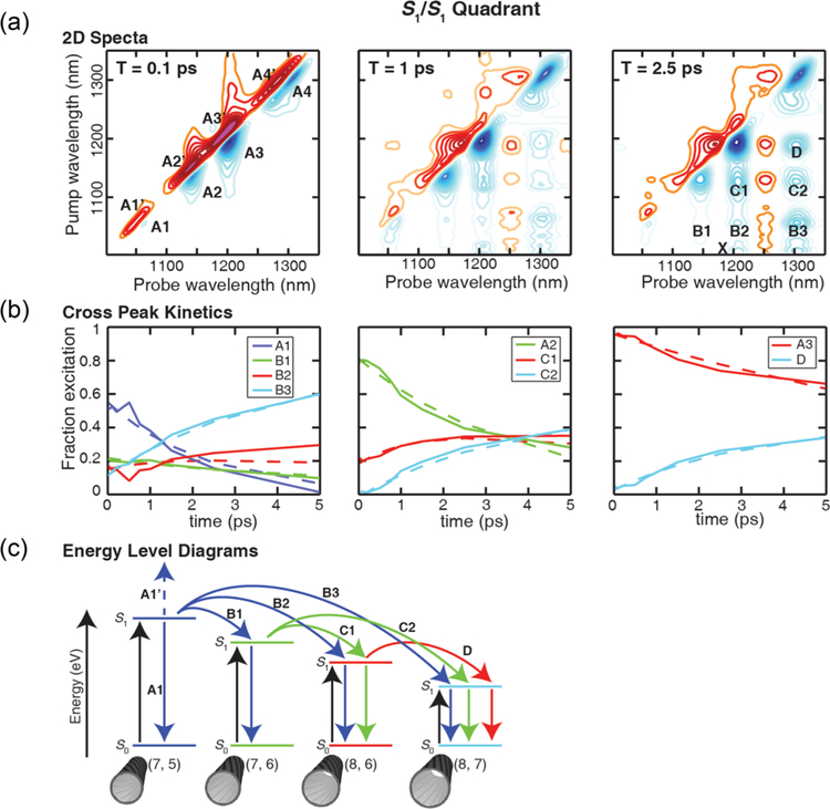
(a) 2D electronic spectra for the indicated pump−probe delays in the S1/S1 quadrant. The simultaneous growth of round cross-peaks indicates energy transfer is uncorrelated and independent of bandgap for S1 excitons. (b) The kinetics of cross-peaks provide rates of energy transfer. Dashed lines are exponential fits to the data, and the measured time constants are all equal to within 1 ps. (c) An energy level diagram showing the energy transfer pathway for each peak in the 2D spectrum. Black arrows denote pumped transitions, while blue, green, and red arrows correspond to excitons initially excited on (7, 5), (7, 6), and (8, 6) nanotubes, respectively. Dashed arrows represent excited state absorption, solid arrows represent ground state bleaches/stimulated emission, and curved arrows denote energy transfer. Reproduced with permission from ref .
References
-
- Hamm P; Zanni MT Concepts and Methods of 2D Infrared Spectroscopy; Cambridge University Press: New York, 2011.
-
- Cho M Two-Dimensional Optical Spectroscopy; Taylor & Francis Group: Boca Raton, FL, 2009.
-
- Mukamel S Principles of Nonlinear Optical Spectroscopy; Oxford University Press: New York, 1995.
-
- Khalil M; Demirdöven N; Tokmakoff, a. Coherent 2D IR Spectroscopy: Molecular Structure and Dynamics in Solution. J. Phys. Chem. A 2003, 107 (27), 5258–5279.
Publication types
MeSH terms
Substances
Grants and funding
LinkOut - more resources
Full Text Sources
Other Literature Sources
