. 2018 Jan 23;71(3):321-335.
doi: 10.1016/j.jacc.2017.11.036.

PET/MR Imaging of Malondialdehyde-Acetaldehyde Epitopes With a Human Antibody Detects Clinically Relevant Atherothrombosis

Affiliations

PET/MR Imaging of Malondialdehyde-Acetaldehyde Epitopes With a Human Antibody Detects Clinically Relevant Atherothrombosis

Max L Senders et al. J Am Coll Cardiol. .

Abstract

Background: Oxidation-specific epitopes (OSEs) are proinflammatory, and elevated levels in plasma predict cardiovascular events.

Objectives: The purpose of this study was to develop novel positron emission tomography (PET) probes to noninvasively image OSE-rich lesions.

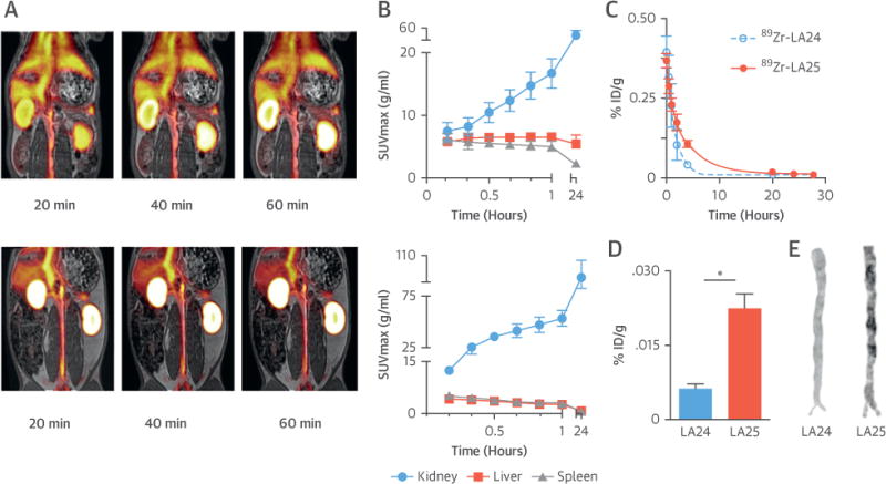
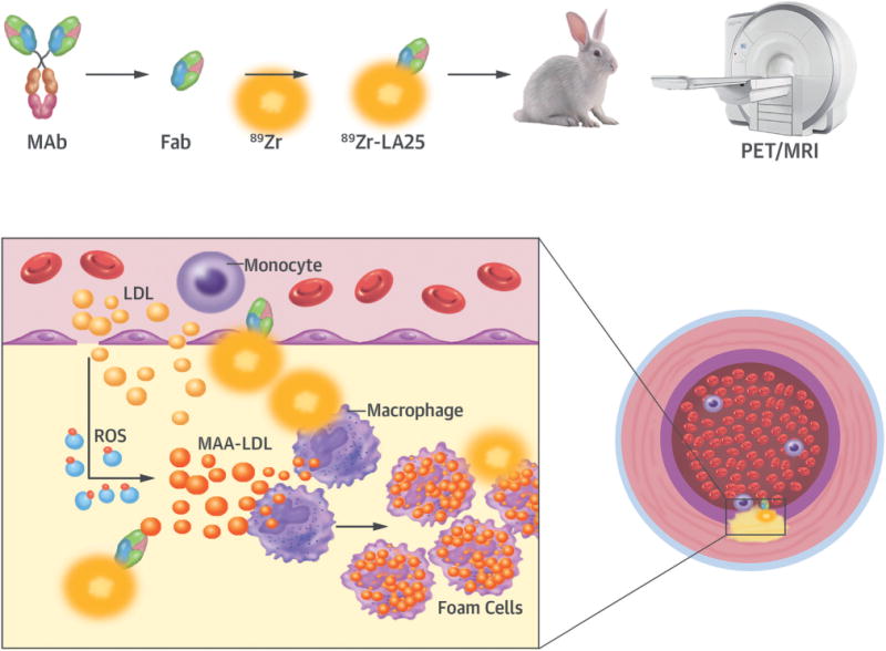
Methods: An antigen-binding fragment (Fab) antibody library was constructed from human fetal cord blood. After multiple rounds of screening against malondialdehyde-acetaldehyde (MAA) epitopes, the Fab LA25 containing minimal nontemplated insertions in the CDR3 region was identified and characterized. In mice, pharmacokinetics, biodistribution, and plaque specificity studies were performed with Zirconium-89 (89Zr)-labeled LA25. In rabbits, 89Zr-LA25 was used in combination with an integrated clinical PET/magnetic resonance (MR) system. 18F-fluorodeoxyglucose PET and dynamic contrast-enhanced MR imaging were used to evaluate vessel wall inflammation and plaque neovascularization, respectively. Extensive ex vivo validation was carried out through a combination of gamma counting, near infrared fluorescence, autoradiography, immunohistochemistry, and immunofluorescence.

Results: LA25 bound specifically to MAA epitopes in advanced and ruptured human atherosclerotic plaques with accompanying thrombi and in debris from distal protection devices. PET/MR imaging 24 h after injection of 89Zr-LA25 showed increased uptake in the abdominal aorta of atherosclerotic rabbits compared with nonatherosclerotic control rabbits, confirmed by ex vivo gamma counting and autoradiography. 18F-fluorodeoxyglucose PET, dynamic contrast-enhanced MR imaging, and near-infrared fluorescence signals were also significantly higher in atherosclerotic rabbit aortas compared with control aortas. Enhanced liver uptake was also noted in atherosclerotic animals, confirmed by the presence of MAA epitopes by immunostaining.

Conclusions: 89Zr-LA25 is a novel PET radiotracer that may allow noninvasive phenotyping of high-risk OSE-rich lesions.

Keywords: PET/MR imaging; atherosclerosis; natural antibodies; oxidation-specific epitopes.


Figures

FIGURE 1
FIGURE 1. Cloning and Characterization of the MAA-Targeted LA25 Antibody Fragment
(A) Schematic depiction of malondialdehyde-acetaldehyde (MAA) low-density lipoprotein (LDL). (B) The configuration of soluble LA25 Fab antibody fragment. The LA25 lambda light-chain and heavy-chain with a hexa-histidine and the influenza hemagglutinin (HA) epitope tag for detection and purification were expressed under the direction of lacZ promoter for phage display or Fab production in E. coli. Proteolysis of the ompA and pelB signal peptides in the periplasm generated the native amino terminus of Fab and facilitated the joining of heavy and light chains together by disulfide bonds as bioactive soluble Fab. (C) Binding of LA25 to a variety of oxidation-specific epitopes. Competition assays for the specificity of LA25 binding to MAA-LDL (D) and MAA-BSA (E). LA25 was incubated in the absence and presence of increasing amounts of indicated competitors, and the extent of binding to plated MAA-BSA and MAA-LDL was determined. Inhibition of MAA-LDL binding to macrophage scavenger receptors by LA25 (F). Data are expressed as a ratio of binding in the presence of competitor (B) divided by absence of competitor (B0). BSA = bovine serum albumin; Cu-OxLDL = copper-oxidized low-density lipoprotein; MDA = malondialdehyde; MSA = mouse serum albumin.
FIGURE 2
FIGURE 2. Immunostaining of Human Pathological Specimens With LA25
Immunostaining with LA25 and control antibody LA24 of coronary artery specimens representing pathological intimal thickening (top row), thick cap fibroatheroma with multiple ruptures and healing phases (middle row) and a ruptured plaque containing thrombus (bottom row), stained with hematoxylin and eosin (H&E), Movat pentachrome, LA25, and LA24 Fab control. Note the presence of thrombus in this section, which also stains with LA25.
FIGURE 3
FIGURE 3. Immunostaining of Sections From Embolized Plaque Debris With LA25
Captured plaque debris from 2 distal protection devices following percutaneous coronary intervention. The material was stained with LA25 for malondialdehyde-acetaldehyde (MAA) epitopes, MDA3 antibody for MDA epitopes, and E06 antibody for OxPL epitopes. No-antibody (control) and LA24 control sections are also shown. MDA = malondialdehyde; OxPL = Oxidized phospholipids.
FIGURE 4
FIGURE 4. 89Zr-LA25 Evaluation in Apoe−/− Mice
(A) Blood time-activity curve for 89Zr-LA25 and -LA24. Gamma counting (B) and autoradiography (C) of aortas from Apoe−/− mice injected with 89Zr-LA24 (blue) and 89Zr-LA25 (red). (D) Radioactivity distribution in selected tissues in Apoe−/− mice 4 h post-injection. (E) From left to right, top row: aortic root sections from an Apoe−/− mouse stained for cell nuclei (DAPI, blue) and macrophages (CD68, red), and autoradiography of an adjacent section after 89Zr-LA25 injection; below: same aortic root section stained for endothelial cells (CD31, green), a merged image (middle), and a 40× magnification (right). (F) Atherosclerotic mouse livers stained with LA24, LA25 (including 40× magnification), and no-antibody control section are shown. *p < 0.05.
FIGURE 5
FIGURE 5. 89Zr-LA25 PET/MR Imaging in Rabbits
(A) Representative coronal fused positron emission tomography (PET)/magnetic resonance (MR) images at 20, 40, and 60 min post-injection (p.i.) of 89Zr-LA25 (top) and 89Zr-LA24 (bottom). (B) Radioactivity quantification in major organs in atherosclerotic rabbits based on PET/MR imaging (10 to 60 min), and 24 h p.i. (C) Pharmacokinetics in atherosclerotic rabbits for 89Zr-LA24 and -LA25, with half-lives of 1.1 and 2.2 h, respectively. Ex vivo radioactivity concentration (D) and autoradiography (E) for 89Zr-LA24 and -LA25 in aortas from rabbits with atherosclerosis 28 h p.i. *p < 0.05. SUV = standardized uptake value.
FIGURE 6
FIGURE 6. Phenotyping of Rabbit Atherosclerotic Plaques by PET/MR Imaging
(A) Representative coronal aortic fused PET/MR imaging 24 h p.i. of 89Zr-LA25, (B) autoradiographs and gamma counting (whole aortas) 28 h p.i. of 89Zr-LA25, (C) MR T2-weighted imaging, (D) 18F-fluorodeoxyglucose (18F-FDG) PET/MR imaging, (E) dynamic contrast enhanced (DCE)-MR imaging, and (F) fluorescently labeled (DiD)-rHDL near infrared fluorescence imaging, of healthy control (white) and atherosclerotic abdominal aortas (black). *p < 0.05. IAUC = area under the normalized signal intensity curve; MWA = mean wall area; SUV = standardized uptake value.
FIGURE 7
FIGURE 7. Ex Vivo Plaque Characterization
Digital autoradiography of atherosclerotic rabbit aorta sections with adjacent slides stained for hematoxylin and eosin, RAM-11, and Oil Red O, with corresponding masks and merged images with autoradiography. On the right, correlations are shown between autoradiography and vessel wall area, RAM-11, and Oil Red O.
CENTRAL ILLUSTRATION
CENTRAL ILLUSTRATION. Targeting Atherothrombosis With 89Zr-LA25
Atherosclerotic lesions are initiated by accumulation of lipoproteins. The modification of lipoproteins generates pro-inflammatory oxidation-specific epitopes, such as malondialdehyde-acetaldehyde (MAA) adducts, which lead to immune cell recruitment to the subintimal space. Accumulation of oxidized lipids results in foam cell formation and, ultimately, complex atherothrombotic lesions. LA25 is a human Fab antibody fragment discovered in the deoxyribonucleic acid (rather than plasma) of newborn babies prior to environmental exposure, consistent with evolutionary conservation. LA25 was found to specifically bind MAA epitopes and engineered to carry the positron emission tomography (PET) isotope 89Zr. Using an integrated PET/magnetic resonance imaging (MRI) system, 89Zr-LA25 was shown to noninvasively image mouse and rabbit atherosclerotic lesions, as well as MAA-rich steatotic livers. Fab = antigen-binding fragment; LDL = low-density lipoprotein; MAb = monclonal antibody; ROS = reactive oxygen species.

Comment in

References

    1. Mozaffarian D, Benjamin EJ, Go AS, et al. Heart disease and stroke statistics–2016 update: A report from the American Heart Association. Circulation. 2016;133:e38–360. - PubMed
    1. Otsuka F, Kramer MC, Woudstra P, et al. Natural progression of atherosclerosis from pathologic intimal thickening to late fibroatheroma in human coronary arteries: a pathology study. Atherosclerosis. 2015;241:772–82. - PMC - PubMed
    1. Arbab-Zadeh A, Fuster V. The myth of the “vulnerable plaque”: transitioning from a focus on individual lesions to atherosclerotic disease burden for coronary artery disease risk assessment. J Am Coll Cardiol. 2015;65:846–55. - PMC - PubMed
    1. Libby P, Pasterkamp G. Requiem for the ‘vulnerable plaque’. Eur Heart J. 2015;36:2984–7. - PubMed
    1. Franck G, Mawson T, Sausen G, et al. Flow perturbation mediates neutrophil recruitment and potentiates endothelial injury via TLR2 in mice: implications for superficial erosion. Circ Res. 2017;121:31–42. - PMC - PubMed

Publication types