

Review
doi: 10.3174/ajnr.A5528.
Epub 2018 Jan 18.
Review of the Imaging Features of Benign Osteoporotic and Malignant Vertebral Compression Fractures
Affiliations
- PMID: 29348133
- PMCID: PMC7655272
- DOI: 10.3174/ajnr.A5528
Item in Clipboard
Review
Review of the Imaging Features of Benign Osteoporotic and Malignant Vertebral Compression Fractures
AJNR Am J Neuroradiol.
2018 Sep.
Abstract
Vertebral compression fractures are very common, especially in the elderly. Benign osteoporotic and malignant vertebral compression fractures have extremely different management and prognostic implications. Although there is an overlap in appearances, characteristic imaging features can aid in the distinction between these 2 types of compression fractures. The aim of this review is to characterize the imaging features of benign and malignant vertebral compression fractures seen with CT, PET, SPECT, and MR imaging.
© 2018 by American Journal of Neuroradiology.
Figures
Abnormal pedicle marrow signal in a malignant VCF. A, Sagittal T1WI of the lumbar spine demonstrates a malignant VCF of L3 with loss of the high T1 normal marrow signal within the pedicle (arrow), indicating tumor infiltration. B, Sagittal T1WI of the lumbar spine demonstrates a typical benign VCF of L1 anteriorly, with preservation of the normal high T1 marrow signal within the pedicle (arrow).
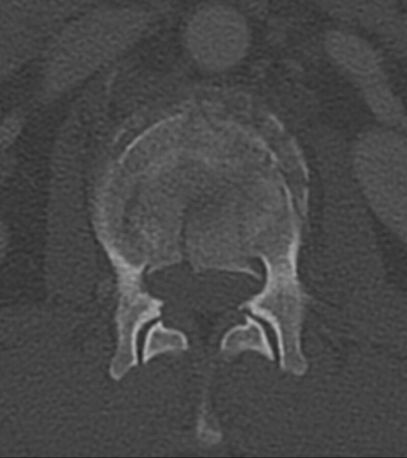
Fracture lines without cortical destruction in a benign VCF. Axial CT with bone windows shows the linear and well-delineated borders of the slightly displaced bone fragments within this benign VCF, an example of the puzzle sign.
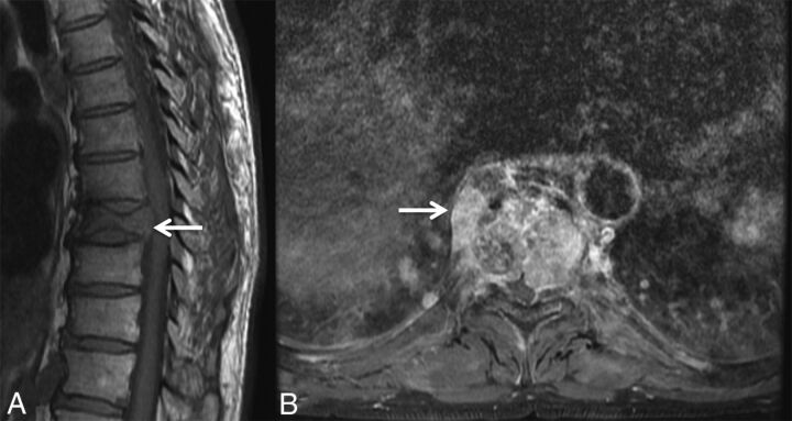
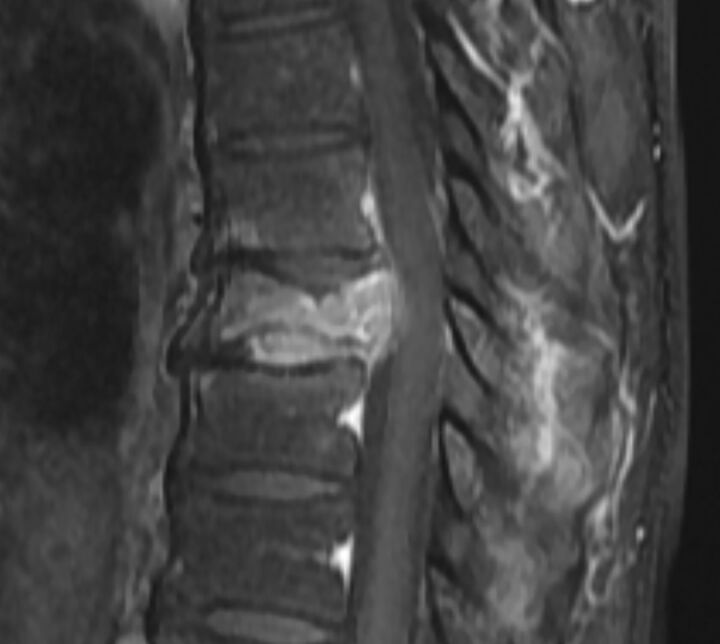
Masslike extension into the paravertebral and epidural space in a malignant VCF. A, Sagittal T1WI of the thoracic spine demonstrates a malignant VCF of T9 with loss of the high T1 normal marrow signal within the vertebral body and convex bowing of the posterior cortex (arrow), both signs indicating a malignant fracture. B, Axial postcontrast T1WI with fat saturation of the T9 fracture demonstrates an irregular enhancing mass (arrow) extending into the right paraspinal soft tissues and the epidural space in this malignant VCF.
Diffuse abnormal marrow signal in a malignant VCF. Sagittal T1WI of the lumbar spine demonstrates a malignant VCF of L2 with marked complete replacement of the normal high T1 vertebral body marrow signal. The diffuse T1 hypointensity indicates tumor infiltration. Note the convex, expanded border of the posterior vertebral body versus the normal posterior concavity of the adjacent vertebral bodies.
Increased enhancement in malignant VCF. Sagittal T1WI postgadolinium with fat saturation of the lumbar spine demonstrates an enhancing malignant VCF of L2. The enhancement is greater than that of the normal adjacent vertebral bodies. Also demonstrated is an expanded posterior convex border.
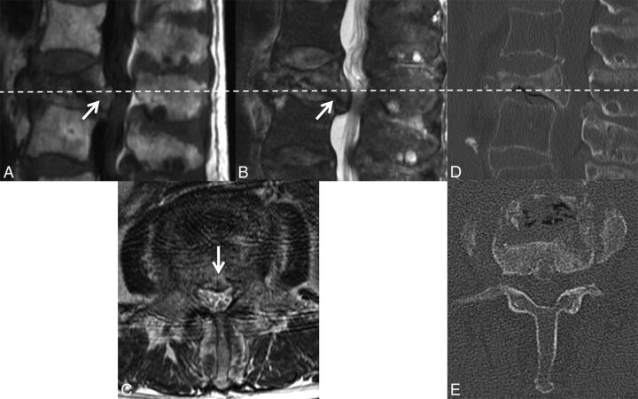
Retropulsion of a bone fragment in a benign VCF. Sagittal T1WI (A) and T2WI (B) with fat saturation of the lumbar spine demonstrate a retropulsed bone fragment (arrow) compressing the thecal sac and narrowing the spinal canal in this benign VCF (C), best seen on the axial T2WI. A similar appearance is demonstrated on the axial (D) and sagittal (E) reformatted thoracic spine CT scans.
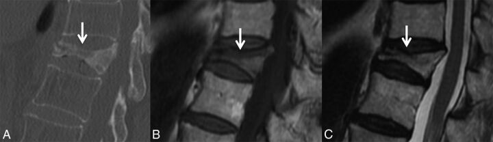
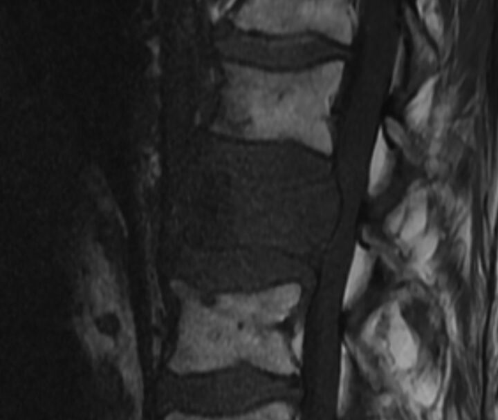
Linear horizontal fracture line in a benign VCF. As seen on the sagittal reformat from a thoracic spine CT in bone windows (A), there is a lucent fracture line (arrow) paralleling the superior endplate of T11. On MR imaging, this fracture is seen as a linear horizontal line (arrow) of T1 and T2 hypointensity through the T11 vertebral body, T1WI (B) and T2WI (C).
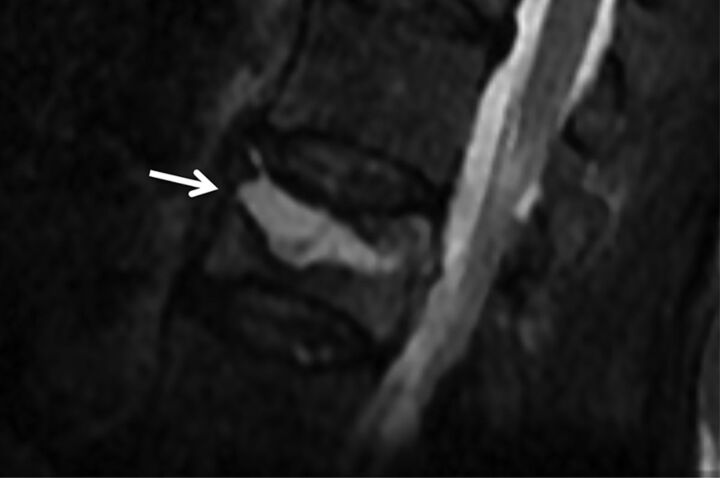
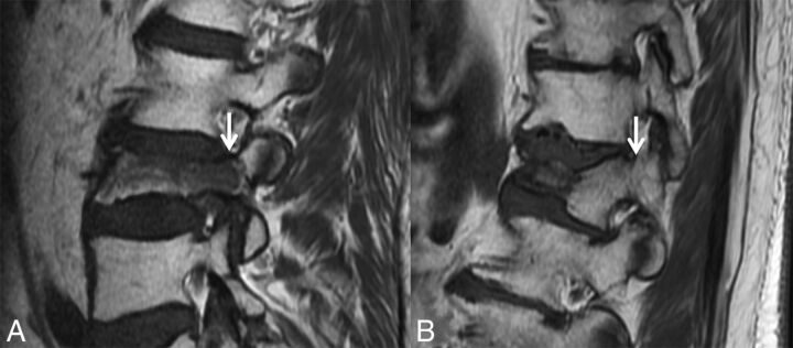
Fluid cleft in a benign VCF. Sagittal T2WI with fat saturation of the lumbar spine demonstrates a triangular fluid cleft (arrow) seen within this benign VCF.
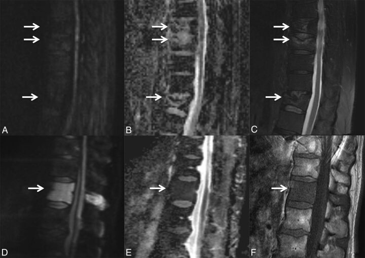
DWI of benign and malignant VCFs. Multiple benign osteoporotic VCFs (A–C, arrows) are seen in the lower thoracic spine. Sagittal DWI (A) and the corresponding ADC map (B) demonstrate the absence of diffusion restriction. Sagittal fat-saturated T2WI (C) demonstrates T2 hyperintensity about the fracture lines compatible with edema from an acute/subacute fracture. In contrast, malignant lymphomatous involvement of T12 (D–F, arrow) demonstrates diffuse diffusion restriction (D) with corresponding low ADC values (E). On the sagittal T1WI (F), there is slight loss of height of the superior and inferior endplates and diffuse T1 hypointensity compatible with marrow replacement.
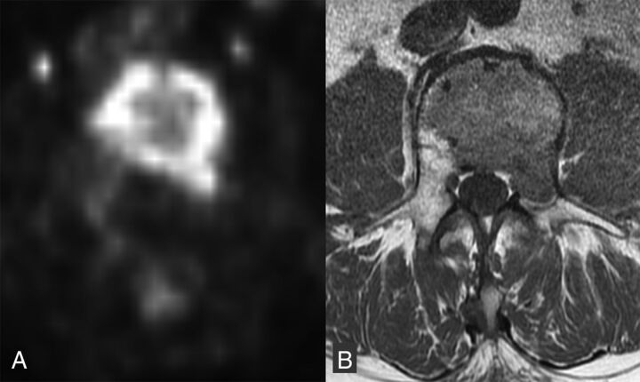
FDG avid malignant VCF. Axial non-attenuation-corrected PET (A) at the level of the malignant lumbar VCF with increased FDG activity throughout the vertebral body and into the left pedicle. Corresponding axial T1WI (B) shows the area of low T1 signal tumor infiltration throughout the vertebral body and left pedicle.
References
-
- Coleman RE. Skeletal complications of malignancy. Cancer 1997;80:1588–94 - PubMed
-
- Porter BA, Shields AF, Olson DO. Magnetic resonance imaging of bone marrow disorders. Radiol Clin North Am 1986;24:269–89 - PubMed
-
- Yamato M, Nishimura G, Kuramochi E, et al. MR appearance at different ages of osteoporotic compression fractures of the vertebrae. Radiat Med 1998;16:329–34 - PubMed
Publication types
MeSH terms
LinkOut - more resources
Full Text Sources
Other Literature Sources
Medical