Comparative Study
. 2018 Feb 23;293(8):2675-2686.
doi: 10.1074/jbc.RA117.000826. Epub 2018 Jan 18.

Thiosulfate sulfurtransferase-like domain-containing 1 protein interacts with thioredoxin

Affiliations
Comparative Study

Thiosulfate sulfurtransferase-like domain-containing 1 protein interacts with thioredoxin

Marouane Libiad et al. J Biol Chem. .

Abstract

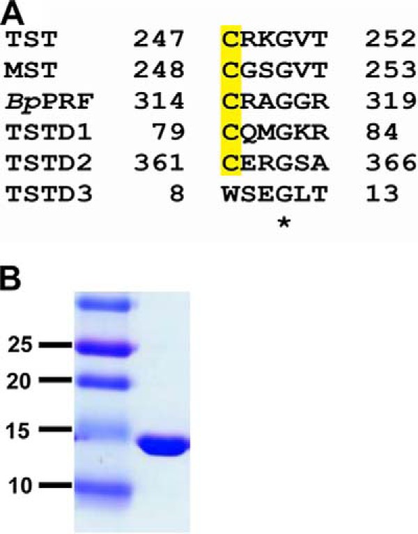
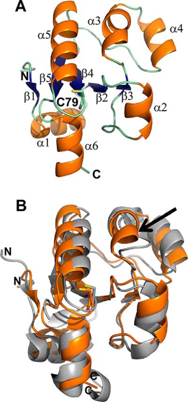
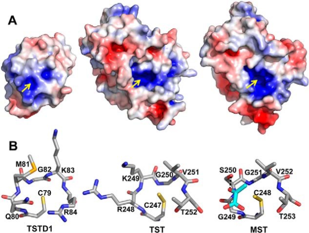
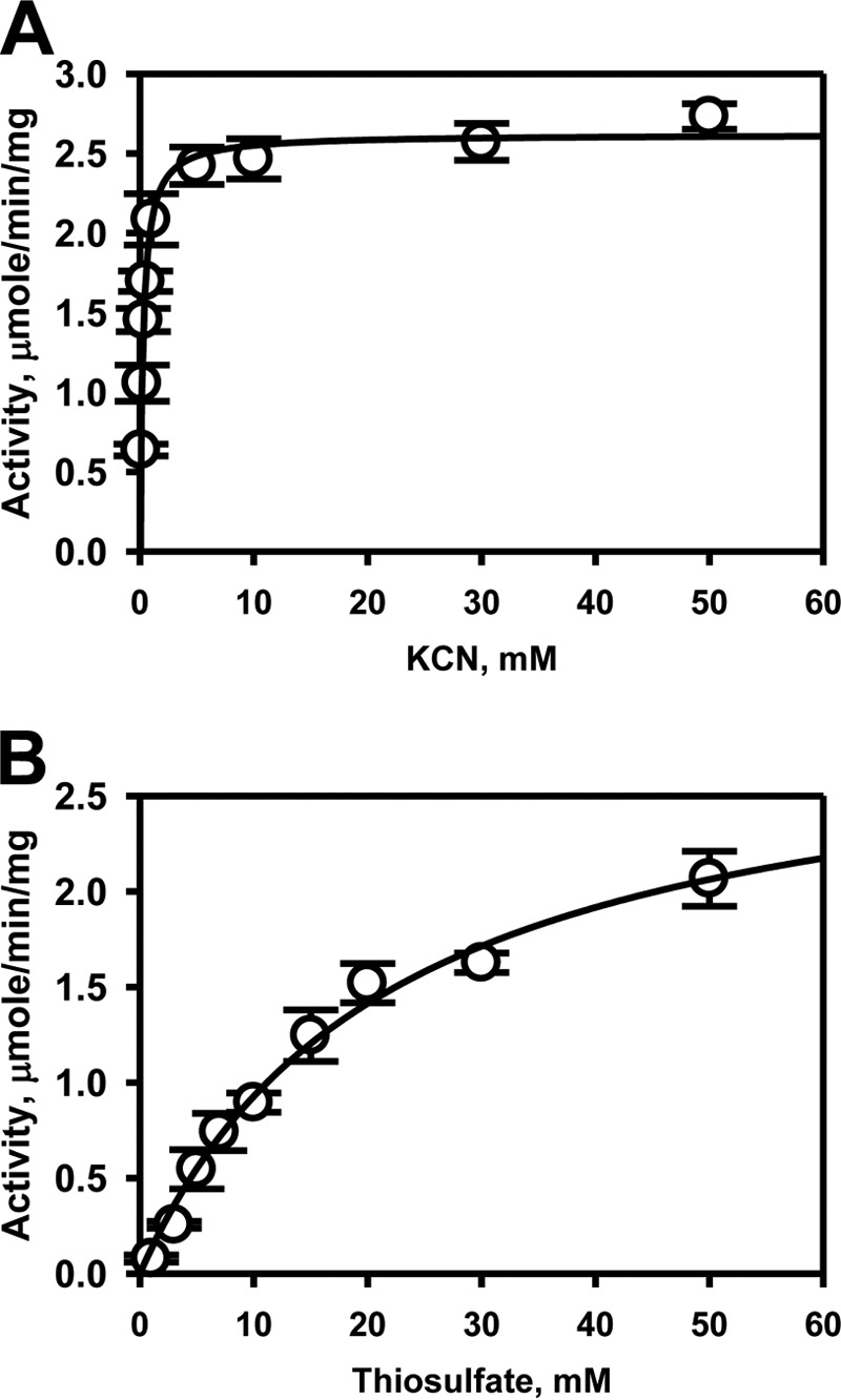
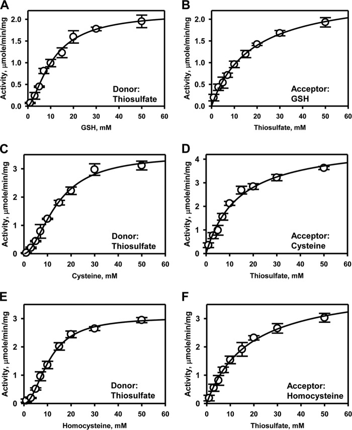
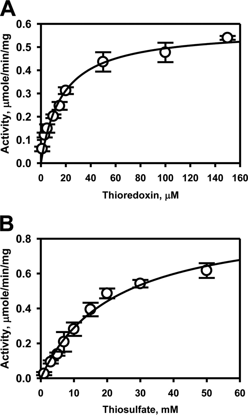
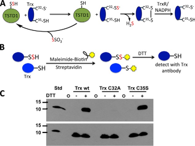
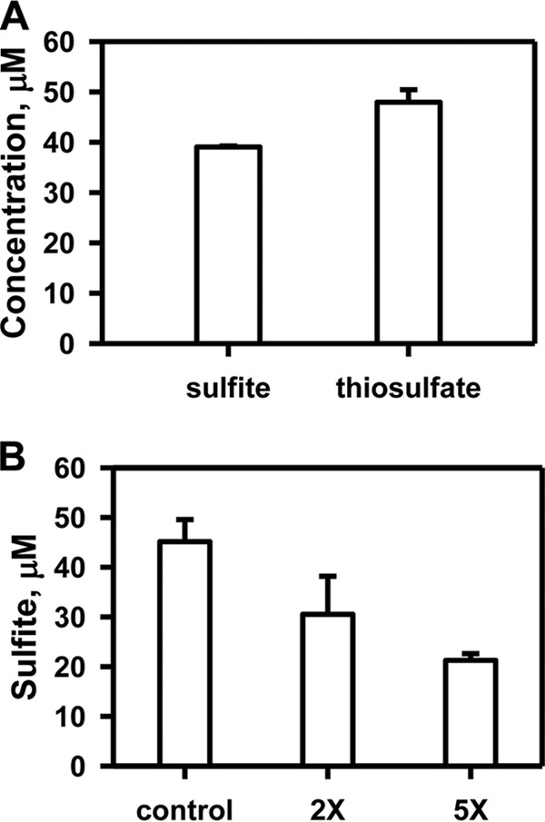
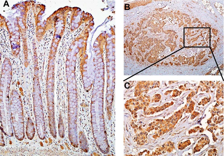
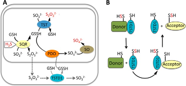
Rhodanese domains are structural modules present in the sulfurtransferase superfamily. These domains can exist as single units, in tandem repeats, or fused to domains with other activities. Despite their prevalence across species, the specific physiological roles of most sulfurtransferases are not known. Mammalian rhodanese and mercaptopyruvate sulfurtransferase are perhaps the best-studied members of this protein superfamily and are involved in hydrogen sulfide metabolism. The relatively unstudied human thiosulfate sulfurtransferase-like domain-containing 1 (TSTD1) protein, a single-domain cytoplasmic sulfurtransferase, was also postulated to play a role in the sulfide oxidation pathway using thiosulfate to form glutathione persulfide, for subsequent processing in the mitochondrial matrix. Prior kinetic analysis of TSTD1 was performed at pH 9.2, raising questions about relevance and the proposed model for TSTD1 function. In this study, we report a 1.04 Å resolution crystal structure of human TSTD1, which displays an exposed active site that is distinct from that of rhodanese and mercaptopyruvate sulfurtransferase. Kinetic studies with a combination of sulfur donors and acceptors reveal that TSTD1 exhibits a low Km for thioredoxin as a sulfane sulfur acceptor and that it utilizes thiosulfate inefficiently as a sulfur donor. The active site exposure and its interaction with thioredoxin suggest that TSTD1 might play a role in sulfide-based signaling. The apical localization of TSTD1 in human colonic crypts, which interfaces with sulfide-releasing microbes, and the overexpression of TSTD1 in colon cancer provide potentially intriguing clues as to its role in sulfide metabolism.

Keywords: crystal structure; enzyme kinetics; hydrogen sulfide; post-translational modification (PTM); sulfur; sulfur transferase.


Conflict of interest statement

The authors declare that they have no conflicts of interest with the contents of this article

Figures

Figure 1.
Figure 1.
Rhodanese (TST) sequence comparison and purity of recombinant human TSTD1. A, sequence alignment of active-site sequences in the human sulfurtransferases: TST, MST, B. phytofirmans PRF (BpPRF); and TSTD1, TSTD2, and TSTD3. The conserved active-site cysteine is highlighted in yellow. B, recombinant human TSTD1 (20 μg) was estimated to be >95% pure by SDS-PAGE analysis and Coomassie Blue staining. Molecular mass markers (in kDa) are shown on the left.
Figure 2.
Figure 2.
Crystal structure of TSTD1. The X-ray crystal structure of TSTD1 was solved at 1.04 Å resolution by SAD phasing with SeMet. A, the structure of the TSTD1 monomer consists of a five-stranded parallel β-sheet core (blue) surrounded by six α-helices (orange; α1–6). The active-site cysteine, Cys-79, is shown in a stick representation. B, comparison of TSTD1 with RDL1. Structural overlay of TSTD1 (orange) with the S. cerevisiae homolog RDL1 (PDB code 3D1P; gray). The active-site cysteine residues are shown in stick representation. The presence of an additional α-helix, α3, in TSTD1 is indicated by the arrow.
Figure 3.
Figure 3.
Comparison of TSTD1 with rhodanese. A, surface electrostatic potential representation of TSTD1 highlighting its exposed active-site pocket, bovine rhodanese (PDB code 1RHD), and MST (PDB code 4JGT). Positive and negative electrostatic potentials are shown in blue and red, respectively, in the range of ±5 kT/e. The yellow arrows point to the active-site cysteines. B, structural comparison of the rhodanese active-site loops of TSTD1, bovine rhodanese, and MST. Pyruvate bound in the active site of MST is shown in cyan.
Figure 4.
Figure 4.
Kinetic analysis of TSTD1 thiosulfate:cyanide sulfurtransferase activity. The dependence of the reaction velocity on cyanide concentration (0.02–50 mm) in the presence of 50 mm thiosulfate (A) and on thiosulfate concentrations (1–50 mm) in the presence of 30 mm cyanide (B) were determined. The data were fitted with the Michaelis–Menten equation. Data represent the mean ± S.D. (error bars) of three independent experiments performed in 300 mm HEPES, pH 7.4, 150 mm NaCl.
Figure 5.
Figure 5.
Kinetic analysis of TSTD1 thiosulfate:thiol sulfurtransferase activity. TSTD1 sulfurtransferase activity was determined in the presence of 50 mm thiosulfate and varying concentrations of GSH (A), cysteine (C), or homocysteine (E). Dependence of the reaction velocity on thiosulfate (1–50 mm) was determined in the presence of 50 mm GSH (B), 50 mm cysteine (D), or 50 mm homocysteine (F), as described under “Experimental Procedures.” The data were fitted with the Michaelis–Menten or Hill equations and represent the mean ± S.D. (error bars) of three independent experiments performed in 300 mm HEPES, pH 7.4, 150 mm NaCl. Hill coefficients for the thiol acceptors GSH, cysteine, and homocysteine are 1.8 ± 0.2, 1.9 ± 0.1, and 2.0 ± 0.1, respectively.
Figure 6.
Figure 6.
Kinetic analysis of TSTD1 thiosulfate-thioredoxin sulfur transfer activity. TSTD1 sulfur transfer activity to thioredoxin was determined in the presence of 50 mm thiosulfate and varying concentrations of thioredoxin (A) and 150 μm thioredoxin and varying concentrations of thiosulfate (B). Reactions were performed as described under “Experimental Procedures” and fitted with the Michaelis–Menten equation. Data represent the mean ± S.D. (error bars) of three independent experiments performed in 300 mm HEPES, pH 7.4, 150 mm NaCl.
Figure 7.
Figure 7.
Mechanism of TSTD1-dependent persulfidation of thioredoxin and its detection by the biotin thiol assay. A, mechanism of sulfur transfer from thiosulfate to thioredoxin (Trx) via TSTD1. Cys-32 is the nucleophilic cysteine, and Cys-35 is the resolving cysteine. Oxidized thioredoxin is recycled by thioredoxin reductase (TrxR) and NADPH. B, strategy for detection of persulfidated thioredoxin using the biotin thiol assay. Reactive and accessible persulfide and thiol groups on thioredoxin are alkylated by maleimide biotin and immobilized on a streptavidin column. Following DTT treatment, only thioredoxin, which had a persulfide group, is released from the column and can be detected by immunoblotting. C, Western blot analysis of TSTD1-dependent persulfidation of thioredoxin from the biotin thiol assay. A representative Western blot is shown in which elution of wildtype thioredoxin (Trx wt) and the C35S but not the C32A mutant was detected following DTT treatment of the loaded streptavidin column. The bottom panel represents an equal loading control for the presence of thioredoxin in each sample before loading on the streptavidin column. The first lane contains purified wildtype thioredoxin (Trx wt STD; 35 ng). The circles represent empty lanes. The position of the molecular mass markers (in kDa) is shown on the left.
Figure 8.
Figure 8.
Product inhibition of TSTD1 in the GSSH-sulfite sulfur transfer reaction. A, analysis of TSTD1 reaction components under single-turnover conditions in the presence of 50 μm GSSH, 50 μm sulfite, and 50 μm enzyme. B, substrate consumption by TSTD1 under single-turnover reaction was performed in the presence of 50 μm each of GSSH, sulfite, and TSTD1 and 0 μm (control), 100 μm (2×), or 250 μm (5×) thiosulfate. Data are representative of three independent experiments each performed in duplicate in 100 mm HEPES, pH 7.4, 150 mm NaCl. Error bars, S.D.
Figure 9.
Figure 9.
Immunohistochemical localization of TSTD1 in human colon. TSTD1 was localized in normal epithelium (A) and in colorectal cancer tissue (B) at ×10 magnification. C, close-up of the area marked in B shown at a magnification of ×40.
Figure 10.
Figure 10.
Alternative proposed roles for TSTD1. A, in one model of the mitochondrial sulfide oxidation pathway denoted by gray arrows, the product of sulfide quinone oxidoreductase (SQR) is thiosulfate (S2O32−), which is further processed in the cytoplasm by TSTD1. The product of this reaction is GSSH, which must go across the mitochondrial membranes to be completely oxidized to sulfate. This pathway is considered to be unlikely because the major product of the SQR reaction at physiologically relevant sulfite and glutathione concentrations is predicted to be GSSH. In the mitochondrially contained sulfide oxidation pathway, H2S is progressively oxidized by SQR, persulfide dioxygenase (PDO), TST, and sulfite oxidase (SO) to give thiosulfate and sulfate, which are the major products of H2S oxidation. The input (H2S) and outputs (S2O32−, SO42−) of the sulfide oxidation pathway are highlighted in red. B, based on the structure of TSTD1 and its ability to transfer sulfane sulfur to thioredoxin, we propose that TSTD1 functions in sulfur relay shuttles between donor and acceptor proteins. Alternatively, thioredoxin could potentially function as a persulfide donor and TSTD1 as an intermediary carrier for sulfane sulfur donation to acceptors.

References

    1. Abe K., and Kimura H. (1996) The possible role of hydrogen sulfide as an endogenous neuromodulator. J. Neurosci. 16, 1066–1071 - PMC - PubMed
    1. Sodha N. R., Clements R. T., Feng J., Liu Y., Bianchi C., Horvath E. M., Szabo C., Stahl G. L., and Sellke F. W. (2009) Hydrogen sulfide therapy attenuates the inflammatory response in a porcine model of myocardial ischemia/reperfusion injury. J. Thorac. Cardiovasc. Surg. 138, 977–984 10.1016/j.jtcvs.2008.08.074 - DOI - PMC - PubMed
    1. Ha C., Tian S., Sun K., Wang D., Lv J., and Wang Y. (2015) Hydrogen sulfide attenuates IL-1β-induced inflammatory signaling and dysfunction of osteoarthritic chondrocytes. Int. J. Mol. Med. 35, 1657–1666 10.3892/ijmm.2015.2183 - DOI - PubMed
    1. Calvert J. W., Jha S., Gundewar S., Elrod J. W., Ramachandran A., Pattillo C. B., Kevil C. G., and Lefer D. J. (2009) Hydrogen sulfide mediates cardioprotection through Nrf2 signaling. Circ. Res. 105, 365–374 10.1161/CIRCRESAHA.109.199919 - DOI - PMC - PubMed
    1. Elrod J. W., Calvert J. W., Morrison J., Doeller J. E., Kraus D. W., Tao L., Jiao X., Scalia R., Kiss L., Szabo C., Kimura H., Chow C. W., and Lefer D. J. (2007) Hydrogen sulfide attenuates myocardial ischemia-reperfusion injury by preservation of mitochondrial function. Proc. Natl. Acad. Sci. U.S.A. 104, 15560–15565 10.1073/pnas.0705891104 - DOI - PMC - PubMed

Publication types

MeSH terms