. 2018 Feb 23;293(8):2744-2754.
doi: 10.1074/jbc.RA117.001611. Epub 2018 Jan 18.

CRISPR RNA and anti-CRISPR protein binding to the Xanthomonas albilineans Csy1-Csy2 heterodimer in the type I-F CRISPR-Cas system

Affiliations

CRISPR RNA and anti-CRISPR protein binding to the Xanthomonas albilineans Csy1-Csy2 heterodimer in the type I-F CRISPR-Cas system

Suji Hong et al. J Biol Chem. .

Erratum in

Abstract

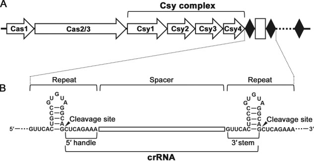
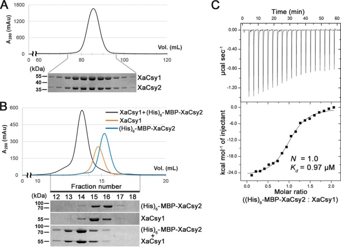
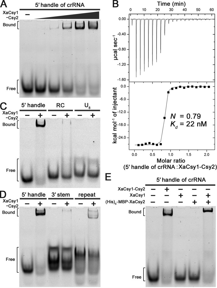
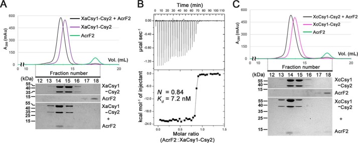
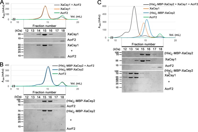
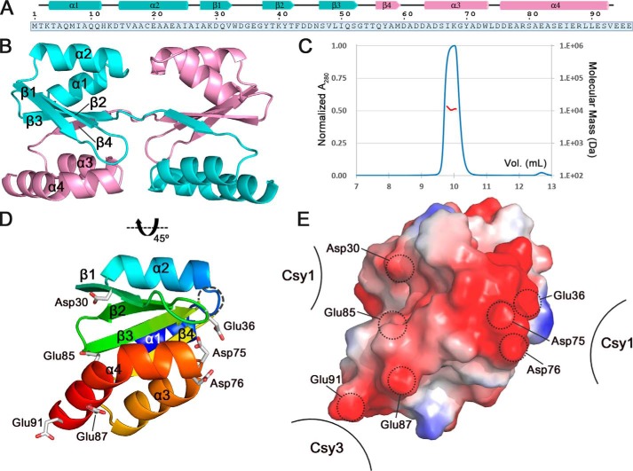
Clustered regularly interspaced short palindromic repeats (CRISPRs) and CRISPR-associated (Cas) proteins provide microbial adaptive immunity against bacteriophages. In type I-F CRISPR-Cas systems, multiple Cas proteins (Csy1-4) compose a surveillance complex (Csy complex) with CRISPR RNA (crRNA) for target recognition. Here, we report the biochemical characterization of the Csy1-Csy2 subcomplex from Xanthomonas albilineans, including the analysis of its interaction with crRNA and AcrF2, an anti-CRISPR (Acr) protein from a phage that infects Pseudomonas aeruginosa The X. albilineans Csy1 and Csy2 proteins (XaCsy1 and XaCsy2, respectively) formed a stable heterodimeric complex that specifically bound the 8-nucleotide (nt) 5'-handle of the crRNA. In contrast, the XaCsy1-XaCsy2 heterodimer exhibited reduced affinity for the 28-nt X. albilineans CRISPR repeat RNA containing the 5'-handle sequence. Chromatographic and calorimetric analyses revealed tight binding between the Acr protein from the P. aeruginosa phage and the heterodimeric subunit of the X. albilineans Csy complex, suggesting that AcrF2 recognizes conserved features of Csy1-Csy2 heterodimers. We found that neither XaCsy1 nor XaCsy2 alone forms a stable complex with AcrF2 and the 5'-handle RNA, indicating that XaCsy1-XaCsy2 heterodimerization is required for binding them. We also solved the crystal structure of AcrF2 to a resolution of 1.34 Å, enabling a more detailed structural analysis of the residues involved in the interactions with the Csy1-Csy2 heterodimer. Our results provide information about the order of events during the formation of the multisubunit crRNA-guided surveillance complex and suggest that the Acr protein inactivating type I-F CRISPR-Cas systems has broad specificity.

Keywords: CRISPR/Cas; RNA-protein interaction; X-ray crystallography; anti-CRISPR; crRNA; crystal structure; protein complex; protein-protein interaction.


Conflict of interest statement

The authors declare that they have no conflicts of interest with the contents of this article

Figures

Figure 1.
Figure 1.
Type I-F CRISPR-Cas system of X. albilineans. A and B, schematic representations of type I-F CRISPR-Cas system (A) and crRNA (B) from X. albilineans. The type I-F CRISPR-Cas system in X. albilineans includes six Cas proteins and a single CRISPR locus consisting of 24 repeats (28 nt; black diamonds) interspaced with 23 spacer sequences (32 nt; white rectangles). A long pre-crRNA is cleaved by Csy4 endoribonuclease into short crRNAs consisting of an 8-nt 5′-handle, a 32-nt spacer, and a 20-nt 3′-stem. The cleavage sites in the repeat regions are also indicated.
Figure 2.
Figure 2.
XaCsy1 interacts with XaCsy2 to form a heterodimeric complex. A, coelution of XaCsy1 and XaCsy2 in SEC using a HiLoad 16/60 Superdex 200 column during the purification of XaCsy1-Csy2 heterodimer. XaCsy1 and XaCsy2 were coexpressed in E. coli cells and purified as described under “Experimental procedures”. The elution fractions of the SEC were analyzed by SDS-PAGE and visualized by Coomassie staining. The protein bands were analyzed by mass spectrometry (Table S1). B, the interaction between XaCsy1 and XaCsy2 in analytical SEC using a Superdex 200 10/300 GL column. Separately purified XaCsy1 (20 μm) and His6-MBP–tagged XaCsy2 (20 μm) were used for the experiment. Elution fractions were analyzed by SDS-PAGE and visualized by Coomassie staining. C, ITC trace for XaCsy1 binding to XaCsy2. The His6-MBP–tagged XaCsy2 was added consecutively to the chamber containing XaCsy1. The experimentally determined N and Kd values are also indicated. mAU, milliabsorbance units.
Figure 3.
Figure 3.
XaCsy1-Csy2 heterodimer binds to the 5′-handle of the crRNA in a sequence specific manner. A, EMSA of the 8-nt 5′-handle of the crRNA was performed with increasing amounts (0.25, 0.5, 1.0, 2.0, and 4.0 μm) of XaCsy1-Csy2 heterodimer. B, ITC analysis for the binding of the 5′-handle of the crRNA to XaCsy1-Csy2 heterodimer. The 5′-handle RNA was injected consecutively into the solution containing XaCsy1-Csy2 heterodimer. C and D, the assays were carried out using an identical amount of XaCsy1-Csy2 heterodimer with various 8-mer RNAs, such as the reverse complement (RC) of the 5′-handle and a poly(U) RNA (U8) (C), and with different parts of the crRNA (D). E, EMSA of the 5′-handle of the crRNA was performed with individual components of the XaCsy1-Csy2 heterodimer using the separately purified XaCsy1 and His6-MBP–tagged XaCsy2 proteins.
Figure 4.
Figure 4.
Binding of AcrF2, an anti-CRISPR protein from a P. aeruginosa phage, to Xanthomonas Csy1-Csy2 heterodimers. A, interaction between AcrF2 and XaCsy1-Csy2 heterodimer in analytical size-exclusion chromatography. Elution fractions were analyzed by SDS-PAGE and visualized by Coomassie staining. B, ITC trace for binding of AcrF2 to the XaCsy1-Csy2 heterodimer. AcrF2 was injected consecutively into the chamber containing XaCsy1-Csy2 heterodimer. C, binding of AcrF2 to Csy1-Csy2 heterodimer from another Xanthomonas bacterium, X. citri. Elution fractions of analytical SEC were analyzed by SDS-PAGE and visualized by Coomassie staining. mAU, milliabsorbance units.
Figure 5.
Figure 5.
Heterodimerization of XaCsy1 and XaCsy2 is required for binding to AcrF2. A and B, analytical SEC experiments for testing the interaction between AcrF2 and individual components of the XaCsy1-Csy2 heterodimer, XaCsy1 (A) and XaCsy2 (B). AcrF2 did not coelute with the separately purified XaCsy1 or His6-MBP–tagged XaCsy2 proteins. C, the formation of the ternary complex containing XaCsy1, XaCsy2, and AcrF2 in analytical size-exclusion chromatography. The AcrF2 binding was recovered when the separately purified XaCsy1 and His6-MBP–tagged XaCsy2 proteins were incubated together with AcrF2. Elution fractions were analyzed by SDS-PAGE and visualized by Coomassie staining. mAU, milliabsorbance units.
Figure 6.
Figure 6.
Crystal structure of AcrF2. A, schematic representation of the secondary structure of AcrF2. The swapped C-terminal region is shown in pink. The amino acid sequence of AcrF2 is shown and numbered below. B, dimeric assembly of AcrF2 formed by swapping the C-terminal regions in the crystal lattice. Chains A and B are shown in cyan and pink, respectively. Secondary structure elements are also indicated. C, the monomeric state of AcrF2 in solution was detected by SEC-MALS analysis. Blue and red lines represent the normalized absorbance at 280 nm and the molecular mass for AcrF2, respectively. The experimentally measured and theoretically calculated molecular masses of AcrF2 are 11.3 and 10.6 kDa, respectively. D, biologically relevant monomeric unit of AcrF2. The monomeric structure of AcrF2 is shown in rainbow format from the N terminus (blue) to the C terminus (red). Carboxyl side chains involved in interactions with the Csy complex are shown in stick representations. E, electrostatic potential surface (red = −75 kT, blue = +75 kT) of the AcrF2 monomer. PyMOL with the Adaptive Poisson-Boltzmann Solver plugin (50) was used to generate the surface. Acidic residues of AcrF2 and their interacting Csy subunits are also indicated.

Similar articles

Cited by

References

    1. Barrangou R., Fremaux C., Deveau H., Richards M., Boyaval P., Moineau S., Romero D. A., and Horvath P. (2007) CRISPR provides acquired resistance against viruses in prokaryotes. Science 315, 1709–1712 10.1126/science.1138140 - DOI - PubMed
    1. Brouns S. J., Jore M. M., Lundgren M., Westra E. R., Slijkhuis R. J., Snijders A. P., Dickman M. J., Makarova K. S., Koonin E. V., and van der Oost J. (2008) Small CRISPR RNAs guide antiviral defense in prokaryotes. Science 321, 960–964 10.1126/science.1159689 - DOI - PMC - PubMed
    1. Wiedenheft B., Sternberg S. H., and Doudna J. A. (2012) RNA-guided genetic silencing systems in bacteria and archaea. Nature 482, 331–338 10.1038/nature10886 - DOI - PubMed
    1. Marraffini L. A. (2015) CRISPR-Cas immunity in prokaryotes. Nature 526, 55–61 10.1038/nature15386 - DOI - PubMed
    1. Bolotin A., Quinquis B., Sorokin A., and Ehrlich S. D. (2005) Clustered regularly interspaced short palindrome repeats (CRISPRs) have spacers of extrachromosomal origin. Microbiology 151, 2551–2561 10.1099/mic.0.28048-0 - DOI - PubMed

Publication types

MeSH terms

LinkOut - more resources