. 2018 Mar 2;293(9):3350-3362.
doi: 10.1074/jbc.RA117.001516. Epub 2018 Jan 18.

The α-arrestin ARRDC3 suppresses breast carcinoma invasion by regulating G protein-coupled receptor lysosomal sorting and signaling

Affiliations

The α-arrestin ARRDC3 suppresses breast carcinoma invasion by regulating G protein-coupled receptor lysosomal sorting and signaling

Aleena K S Arakaki et al. J Biol Chem. .

Abstract

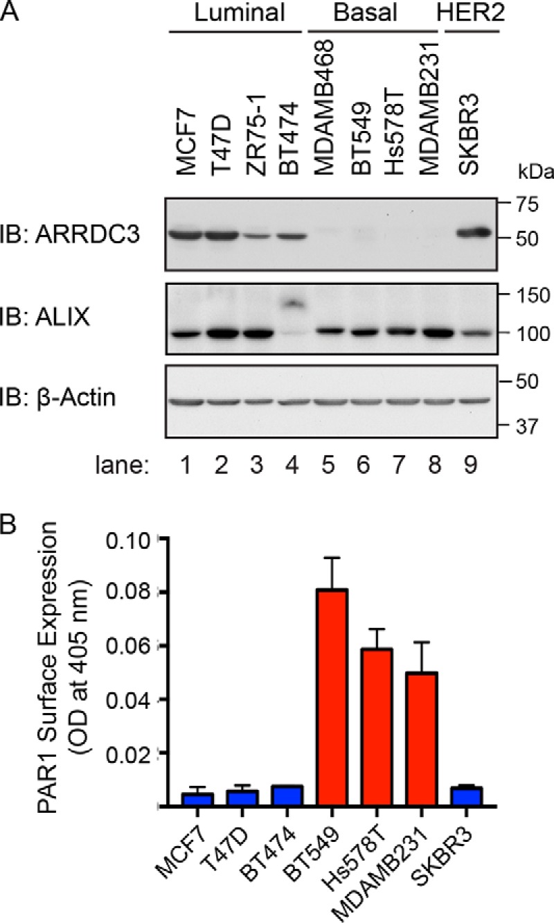
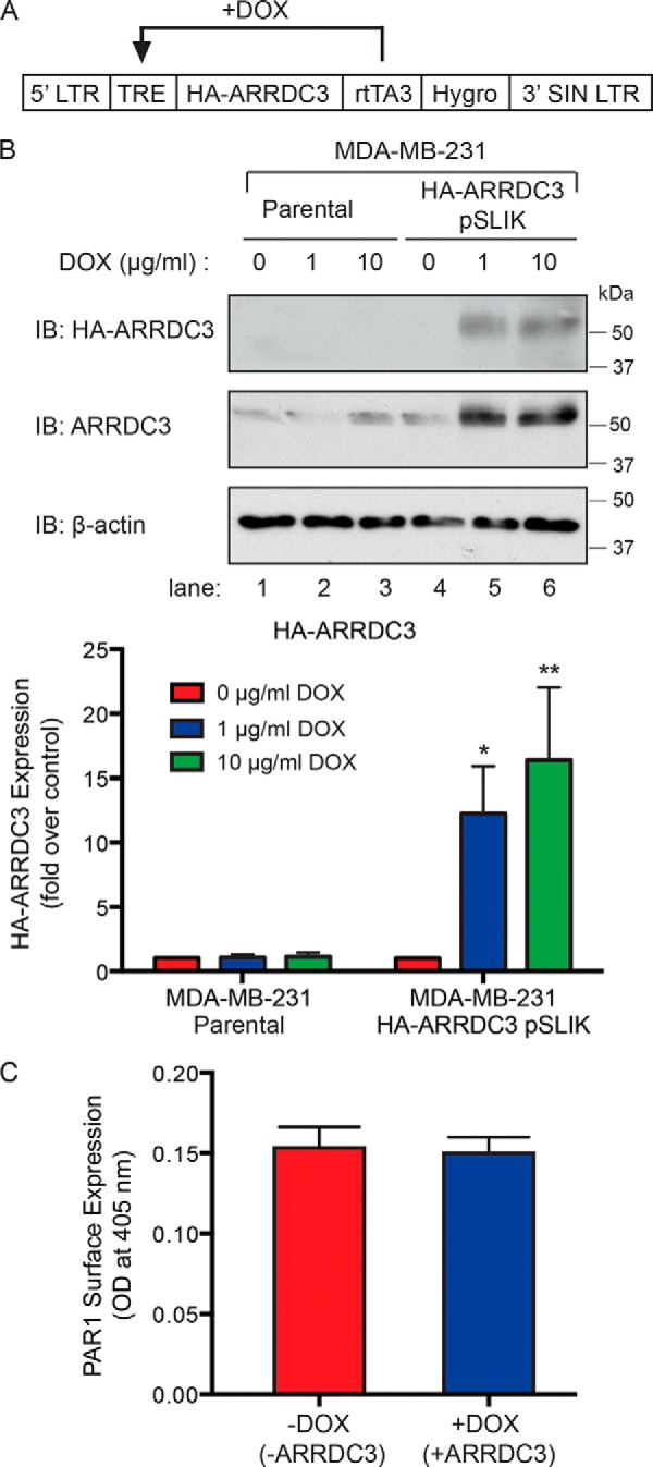
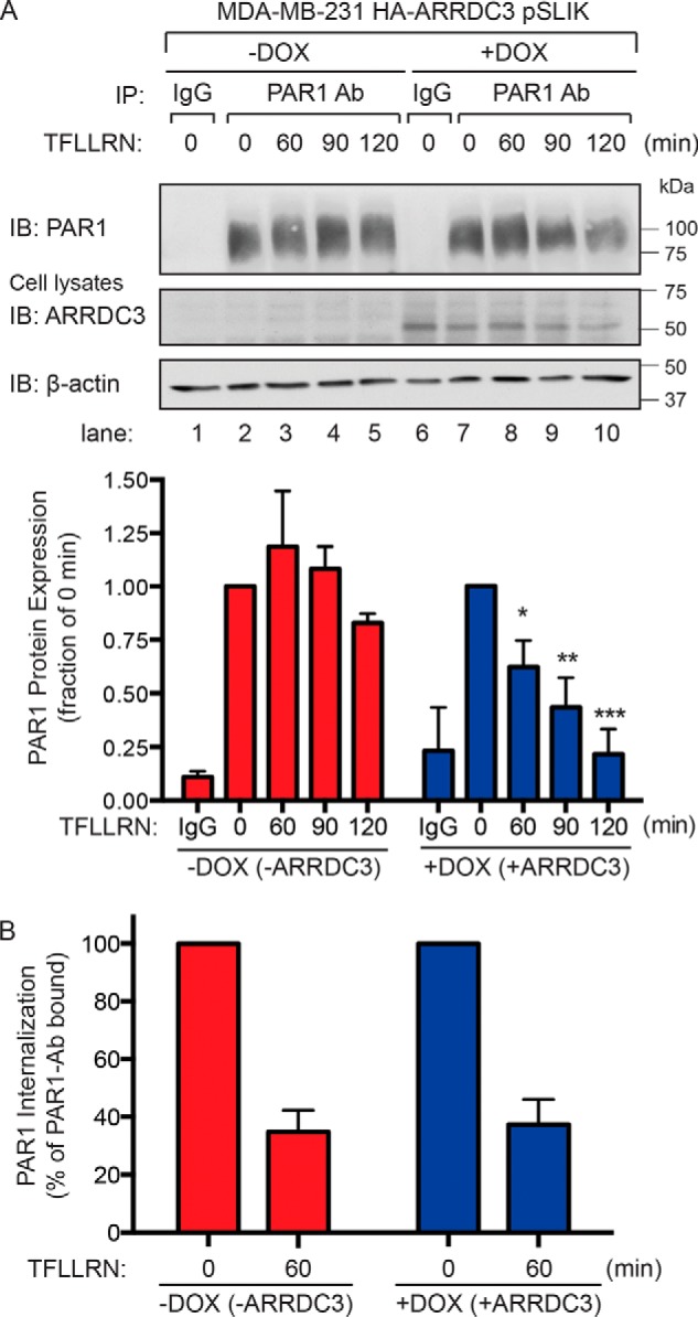
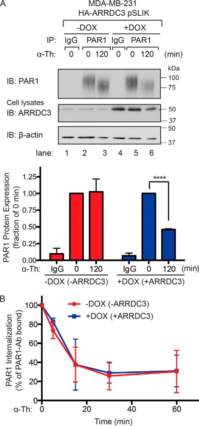
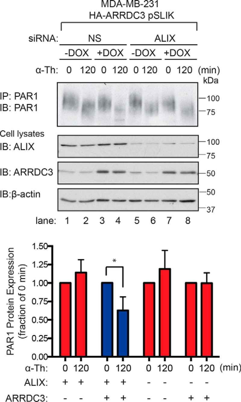
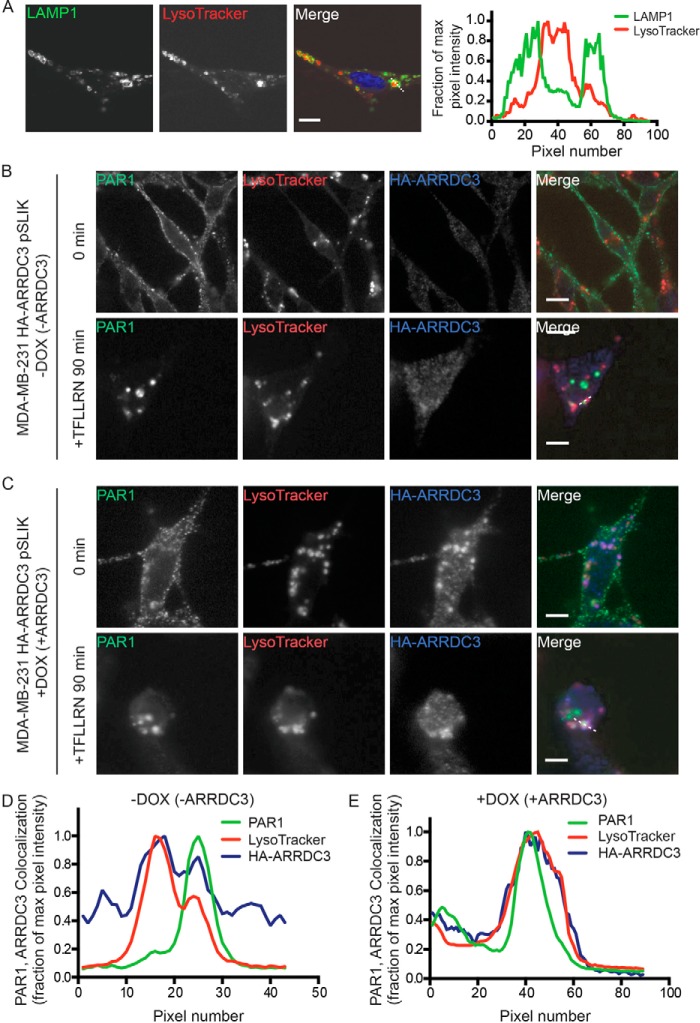
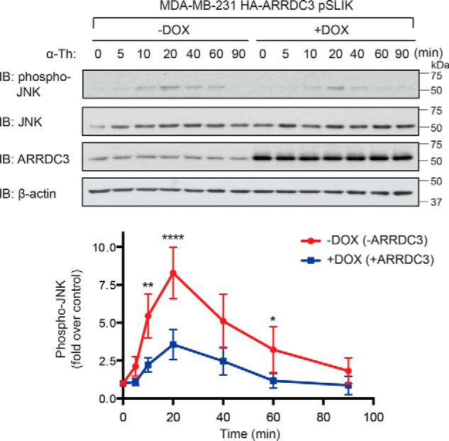
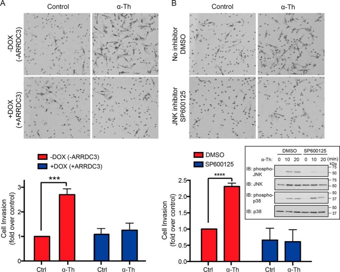
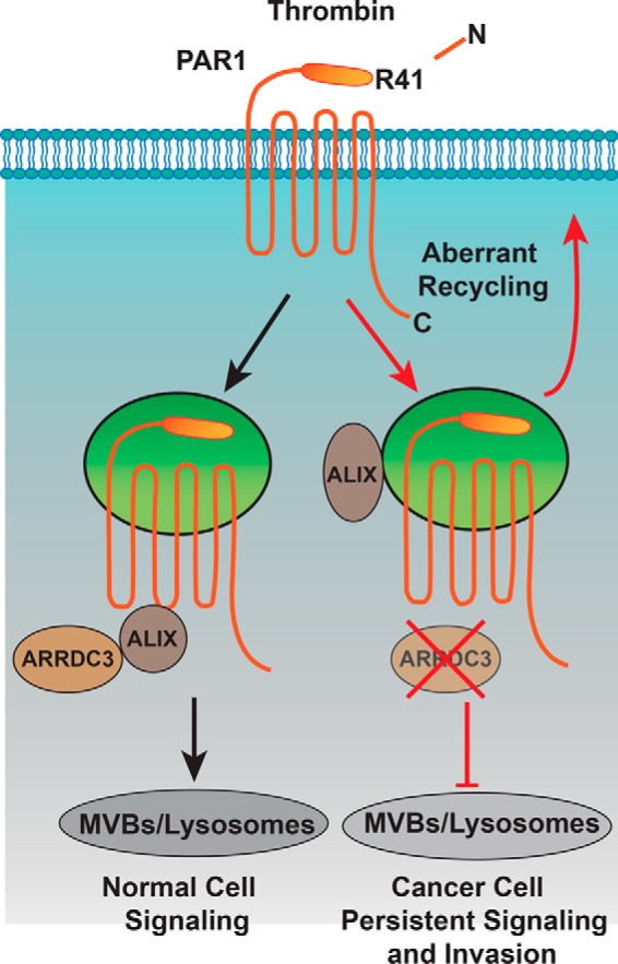
Aberrant G protein-coupled receptor (GPCR) expression and activation has been linked to tumor initiation, progression, invasion, and metastasis. However, compared with other cancer drivers, the exploitation of GPCRs as potential therapeutic targets has been largely ignored, despite the fact that GPCRs are highly druggable. Therefore, to advance the potential status of GPCRs as therapeutic targets, it is important to understand how GPCRs function together with other cancer drivers during tumor progression. We now report that the α-arrestin domain-containing protein-3 (ARRDC3) acts as a tumor suppressor in part by controlling signaling and trafficking of the GPCR, protease-activated receptor-1 (PAR1). In a series of highly invasive basal-like breast carcinomas, we found that expression of ARRDC3 is suppressed whereas PAR1 is aberrantly overexpressed because of defective lysosomal sorting that results in persistent signaling. Using a lentiviral doxycycline-inducible system, we demonstrate that re-expression of ARRDC3 in invasive breast carcinoma is sufficient to restore normal PAR1 trafficking through the ALG-interacting protein X (ALIX)-dependent lysosomal degradative pathway. We also show that ARRDC3 re-expression attenuates PAR1-stimulated persistent signaling of c-Jun N-terminal kinase (JNK) in invasive breast cancer. Remarkably, restoration of ARRDC3 expression significantly reduced activated PAR1-induced breast carcinoma invasion, which was also dependent on JNK signaling. These findings are the first to identify a critical link between the tumor suppressor ARRDC3 and regulation of GPCR trafficking and signaling in breast cancer.

Keywords: GPCR; Rho (Rho GTPase); beta-arrestin; breast cancer; c-Jun N-terminal kinase (JNK); protease; protease-activated receptor; thrombin; trafficking.


Conflict of interest statement

The authors declare that they have no conflicts of interest with the contents of this article

Figures

Figure 1.
Figure 1.
PAR1 and ARRDC3 protein expression are inversely correlated in breast carcinoma cell lines. A, equivalent amounts (20 μg) of cell lysates from various breast cancer cell lines were immunoblotted for ALIX and ARRDC3 expression. β-Actin expression was determined as a loading control. B, PAR1 cell-surface expression was determined by ELISA. Data shown (mean ± S.D., n = 3) are representative of three independent experiments.
Figure 2.
Figure 2.
Induction of ARRDC3 expression in MDA-MB-231 breast carcinoma cell line using pSLIK vector system. A, schematic of the tetracycline-inducible pSLIK system for HA-tagged ARRDC3 expression. rtTA3, reverse tetracycline transactivator; TRE, tetracycline response element; LTR, long terminal repeat. B, cell lysates from MDA-MB-231 parental and HA-ARRDC3 pSLIK–expressing cells were collected after 48 h of incubation with DOX treatment at 0, 1, or 10 μg/ml. Cell lysates were immunoblotted to detect endogenous ARRDC3 or HA-ARRDC3 expression. β-Actin expression was detected as a control. The data shown (mean ± S.D. (error bars), n = 3) were quantified by densitometry and shown as the -fold change in ARRDC3 expression relative to 0 min control following doxycycline incubation. Statistical significance was determined by one-way ANOVA (*, p < 0.05; **, p < 0.01; n = 3). C, expression of PAR1 on the cell surface was determined before and after DOX (1 μg/ml) treatment for 48 h and determined by ELISA. The data (mean ± S.D., n = 3) shown as optical density determined at 405 nm are representative of three independent experiments.
Figure 3.
Figure 3.
Agonist peptide–induced PAR1 lysosomal degradation is restored in cells re-expressing ARRDC3. A, MDA-MB-231 HA-ARRDC3 pSLIK cells were treated with or without 1 μg/ml DOX for 48 h. Cells were then stimulated with 100 μm TFLLRN peptide agonist for the indicated times, lysed, and immunoprecipitated using the anti-PAR1 WEDE antibody or anti-IgG antibody as a control. Immunoprecipitates were immunoblotted with anti-PAR1 rabbit polyclonal antibody. Cell lysates were immunoblotted with anti-HA antibody to detect ARRDC3. β-Actin expression was determined as a control. The data shown (mean ± S.D. (error bars), n = 3) were quantified by densitometry and represented as PAR1 expression relative to unstimulated control (0 min). Statistical significance was determined by one-way ANOVA (*, p < 0.05; **, p < 0.01; ***, p < 0.001; n = 3). B, MDA-MB-231 HA-ARRDC3 pSLIK cells were treated with or without 1 μg/ml DOX for 48 h, labeled with anti-PAR1 WEDE antibody at 4 °C, and then stimulated with or without 100 μm TFLLRN for the indicated times. Cells were then fixed, and the amount of cell-surface PAR1 was determined by ELISA. The data (mean ± S.D., n = 3) are represented as the percentage of PAR1 remaining on the cell surface relative to untreated control (0 min) and representative of three independent experiments.
Figure 4.
Figure 4.
Thrombin-induced PAR1 lysosomal degradation is restored in cells re-expressing ARRDC3. A, MDA-MB-231 HA-ARRDC3 pSLIK cells incubated with or without 1 μg/ml DOX for 48 h were treated with 10 nm α-thrombin for the indicated times, lysed, and immunoprecipitated with anti-PAR1 WEDE antibody or anti-IgG control. Cell lysates were immunoblotted for HA-ARRDC3 and β-actin expression. The data (mean ± S.D. (error bars), n = 3) are represented as the fraction of PAR1 protein remaining relative to 0 min control. Statistical significance was determined using an unpaired t test (****, p < 0.0001; n = 3). B, MDA-MB-231 HA-ARRDC3 pSLIK cells were incubated with or without 1 μg/ml DOX for 48 h, prelabeled with anti-PAR1 WEDE antibody at 4 °C, and then treated with or without 10 nm α-thrombin. The amount of PAR1 remaining on the cell surface was determined by ELISA. The data (mean ± S.D., n = 3) are expressed as the percentage of PAR1 remaining relative to 0 min control and representative of three independent experiments.
Figure 5.
Figure 5.
ALIX is required for ARRDC3-mediated degradation of activated PAR1. MDA-MB-231 HA-ARRDC3 pSLIK cells were transfected with 25 nm nonspecific (NS) or 25 nm of ALIX siRNA using a combination of 12.5 nm ALIX 1 and 12.5 nm ALIX 3 siRNAs and then incubated with or without 1 μg/ml DOX for 48 h. Cells were then stimulated with 10 nm α-thrombin for the indicated times, lysed, and immunoprecipitated using the anti-PAR1 WEDE antibody. Immunoprecipitates were immunoblotted with anti-PAR1 rabbit antibody to detect PAR1 expression. ALIX, HA-ARRDC3, and β-actin expression were determined by immunoblotting cell lysates. The data (mean ± S.D. (error bars), n = 3) are represented as the fraction of PAR1 remaining relative to 0 min control and are representative of three independent experiments. Statistical significance was determined by unpaired t test (*, p < 0.05; n = 3).
Figure 6.
Figure 6.
ARRDC3 expression is required for activated PAR1 lysosomal trafficking. A, MDA-MB-231 HA-ARRDC3 pSLIK cells incubated with 100 nm LysoTracker were fixed, processed, immunostained for LAMP1, and imaged by confocal microscopy. Images are representative of many cells examined in three independent experiments. Scale bars, 10 μm. Line-scan analysis of the white dotted line region is plotted as the fraction of maximum pixel intensity versus pixel number (distance) and demonstrates that LysoTracker (red) accumulates in the lumen of LAMP1-positive lysosomes (green). MDA-MB-231 HA-ARRDC3 pSLIK cells treated without (B) or with (C) 1 μg/ml DOX for 48 h were incubated with 2 mm leupeptin and 100 nm LysoTracker at 37 °C. Cells were then incubated with anti-PAR1 antibody to label the surface cohort, stimulated with 100 μm TFLLRN, fixed, processed, and imaged by confocal microscopy. Images are representative of many cells examined in three independent experiments. Scale bars, 10 μm. Line-scan analysis of the white dotted line region is plotted as described above and indicates that activated PAR1 (green) accumulates in the lumen of LAMP1-positive lysosomes (red) in the presence of ARRDC3 (blue) (E) but not in lysosomes in the absence of ARRDC3 expression (D).
Figure 7.
Figure 7.
ARRDC3 re-expression attenuates PAR1-stimulated JNK signaling. MDA-MB-231 HA-ARRDC3 pSLIK cells treated with or without 1 μg/ml DOX for 48 h were stimulated with 10 nm α-thrombin for the indicated times. Cells were lysed and immunoblotted for phospho-JNK1/2, total JNK, HA-ARRDC3, and β-actin expression. The data shown (mean ± S.D. (error bars), n = 3) were quantified by densitometry and are represented as the -fold increase in JNK1/2 phosphorylation relative to 0 min control. Statistical significance was determined by one-way ANOVA (**, p < 0.01; ****, p < 0.0001; n = 3).
Figure 8.
Figure 8.
PAR1-stimulated breast carcinoma invasion is suppressed by ARRDC3 and JNK inhibition. A, MDA-MB-231 HA-ARRDC3 pSLIK cells treated with or without 1 μg/ml DOX for 48 h were serum-starved cells, seeded onto transwells coated with Matrigel, and incubated with or without 1 pm α-thrombin (α-Th) for 5 h at 37 °C. Cells were fixed, stained, and imaged. Images shown are representative of three independent experiments. The data (mean ± S.D. (error bars), n = 3) were quantified from nine different fields of view at ×10 magnification for each condition and are represented as the -fold change over untreated control cells. Statistical significance was determined by unpaired t test (***, p < 0.001; n = 3). B, MDA-MB-231 HA-ARRDC3 pSLIK cells were preincubated with DMSO or 20 μm SP600125 JNK inhibitor for 2 h at 37 °C, seeded onto transwells coated with Matrigel, and then treated with 1 pm α-Th. Cells were processed, and statistical significance was determined by unpaired t test (****, p < 0.0001; n = 3). The inset shows effects of DMSO and SP600125 on α-Th–stimulated phosphorylation of JNK and p38; cell lysates were immunoblotted for total JNK and p38 as a control (Ctrl).
Figure 9.
Figure 9.
Model of ARRDC3 and PAR1 trafficking. Thrombin binds to and cleaves the PAR1 N terminus at arginine 41, exposing a new N-terminal domain that acts like a tethered ligand. Due to the irreversible proteolytic mechanism of PAR1 activation, internalization and lysosomal sorting is critical for termination of G protein signaling. Unlike most classic GPCRs, activated PAR1 is rapidly sorted from endosomes to lysosomes through a non-canonical pathway mediated by ARRDC3 and ALIX. In invasive breast cancer, activated PAR1 is internalized and recycled and fails to sort to lysosomes for degradation and consequently signals persistently, which promotes tumor cell invasion and growth. We discovered that loss of ARRDC3 of expression in invasive breast cancer is responsible for defective PAR1 trafficking that results in persistent signaling and cellular invasion.

References

    1. Singh A., Nunes J. J., and Ateeq B. (2015) Role and therapeutic potential of G-protein coupled receptors in breast cancer progression and metastases. Eur. J. Pharmacol. 763, 178–183 10.1016/j.ejphar.2015.05.011 - DOI - PMC - PubMed
    1. Bar-Shavit R., Maoz M., Kancharla A., Nag J. K., Agranovich D., Grisaru-Granovsky S., and Uziely B. (2016) G protein-coupled receptors in cancer. Int. J. Mol. Sci. 17, E1320 - PMC - PubMed
    1. O'Hayre M., Vázquez-Prado J., Kufareva I., Stawiski E. W., Handel T. M., Seshagiri S., and Gutkind J. S. (2013) The emerging mutational landscape of G proteins and G-protein-coupled receptors in cancer. Nat. Rev. Cancer 13, 412–424 10.1038/nrc3521 - DOI - PMC - PubMed
    1. Prickett T. D., Wei X., Cardenas-Navia I., Teer J. K., Lin J. C., Walia V., Gartner J., Jiang J., Cherukuri P. F., Molinolo A., Davies M. A., Gershenwald J. E., Stemke-Hale K., Rosenberg S. A., Margulies E. H., and Samuels Y. (2011) Exon capture analysis of G protein-coupled receptors identifies activating mutations in GRM3 in melanoma. Nat. Genet. 43, 1119–1126 10.1038/ng.950 - DOI - PMC - PubMed
    1. Kan Z., Jaiswal B. S., Stinson J., Janakiraman V., Bhatt D., Stern H. M., Yue P., Haverty P. M., Bourgon R., Zheng J., Moorhead M., Chaudhuri S., Tomsho L. P., Peters B. A., Pujara K., et al. (2010) Diverse somatic mutation patterns and pathway alterations in human cancers. Nature 466, 869–873 10.1038/nature09208 - DOI - PubMed

Publication types

MeSH terms