Review
. 2018 Jan 19;359(6373):eaan1133.
doi: 10.1126/science.aan1133.

Toward dynamic structural biology: Two decades of single-molecule Förster resonance energy transfer

Affiliations
Review

Toward dynamic structural biology: Two decades of single-molecule Förster resonance energy transfer

Eitan Lerner et al. Science. .

Abstract

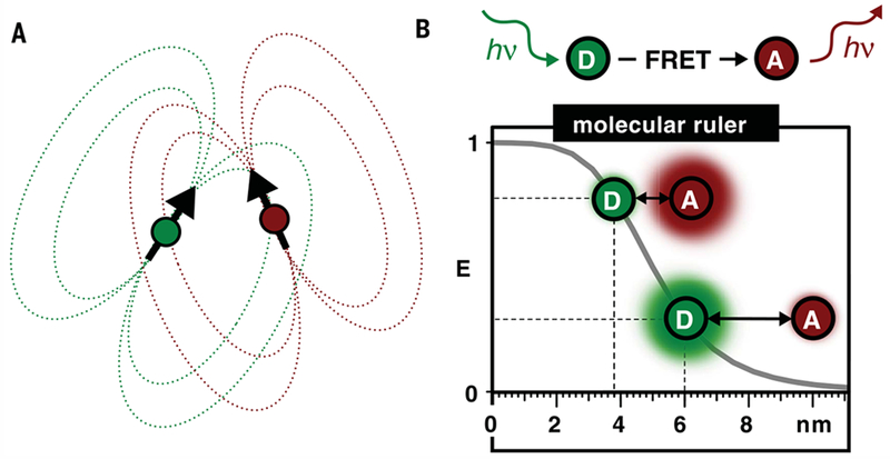
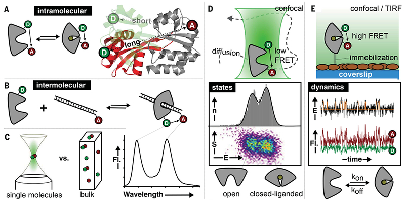
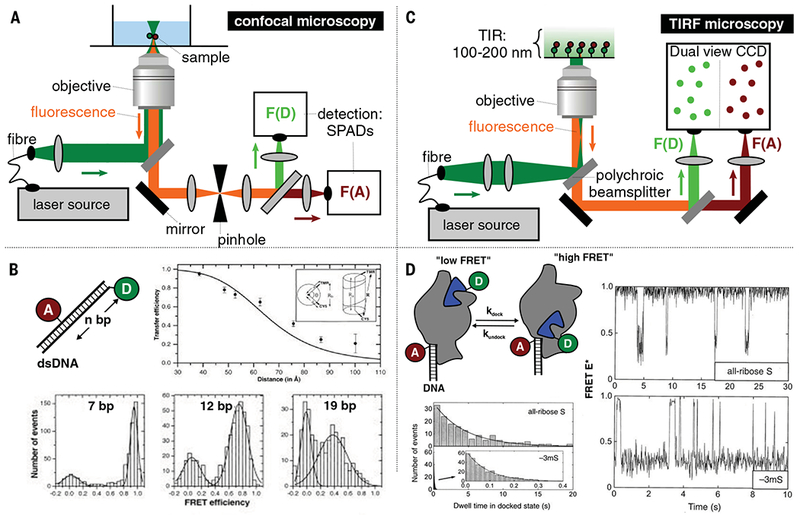
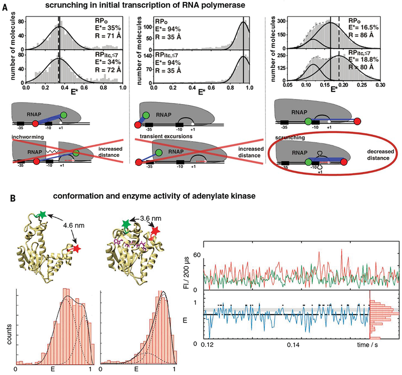
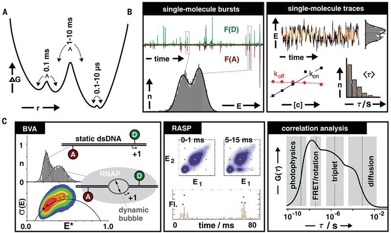
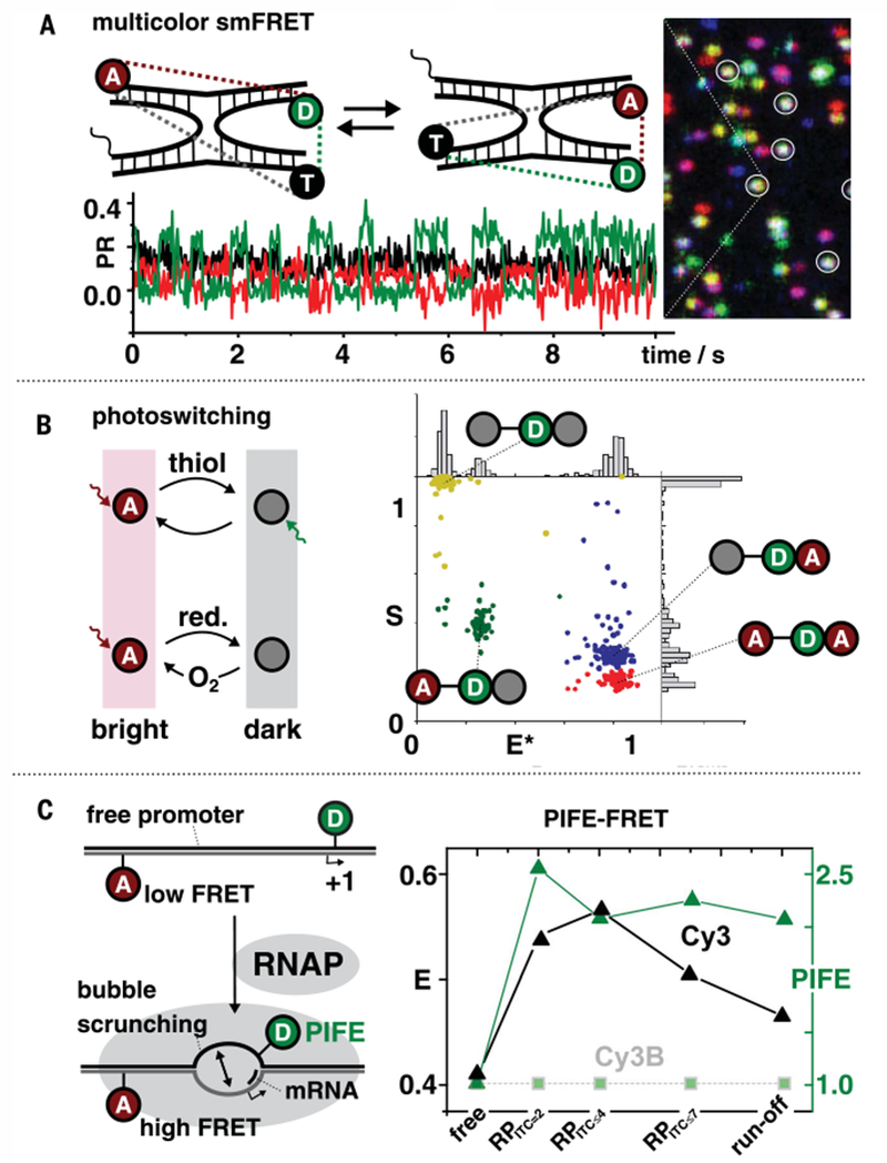
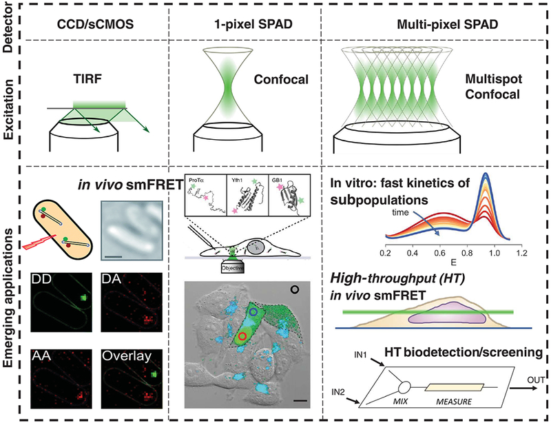
Classical structural biology can only provide static snapshots of biomacromolecules. Single-molecule Förster resonance energy transfer (smFRET) paved the way for studying dynamics in macromolecular structures under biologically relevant conditions. Since its first implementation in 1996, smFRET experiments have confirmed previously hypothesized mechanisms and provided new insights into many fundamental biological processes, such as DNA maintenance and repair, transcription, translation, and membrane transport. We review 22 years of contributions of smFRET to our understanding of basic mechanisms in biochemistry, molecular biology, and structural biology. Additionally, building on current state-of-the-art implementations of smFRET, we highlight possible future directions for smFRET in applications such as biosensing, high-throughput screening, and molecular diagnostics.


Figures

Fig. 1.
Fig. 1.. The concept of FRET.
(A and B) An electromagnetic transmitter-receiver (A) is a macroscopic analog for the molecular dipole-dipole coulombic interaction between donor and acceptor (D and A) fluorophores (B). The dependence of the efficiency of energy transfer from D to A on their distance provides a molecular ruler with a high dynamic range on the 3- to 9-nm scale.
Fig. 2.
Fig. 2.. Principle and use of FRET for elucidating biomolecular reaction mechanisms and structural dynamics.
(A to C) Principle of intra-molecular (A) and intermolecular (B) FRET assays and their readout (C) in single-molecule and bulk fluorescence (Fl.) experiments. The bulk experiments always show an average value [i.e., donor (D) and acceptor (A) intensity of, e.g., hypothetical 50/50], whereas smFRET can determine (dynamically interconverting) states directly. The crystal structure overlay of substrate-binding domains of an ABC transporter in (A) shows open (red) and closed (green) conformations. (D and E) smFRET with diffusing molecules (D) or immobilized molecules (E) including accessible biophysical parameters (i.e., conformational states and dynamical changes). For characterization of conformational states, histograms of FRET efficiency E with frequency n are used; dynamics are directly seen via temporal evolution of E obtained via ratio of acceptor (A) fluorescence to fluorescence from both donor (D) and acceptor after donor excitation. [(A) and (D) adapted, with permission, from (154)]
Fig. 3.
Fig. 3.. Pioneering implementations of smFRET.
(A) Schematic of a confocal microscope setup used for the acquisition of diffusion-based smFRET data; F(D) and F(A) indicate the donor and acceptor detection channels, respectively. (B) Example of data obtained with such a setup. The different histograms show the FRET efficiency distributions obtained for DNA samples differing by the distance between donor and acceptor labels; bp, base pairs. [Adapted, with permission, from (28)] (C) Schematic of a total internal reflection fluorescence (TIRF) setup allowing the study of smFRET on surface-immobilized molecules. (D) Example of data obtained with such a setup, showing the real-time dynamics of RNA catalysis and folding. FRET trajectories were retrieved for individual RNA molecules (right) and histograms of dwell times reported on the time scale of the dynamics (lower left). [Adapted from (6)]
Fig. 4.
Fig. 4.. Typical examples of smFRET studies.
(A) Transcription initiation involves a DNA scrunching mechanism. The results of three experiments differing by the location of the donor and acceptor dyes are shown (see text). The cartoons indicate which model is or is not compatible with the results. [Adapted from (60)] (B) Intrinsic domain motions between conformations in adenylate kinase (AK). The experiment tracks the distance between substrate-binding domains (donor and acceptor dyes as green and red stars, respectively) in the AK enzyme in apo form (left histogram) and when bound to the substrate-mimicking inhibitor Ap5A (right histogram). FRET efficiency histograms (left) and single-molecule time traces (right) show that in apo conformation, AK dynamically switches between two conformations, one of which is similar to the substrate-bound state. [Adapted, with permission, from (62)]
Fig. 5.
Fig. 5.. Biomolecular dynamics accessible by smFRET.
(A) Hypothetical energy landscape with Gibbs free energy projected onto a single reaction coordinate r showing different local minima (states) separated by energy barriers of different heights, giving rise to conformational transitions over different time scales. (B) smFRET data from diffusing molecules (bursts, left) and immobilized molecules (time traces, right) can be analyzed by various methods with differing temporal resolutions to study conformational transitions over different time scales. Conformational dynamics slower than ~0.1 s can be studied by analysis of single-molecule traces and dwell times in each FRET-associated state. (C) Examples of data analysis techniques using details of burst properties and photon statistics: Burst variance analysis (BVA) identifies bursts with variance of the FRET efficiency larger than expected from shot noise; recurrence analysis (RASP) identifies whether the FRET efficiency has changed between consecutive bursts of the same molecule; and correlation techniques identify time scales (including <100 μs) at which fluorescence-related processes occur, including changes in FRET efficiency. [Reproduced, with permission, from (87, 88)]
Fig. 6.
Fig. 6.. smFRET-based approaches to study molecular coordination.
(A) Multicolor smFRET studying coordinated movement of a Holliday junction via proximity ratio PR: donor-transmitter D-T (green trace), transmitter-acceptor T-A (black), and donor-acceptor D-A (red). [Adapted, with permission, from (97)] (B) Photoswitchable FRET relies on temporal separation of donor-acceptor interactions via photoswitching and isolation of molecular species with one distinct donor-acceptor pair at any given time point. [Adapted, with permission, from (100)] (C) PIFE-FRET uses a standard two-color assay with donor and acceptor (D-A) but adds information on protein binding via use of an environmentally sensitive donor (Cy3; Cy3B is used as the control dye that is insensitive to changes in the environment). [Adapted, with permission, from (104)]
Fig. 7.
Fig. 7.. Emerging applications and future directions of smFRET.
Top rows show current detector and excitation formats for smFRET; the bottom row shows emerging developments that go beyond existing capabilities. smFRET measurements have been demonstrated in live bacteria using TIRF with probes internalized via electroporation [left; adapted from (135)] and in eukaryotic cells using confocal excitation and microinjected molecules [center; after (70)]. Multipixel SPADs (right) allow fast detection schemes and will allow retrieval of FRET trajectories of single molecules in vivo (scanning different z-layers via light-sheet microscopy) and in vitro (nonequilibrium kinetics via smFRET using mixers or continuous-flow microfluidic devices) [adapted from (143)].

Similar articles

Cited by

References

    1. Brünger AT, X-ray crystallography and NMR reveal complementary views of structure and dynamics. Nat. Struct. Biol 4 (suppl.), 862–865 (1997). pmid: - PubMed
    1. Zewail AH, Diffraction, crystallography and microscopy beyond three dimensions: Structural dynamics in space and time. Philos. Trans. R. Soc. A 363, 315–329 (2005). doi: 10.1098/rsta.2004.1513; pmid: - DOI - PubMed
    1. Bharat TA, Russo CJ, Löwe J, Passmore LA, Scheres SH, Advances in single-particle electron cryomicroscopy structure determination applied to subtomogram averaging. Structure 23, 1743–1753 (2015). doi: 10.1016/j.str.2015.06.026; pmid: - DOI - PMC - PubMed
    1. Cheng Y, Single-particle cryo-EM at crystallographic resolution. Cell 161, 450–457 (2015). doi: 10.1016/j.cell.2015.03.049; pmid: - DOI - PMC - PubMed
    1. Hennig M, Ruf A, Huber W, Combining biophysical screening and X-ray crystallography for fragment-based drug discovery. Top. Curr. Chem 317, 115–143 (2011). doi: 10.1007/128_2011_225; pmid: - DOI - PubMed

Publication types

MeSH terms