. 2018 Feb 23;359(6378):940-944.
doi: 10.1126/science.aar5700. Epub 2018 Jan 18.

Structures of human PRC2 with its cofactors AEBP2 and JARID2

Affiliations

Structures of human PRC2 with its cofactors AEBP2 and JARID2

Vignesh Kasinath et al. Science. .

Abstract

Transcriptionally repressive histone H3 lysine 27 methylation by Polycomb repressive complex 2 (PRC2) is essential for cellular differentiation and development. Here we report cryo-electron microscopy structures of human PRC2 in a basal state and two distinct active states while in complex with its cofactors JARID2 and AEBP2. Both cofactors mimic the binding of histone H3 tails. JARID2, methylated by PRC2, mimics a methylated H3 tail to stimulate PRC2 activity, whereas AEBP2 interacts with the RBAP48 subunit, mimicking an unmodified H3 tail. SUZ12 interacts with all other subunits within the assembly and thus contributes to the stability of the complex. Our analysis defines the complete architecture of a functionally relevant PRC2 and provides a structural framework to understand its regulation by cofactors, histone tails, and RNA.


Figures

Fig. 1
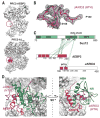
Fig. 1. Cryo-EM structure of PRC2-JARID2-AEBP2
(A) Schematic representation of the proteins in the PRC2-JARID2-AEBP2 complex. Dashed boxes highlight regions described in our study for which structural information was previously unavailable. (B) XL-MS results for PRC2-AJ119 (left) and PRC2-AJ106 (right). (C) Cryo-EM densities for the two conformational states (compact active and extended active) of PRC2-AJ106, with differences in SBD and SANT1 highlighted by the red dashed circles. SRM is ordered and visible only in the compact active state (top). The JARID2 K116me3 fragment is bound in both states. (D) Atomic model of the compact active state, with EZH2 (SANT1, SBD, BAM, and SRM) in gold, EZH2 (SET) in dark blue, EZH2 (SANT2) in pink, EED in light blue, RBAP48 in violet, AEBP2 in red, JARID2 in magenta, SUZ12 (ABH and BSD) in mint green, SUZ12 (NR) in dark green, and SUZ12 (VEFS) in bright green. This color-coding also applies to (B) and (C).
Fig. 2
Fig. 2. Cofactors AEBP2 and JARID2 mimic histone H3 tails
(A) Interaction of JARID2 K116me3 (magenta) with EED (light blue) in the two conformational states sampled by PRC2-AJ106. JARID2 K116me3 sits in the middle of an aromatic cage (F97, Y148, and Y365) with hydrogen-bond interactions (R414:F117 and W364:R115) that stabilize the JARID2 peptide backbone. In the compact active state, additional interaction between EZH2 D136 and the backbone amide of JARID2 K116 helps position the EZH2 SRM helix (gold) next to the EZH2 SETdomain. (B) The EZH2 SETdomain of PRC2-AJ106 is in a similar conformation in both states (compact active, orange; extended active, blue; Cα root mean square deviation = 0.74 Å; fig. S4B) and is bound to a substrate peptide (cyan). Inset, density of substrate observed in our cryo-EM reconstruction shown with a model for JARID2 residues 114 to 118. (C) Cryo-EM density of the PRC2-AJ119 EED and EZH2 SETregions with ribbon representations of EED (light blue) and SET (dark blue), showing the absence of both the stimulatory fragment bound to EED and the substrate bound to the SETdomain (red dashed circles). (D) AEBP2 (residues 232 to 295, red), shown in surface representation, connects RBAP48, the SUZ12 N-terminal region, and the EZH2 SETdomain. (E) Map versus model of the AEBP2 region that binds RBAP48 at the histone H3 binding site. (F) View of the RBAP48 WD40 domain showing, in stick representation, the conserved amino acids in RBAP48 (violet) interacting with K294 and R295 of AEBP2 (red). The overlay of the crystal structure of the Drosophila homolog of RBAP48 (NURF55) bound to unmodified histone H3 peptide [PDB ID, 2YBA (16); green] highlights the similarity of the binding mode between AEBP2 and histone H3, with all key residues in identical conformations; only the AEBP2 (H3) residues R295 (R2) and K294 (K4) are shown for clarity. Single-letter abbreviations for the amino acid residues are as follows: A, Ala; C, Cys; D, Asp; E, Glu; F, Phe; G, Gly; H, His; I, Ile; K, Lys; L, Leu; M, Met; N, Asn; P, Pro; Q, Gln; R, Arg; S, Ser; T, Thr; V, Val; W, Trp; and Y, Tyr.
Fig. 3
Fig. 3. SUZ12 interacts with all subunits of PRC2
(A) Schematic representation of the different domains of SUZ12 (shades of green; top) and cryo-EM density of SUZ12 showing the location of each domain within PRC2 (bottom). (B) Orthogonal views of the SUZ12 AEBP2-binding helix (ABH) and neck region (NR), defining the part of SUZ12 at the center of the complex. The insets show the relative location of these regions within the full PRC2 complex. (C) Cryo-EM density of the β-sheet–rich domain (BSD) making up the “foot” of the complex, with a fitted polyalanine model. Top insets, position of BSD within PRC2. (D) Map versus model for the SUZ12 zinc-finger domain, which interacts with AEBP2 (residues 246 to 266), the helix-turn-helix (HTH) of JARID2 (Fig. 4), and the ABH of SUZ12. (E) Close-up of the neck region of PRC2 showing, in stick representation, the amino acids contributing to the hydrophobic interactions between the SUZ12 NR, the SAL of EZH2, EED, and the SUZ12 VEFS. Dotted lines indicate potential hydrogen bonds. Mutations in R508 and N562 (asterisks) have been observed in a number of cancers.
Fig. 4
Fig. 4. JARID2-AEBP2-SUZ12 interactions contribute to the stability of PRC2
(A) The density region marked by the red dashed circle is only present in JARID2-containing complexes [PRC2-AEBP2 (top) and PRC2-AJ106 (bottom, with the JARID2 segment in magenta)]. (B) Map versus model for JARID2 residues 140 to 166. The model built into the density agrees well with the HTH secondary structure prediction for this region. (C) Cross-links of the SUZ12 NR with AEBP2 and JARID2 (residues 140 to 166) confirm the relative localization of each of these regions in the cryo-EM map. (D) The JARID2 HTH (magenta) is wedged between AEBP2 (red) and the SUZ12 ABH (mint green) and NR (green).

Comment in

References

    1. Cao R, et al. Science. 2002;298:1039–1043. - PubMed
    1. Talbert PB, Henikoff S. Nat Rev Genet. 2006;7:793–803. - PubMed
    1. Margueron R, Reinberg D. Nature. 2011;469:343–349. - PMC - PubMed
    1. Jiao L, Liu X. Science. 2015;350:aac4383. - PMC - PubMed
    1. Margueron R, et al. Nature. 2009;461:762–767. - PMC - PubMed

Publication types