. 2018 Jan 18;8(1):1115.
doi: 10.1038/s41598-018-19521-9.

Macrophage sensing of single-walled carbon nanotubes via Toll-like receptors

Affiliations

Macrophage sensing of single-walled carbon nanotubes via Toll-like receptors

Sourav P Mukherjee et al. Sci Rep. .

Abstract

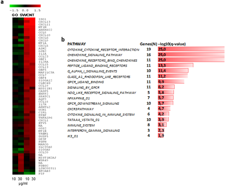
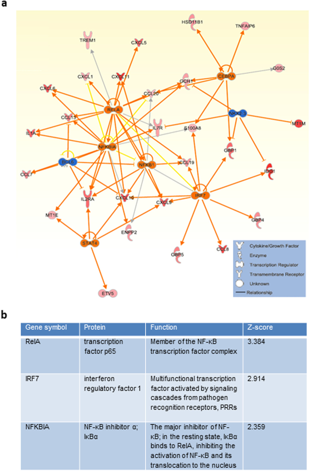
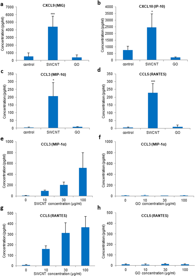
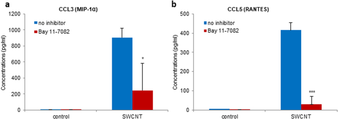
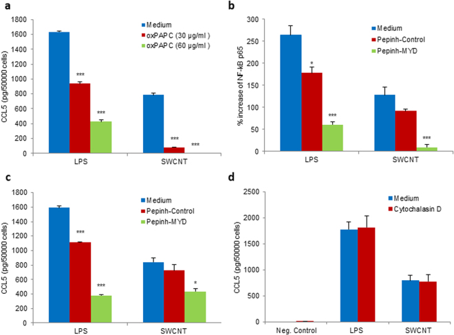
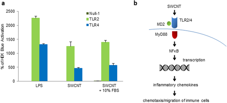
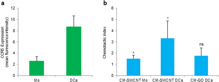
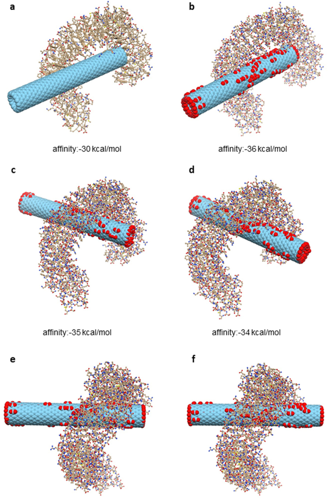
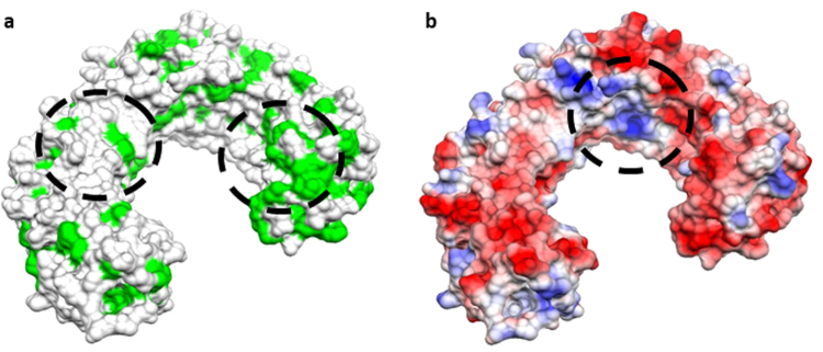
Carbon-based nanomaterials including carbon nanotubes (CNTs) have been shown to trigger inflammation. However, how these materials are 'sensed' by immune cells is not known. Here we compared the effects of two carbon-based nanomaterials, single-walled CNTs (SWCNTs) and graphene oxide (GO), on primary human monocyte-derived macrophages. Genome-wide transcriptomics assessment was performed at sub-cytotoxic doses. Pathway analysis of the microarray data revealed pronounced effects on chemokine-encoding genes in macrophages exposed to SWCNTs, but not in response to GO, and these results were validated by multiplex array-based cytokine and chemokine profiling. Conditioned medium from SWCNT-exposed cells acted as a chemoattractant for dendritic cells. Chemokine secretion was reduced upon inhibition of NF-κB, as predicted by upstream regulator analysis of the transcriptomics data, and Toll-like receptors (TLRs) and their adaptor molecule, MyD88 were shown to be important for CCL5 secretion. Moreover, a specific role for TLR2/4 was confirmed by using reporter cell lines. Computational studies to elucidate how SWCNTs may interact with TLR4 in the absence of a protein corona suggested that binding is guided mainly by hydrophobic interactions. Taken together, these results imply that CNTs may be 'sensed' as pathogens by immune cells.


Conflict of interest statement

The authors declare that they have no competing interests.

Figures

Figure 1
Figure 1
Transcriptomics analysis of macrophages exposed to carbon-based nanomaterials. Global gene expression profiling of human monocyte-derived macrophages (HMDM) was conducted after 24 h exposure to single-walled carbon nanotubes (SWCNTs) or graphene oxide (GO) (see Supplementary Table 1). (a) Heatmap of differently expressed genes in response to 10 and 30 µg/mL GO (line 1 and 2, respectively) or 10 and 30 µg/mL SWCNTs (line 3 and 4, respectively). Upregulated transcripts are presented in red and downregulated transcripts in green (log2 fold change > 0.75). The genes affected upon exposure to SWCNTs (n = 52) and GO (n = 7) did not overlap. (b) Canonical pathways modulated in HMDM after exposure to SWCNTs (see Supplementary Table 2); ranking was performed according to multiple testing corrected p-value.
Figure 2
Figure 2
Upstream regulator analysis of the transcriptomics results. (a) The NF-κB network was identified as a potential upstream regulator of SWCNT-triggered responses in HMDM according to upstream regulator analysis (p < 0.01; Z-score > 2 S.D.). (b) Upstream regulator analysis of the data identified the modulation of NF-κB network members in HMDM exposed to SWCNTs for 24 h. Data were analyzed through the use of IPA (QIAGEN Inc., www.qiagenbioinformatics.com/products/ingenuity-pathway-analysis).
Figure 3
Figure 3
SWCNTs, but not GO, trigger macrophage secretion of chemokines. Secretion of chemokines by primary human macrophages (HMDM) after a 24 h exposure to SWCNTs or GO as measured by a multi-plex immunoassay. (ad) Exposure of HMDM to 30 µg/mL SWCNTs showed a significant increase in CXCL9, CXCL10, CCL3/MIP-1α, and CCL5/RANTES, while there was no response in cells exposed to GO at the same concentration. (e,g) Dose-dependent secretion of CCL3/MIP-1α, and CCL5/RANTES in cells exposed to SWCNTs, while there was no response to GO at any of the concentrations tested (f,h). Data are shown as mean values ± S.D. of three independent experiments using cells from different donors; p-values by Student’s t-test, * < 0.05; *** < 0.001.
Figure 4
Figure 4
SWCNT-triggered chemokine secretion is NF-κB-dependent. Pretreatment with the NF-κB inhibitor, Bay 11-7082 (10 µM) of HMDM exposed to 30 µg/mL SWCNTs or medium alone reduced the secretion of CCL3 (a) and completely blocked the secretion of CCL5 (b), by multi-plex assay. Data are mean values ± S.D. of three independent experiments using cells from different donors; p-values by Student’s t-test, * < 0.05; *** < 0.001.
Figure 5
Figure 5
TLR2/4- and MyD88-dependent secretion of chemokines. (a) Significant reduction of CCL5 secretion in HMDM after 12 h exposure to SWCNT (30 µg/mL) in the presence of the TLR2/4 inhibitor, oxPAPC (30 or 60 µg/mL). oxPAPC also blocked CCL5 secretion triggered by LPS (0.1 µg/mL) in a dose-dependent manner. Furthermore, the MyD88 inhibitor, Pephinh-MYD (25 µM), but not Pepinh-Control (25 µM), reduced NF-kB p65 phosphorylation (b) and CCL5 expression (c) in cells exposed to SWCNT (30 µg/mL) for 12 h. Pephinh-MYD (25 µM) also reduced NF-kB activation and CCL5 secretion by LPS (0.1 µg/mL). NF-kB p65 phosphorylation and CCL5 expression was determined by ELISA. (d) Cytochalasin D (10 μM), an inhibitor of actin polymerization, does not affect CCL5 secretion in HMDM exposed for 12 h to SWCNT (30 µg/mL). LPS (0.1 µg/mL) was included as a control. CCL5 levels were determined by ELISA. Data shown in panels a to d are reported as mean values ± S.D. of at least three independent experiments using cells from different donors. p* < 0.05; ** < 0.01; *** < 0.001 (one-way ANOVA with post-hoc tukey’s test).
Figure 6
Figure 6
SWCNTs trigger TLR2 and TLR4 activation. (a) HEK 293 cells co-transfected with human TLR2 (HEK-Blue™ hTLR2) or TLR4 (HEK-Blue™ hTLR4 cells) and an NF-κB/AP-1-secreted embryonic alkaline phosphatase (SEAP) reporter gene were exposed to SWCNT (30 µg/mL) for 12 h in the presence or absence of 10% FBS. LPS (0.1 µg/mL) was included as a positive control. SWCNTs activated TLR2/4 independently of the presence of serum in the culture medium. Data are shown as mean values ± S.D. of three independent experiments. (b) Schematic diagram showing the ‘sensing’ of SWCNTs by HMDMs via TLR receptors resulting in MyD88-dependent activation of NF-kB leading to nuclear translocation of NF-kB with transcription and secretion of CCL5. The secreted chemokine(s) induce chemotaxis of immune cells bearing the corresponding receptor(s).
Figure 7
Figure 7
Macrophage secreted factors promote migration of DCs. (a) Expression of the chemokine receptor CCR5 in primary human monocytes (Mo) and monocyte-derived dendritic cells (DCs) was determined by flow cytometry. The average expression of CCR5 in cells from three different donors is depicted. (b) Migration of monocytes (Mo) and DCs in response to conditioned medium (CM) of human monocyte-derived macrophages (HMDM) exposed to 100 µg/mL SWCNT (CM-SWCNT) or 100 µg/mL GO (CM-GO). Cell migration (3 h period) was determined by using transwell chemotaxis microchambers. Data are shown as mean values ± S.D. of three independent experiments; p* < 0.05, Student’s t-test.
Figure 8
Figure 8
Molecular docking of CNTs and TLR4. Results of docking simulations of pristine and carboxylated CNTs and TLR4. (a) The best binding mode for pristine CNT. (b) best binding mode for carboxylated CNT. (c) lateral view of the second top binding pose for carboxylated CNT. (d) lateral view of the third top binding pose for the same CNT. (e) top view of the same configuration as in (c). (f) top view of the same configuration as in (d). The mechanism involves interactions of the target protein with both the tip and side-wall of CNTs.
Figure 9
Figure 9
Identification of interaction sites. The molecular surface of TLR4 is shown with two different coloring schemes: (a) hydrophobicity scale (hydrophobic residues are depicted in green) and (b) electrostatic potential (coloring scale runs from red to blue in the interval of values [−16.89:16.89] kT/e). Circled areas represent the regions of TLR4 that directly interact with CNTs by hydrophobic contact (a) and by electrostatic interaction (b).

References

    1. Bhattacharya K, et al. Biological interactions of carbon-based nanomaterials: from coronation to degradation. Nanomedicine. 2016;12:333–351. doi: 10.1016/j.nano.2015.11.011. - DOI - PMC - PubMed
    1. Boraschi, D., Fadeel, B. & Duschl, A. Immune System. In: Adverse Effects of Engineered Nanomaterials: Exposure, Toxicology and Impact on Human Health (Second Edition). Eds. Fadeel, B., Pietroiusti, A., Shvedova, A. pp. 313–337. Elsevier (2017).
    1. Fadeel B. Clear and present danger? Engineered nanoparticles and the immune system. Swiss Med Wkly. 2012;142:w13609. - PubMed
    1. Deng ZJ, Liang M, Monteiro M, Toth I, Minchin RF. Nanoparticle-induced unfolding of fibrinogen promotes Mac-1 receptor activation and inflammation. Nat Nanotechnol. 2011;6:39–44. doi: 10.1038/nnano.2010.250. - DOI - PubMed
    1. Castranova V, Schulte PA, Zumwalde RD. Occupational nanosafety considerations for carbon nanotubes and carbon nanofibers. Acc Chem Res. 2013;46:642–649. doi: 10.1021/ar300004a. - DOI - PMC - PubMed

Publication types