. 2018 Jun;37(25):3384-3398.
doi: 10.1038/s41388-017-0110-2. Epub 2018 Jan 18.

Piperlongumine and p53-reactivator APR-246 selectively induce cell death in HNSCC by targeting GSTP1

Affiliations

Piperlongumine and p53-reactivator APR-246 selectively induce cell death in HNSCC by targeting GSTP1

Wei Hang et al. Oncogene. 2018 Jun.

Abstract

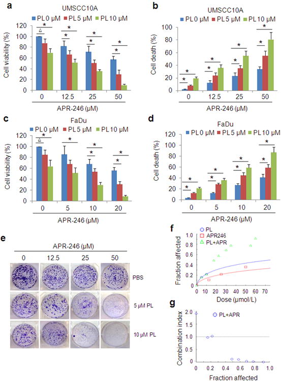
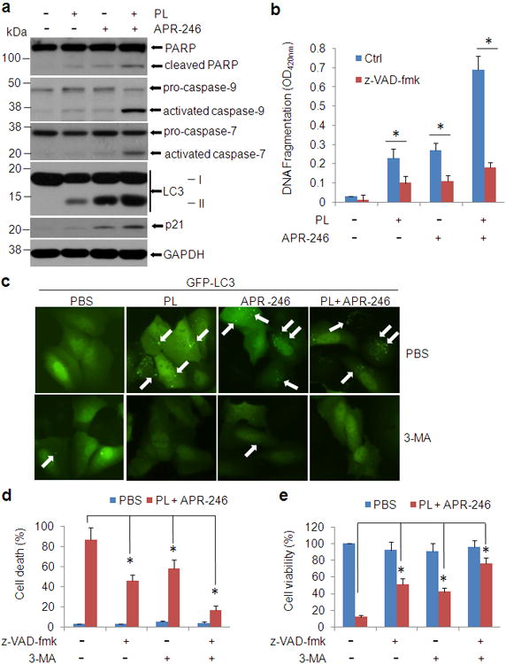
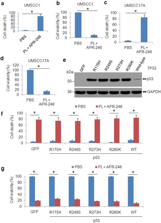
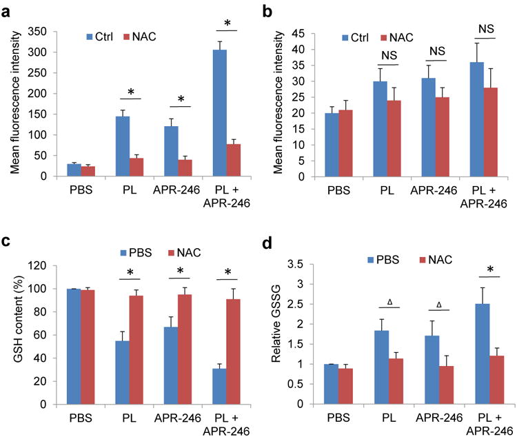
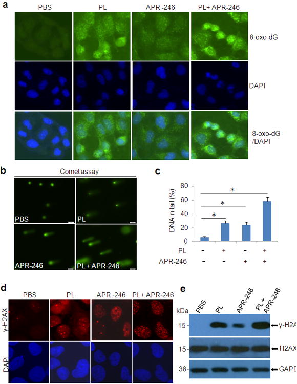
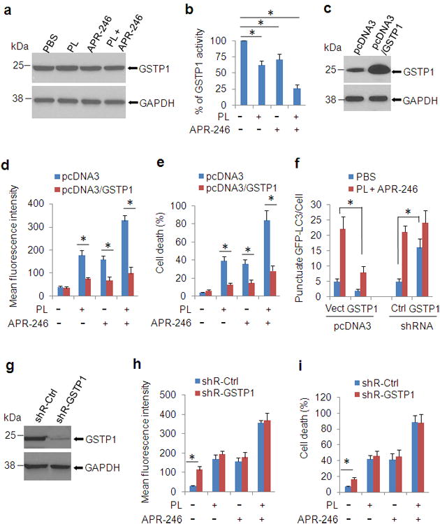
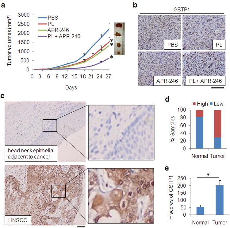
TP53 mutations frequently occur in head and neck squamous cell carcinoma (HNSCC) patients without human papillomavirus infection. The recurrence rate for these patients is distinctly high. It has been actively explored to identify agents that target TP53 mutations and restore wild-type (WT) TP53 activities in HNSCC. PRIMA-1 (p53-reactivation and induction of massive apoptosis-1) and its methylated analogue PRIMA-1Met (also called APR-246) were found to be able to reestablish the DNA-binding activity of p53 mutants and reinstate the functions of WT p53. Herein we report that piperlongumine (PL), an alkaloid isolated from Piper longum L., synergizes with APR-246 to selectively induce apoptosis and autophagic cell death in HNSCC cells, whereas primary and immortalized mouse embryonic fibroblasts and spontaneously immortalized non-tumorigenic human skin keratinocytes (HaCat) are spared from the damage by the co-treatment. Interestingly, PL-sensitized HNSCC cells to APR-246 are TP53 mutation-independent. Instead, we demonstrated that glutathione S-transferase pi 1 (GSTP1), a GST family member that catalyzes the conjugation of GSH with electrophilic compounds to fulfill its detoxification function, is highly expressed in HNSCC tissues. Administration of PL and APR-246 significantly suppresses GSTP1 activity, resulting in the accumulation of ROS, depletion of GSH, elevation of GSSG, and DNA damage. Ectopic expression of GSTP1 or pre-treatment with antioxidant N-acetyl-L-cysteine (NAC) abrogates the ROS elevation and decreases DNA damage, apoptosis, and autophagic cell death prompted by PL/APR-246. In addition, administration of PL and APR-246 impedes UMSCC10A xenograft tumor growth in SCID mice. Taken together, our data suggest that HNSCC cells are selectively sensitive to the combination of PL and APR-246 due to a remarkably synergistic effect of the co-treatment in the induction of ROS by suppression of GSTP1.


Conflict of interest statement

Conflict of Interest: The authors have no conflict of interest to report.

Figures

Figure 1
Figure 1. PL and APR-246 synergistically induce HNSCC cell death
(a-d) UMSCC10A and FaDu cells were treated with 0-10 μM PL in the presence or absence of APR-246 (0-50 μM) for 72 h. Cell viability was determined by the MTT assay. Cell death was measured with the trypan blue exclusion assay. The assays were performed in triplicate samples, and the results are representative of three independent experiments (ΔP < 0.05; * P < 0.01). (e-g) UMSCC10A cells were treated with various concentrations of PL and/or APR-246 for 16 h. Clonogenic survival assays were performed. (e) Representative images of the clonogenic survival assays are presented. (f, g) Assessment of the synergy between PL and APR-246 in the cells using the CompuSyn software (ComboSyn, Inc. Paramus, NJ 2007). Drug interactions are expressed as fraction affected (FA) curves (f) and combination index (CI) plots (g). The CI values were automatically generated by the software over a range of FA levels from growth inhibition percentages. A CI < 1.0 was considered as synergy. Synergy was remarkable at relevant FA values being 50% or greater (g).
Figure 2
Figure 2. PL enhances APR-246-induced apoptosis and autophagy in HNSCC cells
(a) UMSCC10A cells were treated with 10 μM PL and/or 25 μM APR-246 for 24 h. After the treatments, whole cell extracts were collected for the western blot analysis. Thirty μg proteins were loaded in each lane. GAPDH serves as a loading control. (b) UMSCC10A cells were treated with 10 μM PL and/or 25μM APR-246 in the presence or absence of 20 μM z-VAD-fmk for 72 h. After the treatment, cell apoptosis was quantified using a cell death ELISA kit (Roche Diagnostics) showing enrichment of nucleosomes in the cytoplasmic fraction of the cells. Values represent the mean ± S.D. * P < 0.01, n = 3. (c) UMSCC10A cells were transfected with pcDNA3/GFP-LC3 for 24 h. The cells were then treated with 10 μM PL and/or 25μM APR-246 for additional 24 h in the presence or absence of 1 mM 3-MA. Autophagy (GFP-LC3 punctuates) wasmonitored under an Olympus IX51 fluorescence microscope. White arrows show the autophagosomes/autolysomes. (d, e) UMSCC10A cells were co-treated with 10 μM PL and 25 μM APR-246 in the presence or absence of 20 μM z-VAD-fmk or 1 mM 3-MA for 72 h. After the treatment, cell death (d) and cell viability (e) were determined as described in Figure 1. Values represent the mean ± S.D. * P < 0.01 as compared with respective treatment without z-VAD and 3-MA, n = 3.
Figure 3
Figure 3. PL- and APR-246-induced cell death is independent of TP53
(a-d) UMSCC1 (null TP53) cells (a, b) and UMSCC17A (wild-type TP53) cells (c, d) were exposed to 10 μM PL and 25 μM APR-246 for 72 h. After the treatment, cell death and cell viability were determined as described in Figure 1. (e-g) UMSCC1 cells were infected with lentiviruses expressing TP53 mutants R175H, R249S, R273H, R280K, and wild-type TP53, or GFP (control). Cell transduction efficiency was about 60% with the fluorescence microscopy analysis at 48 h after the infection. Immunoblot was used for the detection of p53 expression in the transduced cells (e). Cell death (f) and cell viability (g) in the cells treated with 10 μM PL and 25μM APR-246 for additional 72 h were determined as described in Figure 1. The data represent the mean ± S.D. * P < 0.01, n = 3.
Figure 4
Figure 4. PL and APR-246 promote the accumulation of ROS, the depletion of GSH, and the elevation of GSSG in HNSCC cells, which are reversed by the administration of antioxidant NAC
UMSCC10A cells (a, c, d) and MEFs (b) were treated with 10 μM PL and/or 25μM APR-246 in the presence or absence of 5 mM NAC for 24 h. (a, b) ROS in the cells. One hour prior to the termination of the treatment, 100 ng/ml dihydroethidium was added to the medium. The cells were collected, washed and analyzed by flow cytometry with the red laser channel (FL-3) using a FACscan analyzer. (c) GSH contents in the cells. GSH was analyzed using a commercial kit from Abcam. The values were then normalized to mg protein. The number in PBS-treated cells was set as “100%”. (d) GSSG levels in the cells. GSSG was analyzed using a commercial kit from Abcam. The values were then normalized to mg protein. The number in PBS-treated cells was set as “1”. Means ± SD for three independent experiments were presented. *P < 0.01;Δ P < 0.05; NS = Not Significant.
Figure 5
Figure 5. PL/APR-246 treatment promotes DNA damage
(a) 8-oxo-dG in UMSCC10A cells treated with PL and/or APR-246. UMSCC10A cells growing on fibronectin-coated coverslips were treated with 10 μM PL and/or 25 μM APR-246 in serum-free and phenol-red-free medium for 16 h at 37°C, fixed in absolute methanol (- 20°C, 20 min) and permeabilized with 0.1% Triton X-100 at room temperature for 15 min. After blocking, the cells were stained with FITC-conjugated avidin for 1 h at 37°C. Fluorescence is captured with an Olympus IX51 fluorescence microscope. (b, c)Comet assay showing elevated DNA damage in cells treated with PL and APR-246. UMSCC10A cells were treated with 10 μM PL and/or 25μM APR-246 for 16 h. The cells were then trypsinized and washed with PBS. Two thousand cells were mixed with 100 μl low melting agarose for alkaline comet assay. Cells in the gel were stained and visualized with epifluorescence microscopy (b). (c) Percentage of DNAs in the tail (damaged DNA) was calculated. *P < 0.01, n = 3. (d, e) Expression of γ-H2AX in PL/APR-246-treated cells. UMSCC10A cells were treated with 10 μM PL and/or 25 μM APR-246 for 16 h. After the treatment, the cells were collected for the immunofluorescent analysis of γ-H2AX foci formation (d) or immunoblot analysis of γ-H2AX and H2AX expression (e).
Figure 6
Figure 6. Treatment of PL/APR-246 inactivates GSTP1, manipulations of which affect the function of PL/APR-246
(a, b) PL/APR-246 treatment reduces the activity of GSTP1. UMSCC10A cells were treated with 10 μM PL and/or 25 μM APR-246 for 24 h. After the treatment, the cells were collected for the immunoblot analysis of GSTP1 expression (a) or the measurement of GSTP1 activity (b). Measurements of the GSTP1 activity were performed using a commercial kit from Abcam following the manufacturer's instructions. The values were then normalized to mg protein. The number in PBS-treated cells was set as “100%”. The data represent means ± SD from three independent experiments (*P < 0.01). (c-e) Overexpression of GSTP1 reduces PL- and APR-246- induced ROS escalation and cell death. UMSCC10A/pcDNA3-GSTP1 cells were exposed to 10 μM PL and/or 25 μM APR-246 for 48 h. The cells were collected for the determination of GSTP1 expression by western blot (c), ROS determination (d), or cell death measurement (e). (f) Manipulations of GSTP1 affect autophagy induction by the cotreatment of PL and APR-246. UMSCC10A cells with GSTP1 overexpression or knockdown were transfected with pcDNA3/GFP-LC3 for 24 h. The cells were then treated with 10 μM PL and 25 μM APR-246 for additional 24 h. Autolysosomes/autophagosomes (GFP-LC3 punctuates) were monitored under an Olympus IX51 fluorescence microscope. The average punctate GFP-LC3 fluorescence dots were calculated in 200 cells. Data are shown as mean ± S.D. (g - i) Knockdown of GSTP1 increases intracellular ROS levels, but only marginally escalates ROS in PL- and APR-246-treated cells. Scramble shRNA and shR-GSTP1 were packaged with lentiviral system and transduced into UMSCC10A cells. After selection with puromycin for 2 weeks, the cells were verified for the knock-down by western blot (f). The cells were then exposed to 10 μM PL and/or 25μM APR-246 for 72 h for intracellular ROS determination (h) or cell death detection (i). ROS were measured as described in Figure 4. Cell death was determined as described in Figure 1b and d. Means ± SD for three independent experiments were presented. *P < 0.01.
Figure 7
Figure 7. GSTP1 is highly expressed in HNSCC tissues. Application of PL and APR-246 reduces xenograft tumor growth of HNSCC cells
(a, b) 5 × 10⁶ UMSCC10A cells were inoculated subcutaneously under the flank of SCID mice. Three days later, the mice were randomized into4 groups (n = 10 / group) receiving PBS (control), PL (4 mg/kg/day), APR-246 (100 mg/kg/day), or PL (4 mg/kg/day) + APR-246 (100 mg/kg/day) by i. p. Treatment was performed daily for 24 days. (a) Tumor volumes were measured every 3 days. *P < 0.01 as compared with control treatment group. (b) The tumors were removed from euthanized mice. IHC was used to detect GSTP1. Scale bar = 100 μm. (c - e) HNSCC tissues from healthy (n = 28) and HNSCC (n = 194) subjects were assessed for the expression of GSTP1 by IHC. (c) Representative IHC staining of GSTP1 in a normal head and neck epithelial tissue and in an HNSCC tissue. Scale bar = 100 μm. (d) Quantification of GSTP1 expression in human head and neck tissues. Low: overall negative or weak staining; High: overall moderate or strong staining. The Pearson's chi-square test was used to analyze the distribution difference of GSTP1 between healthy and HNSCC tissues (P < 0.01). (e) H-scores of GSTP1 in head and neck tissues (*P< 0.01).

Similar articles

Cited by

References

    1. Trachootham D, Alexandre J, Huang P. Targeting cancer cells by ROS-mediated mechanisms: a radical therapeutic approach? Nat Rev Drug Discov. 2009;8:579–591. - PubMed
    1. Cosenza M, Civallero M, Marcheselli L, Sacchi S, Pozzi S. Ricolinostat, a selective HDAC6 inhibitor, shows anti-lymphoma cell activity alone and in combination with bendamustine. Apoptosis. 2017;22:827–840. - PMC - PubMed
    1. Huang X, Motea EA, Moore ZR, Yao J, Dong Y, Chakrabarti G, et al. Leveraging an NQO1 Bioactivatable Drug for Tumor-Selective Use of Poly(ADP-ribose) Polymerase Inhibitors. Cancer Cell. 2016;30:940–952. - PMC - PubMed
    1. Huang BK, Langford TF, Sikes HD. Using Sensors and Generators of H2O2 to Elucidate the Toxicity Mechanism of Piperlongumine and Phenethyl Isothiocyanate. Antioxid Redox Signal. 2016;24:924–938. - PMC - PubMed
    1. Jutooru I, Guthrie AS, Chadalapaka G, Pathi S, Kim K, Burghardt R, et al. Mechanism of action of phenethylisothiocyanate and other reactive oxygen species-inducing anticancer agents. Mol Cell Biol. 2014;34:2382–2395. - PMC - PubMed

Publication types

MeSH terms