. 2018 Jan 18;9(2):31.
doi: 10.1038/s41419-017-0183-4.

Efficacy of MEK inhibition in a K-Ras-driven cholangiocarcinoma preclinical model

Affiliations

Efficacy of MEK inhibition in a K-Ras-driven cholangiocarcinoma preclinical model

Mingjie Dong et al. Cell Death Dis. .

Abstract

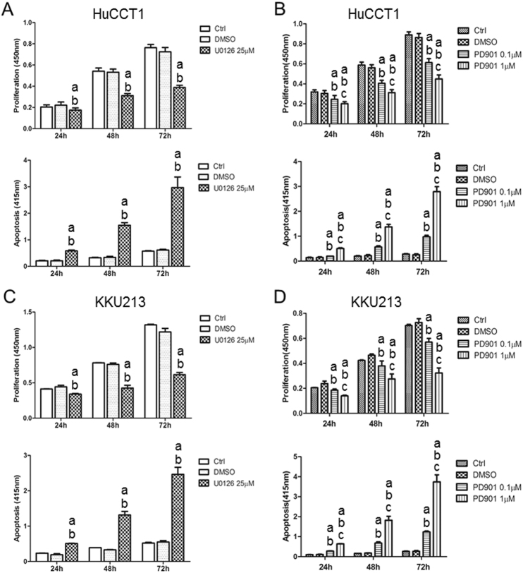
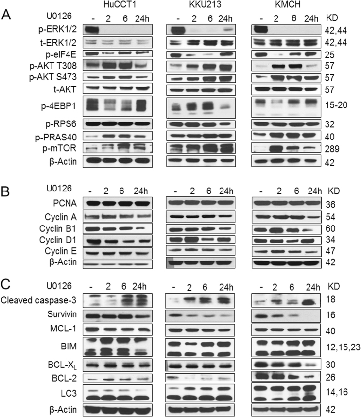
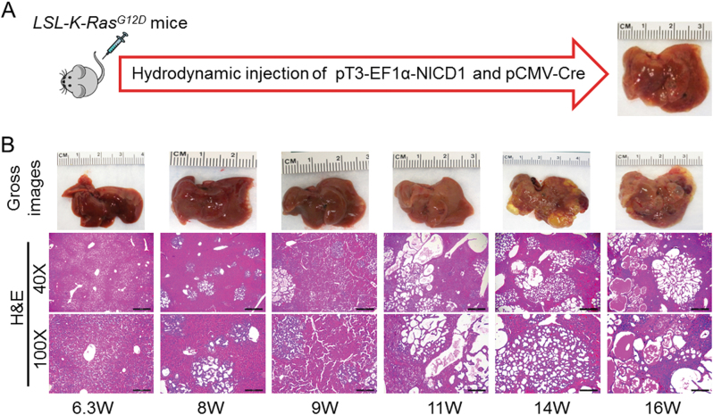
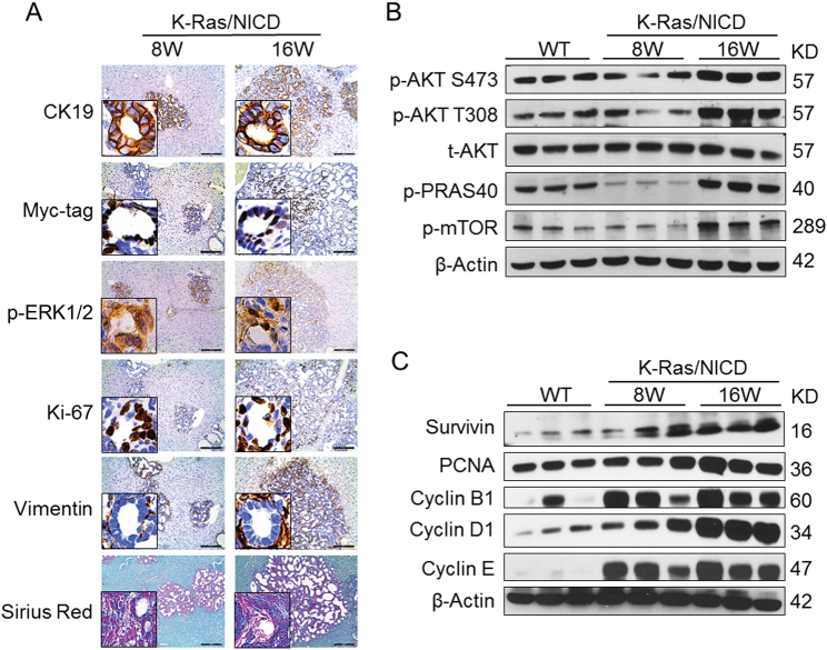
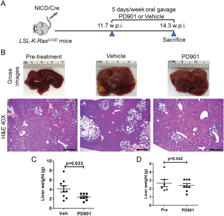
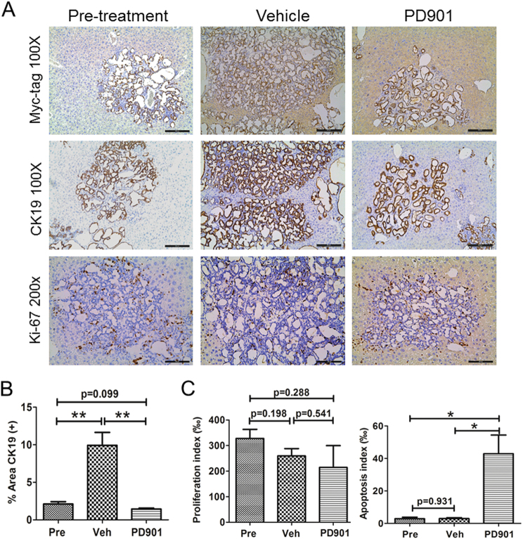
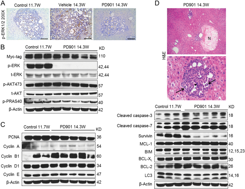
Intrahepatic cholangiocarcinoma (iCCA) is a deadly malignancy with limited treatment options. Gain-of-function mutations in K-Ras is a very frequent alteration, occurring in ~15 to 25% of human iCCA patients. Here, we established a new iCCA model by expressing activated forms of Notch1 (NICD) and K-Ras (K-RasV12D) in the mouse liver (K-Ras/NICD mice). Furthermore, we investigated the therapeutic potential of MEK inhibitors in vitro and in vivo using human CCA cell lines and K-Ras/NICD mice, respectively. Treatment with U0126, PD901, and Selumetinib MEK inhibitors triggered growth restraint in all CCA cell lines tested, with the most pronounced growth suppressive effects being observed in K-Ras mutant cells. Growth inhibition was due to reduction in proliferation and massive apoptosis. Furthermore, treatment of K-Ras/NICD tumor-bearing mice with PD901 resulted in stable disease. At the molecular level, PD901 efficiently inhibited ERK activation in K-Ras/NICD tumor cells, mainly leading to increased apoptosis. Altogether, our study demonstrates that K-Ras/NICD mice represent a novel and useful preclinical model to study K-Ras-driven iCCA development and the effectiveness of MEK inhibitors in counteracting this process. Our data support the usefulness of MEK inhibitors for the treatment of human iCCA.


Conflict of interest statement

The authors declare that they have no conflict of interest.

Figures

Fig. 1
Fig. 1. Effect of the MEK inhibitors U0126 and PD901 on the cell proliferation and apoptosis of K-Ras mutant HuCCT1 and KKU213 human CCA cell lines.
a, b Cell proliferation and apoptosis rates of HuCCT1 cells treated with U0126 (a) or PD901 (b). c, d Cell proliferation and apoptosis rates of KKU213 cells treated with U0126 (c) or PD901 (d). Tukey Kramer test, p < 0.05, a vs. untreated cells; b vs. DMSO-treated cells; c vs. PD901 0.1 µM
Fig. 2
Fig. 2. Biochemical analysis of U0126-treated human CCA cell lines.
K-Ras mutant HuCCT1 and KKU213 cells as well as K-Ras wild-type KMCH cells were treated with U0126 at IC50 concentration, and analyzed at 0, 2, 6, and 24 h post treatment for a Ras/MAPK and AKT/mTOR pathways; b cell proliferation proteins; and c cell apoptosis-related proteins
Fig. 3
Fig. 3. Activated Notch1 (NICD) synergizes with K-RasG12D to promote iCCA development in mice.
a Study design. b Gross images and H&E staining of K-Ras/NICD mouse liver at various time points. W: weeks post injection. Scale bars: 500 μm in x40, 200 μm in x100
Fig. 4
Fig. 4. Molecular and biochemical features of K-Ras/NICD cholangiocellular tumors.
a Immunohistochemical staining of K-Ras/NICD tumors at 8 weeks (8 W) and 16 weeks (16 W) post-hydrodynamic injection. b Western blotting of AKT/mTOR pathway genes in wild-type normal liver (WT) and K-Ras/NICD tumors at 8 W and 16 W post-hydrodynamic injection. c Western blotting of cell proliferation and apoptosis-related genes in WT normal liver and K-Ras/NICD tumors at 8 W and 16 W post-hydrodynamic injection
Fig. 5
Fig. 5. Treatment with the MEK inhibitor PD901 leads to stable disease in K-Ras/NICD mice.
a Study design. b Gross images and H&E staining of K-Ras/NICD mouse liver at 11.7 weeks post injection (pre-treatment); vehicle treated at 14.3 weeks post injection as well as PD901 treated at 14.3 weeks post injection. Scale bars: 500 μm; c Liver weight comparison in vehicle and PD901-treated K-Ras/NICD mice; d Liver weight comparison in pretreated and PD901-treated K-Ras/NICD mice. Pre Pre-treatment, Veh Vehicle
Fig. 6
Fig. 6. PD901 treatment triggers apoptosis in K-Ras/NICD mouse lesions.
a Immunohistochemical staining of pretreated, vehicle-treated, and PD901-treated K-Ras/NICD mice; b Quantification of CK19(+) area as a measurement of tumor burden in three mouse cohorts. c Proliferation index and apoptosis index. Scale bars: 200 μm for Myc-tag and CK19 staining; 100 μm in Ki67 staining. Student’s t test, *p < 0.05; **p < 0.01. Pre Pre-treatment, Veh Vehicle
Fig. 7
Fig. 7. Biochemical analysis of PD901-treated K-Ras/NICD mice.
a Immunohistochemical staining of p-ERK in pretreated, vehicle-treated, and PD901-treated K-Ras/NICD mice; b Western blotting analysis of MAPK and AKT pathways in pretreated and PD901-treated K-Ras/NICD mice; c Western blotting analysis of cell proliferation proteins; d histopathological analysis of PD901-treated K-Ras/NICD livers (upper and middle panel) and western blot analysis of apoptosis-related proteins (lower panel). Treatment with PD901 resulted in complete tumor necrosis (upper panel; indicated as N) as well as by infiltration of neoplastic lesions by inflammatory cells (middle panel; arrowhead indicates granulocytes) and induction of apoptosis (middle panel; apoptotic debris are indicated by arrows)
Fig. 8
Fig. 8. Ubiquitous activation of the MAPK pathway in human intrahepatic cholangiocarcinoma (iCCA) with or without K-Ras mutations.
Immunohistochemical pattern of phosphorylated/activated (p)-ERK1/2, a surrogate marker of MAPK pathway activation, in two human iCCA specimens, exhibiting mutant K-RasG12D allele (a) and wild-type K-Ras allele (b), respectively. The two iCCA samples are depicted in two magnifications (x40 and x100) and show strong nuclear immunoreactivity for p-ERK1/2 in the tumor part (T) when compared with non-tumorous surrounding liver tissues (ST). H&E hematoxylin and eosin staining. Scale bar: 500 μm in x40, 200 μm in x100

References

    1. Razumilava N, Gores GJ. Cholangiocarcinoma. Lancet. 2014;383:2168–2179. doi: 10.1016/S0140-6736(13)61903-0. - DOI - PMC - PubMed
    1. Patel T. Cholangiocarcinoma--controversies and challenges. Nat. Rev. Gastroenterol. Hepatol. 2011;8:189–200. doi: 10.1038/nrgastro.2011.20. - DOI - PMC - PubMed
    1. Marcano-Bonilla L, Mohamed EA, Mounajjed T, Roberts LR. Biliary tract cancers: epidemiology, molecular pathogenesis and genetic risk associations. Chin. Clin. Oncol. 2016;5:61. doi: 10.21037/cco.2016.10.09. - DOI - PubMed
    1. Bergquist A, von Seth E. Epidemiology of cholangiocarcinoma. Best Pract. Res. Clin. Gastroenterol. 2015;29:221–232. doi: 10.1016/j.bpg.2015.02.003. - DOI - PubMed
    1. Valle J, et al. Cisplatin plus gemcitabine versus gemcitabine for biliary tract cancer. N. Engl. J. Med. 2010;362:1273–1281. doi: 10.1056/NEJMoa0908721. - DOI - PubMed

Publication types