. 2018 Jan 18;8(1):1030.
doi: 10.1038/s41598-018-19528-2.

Motor signatures of emotional reactivity in frontotemporal dementia

Affiliations

Motor signatures of emotional reactivity in frontotemporal dementia

Charles R Marshall et al. Sci Rep. .

Abstract

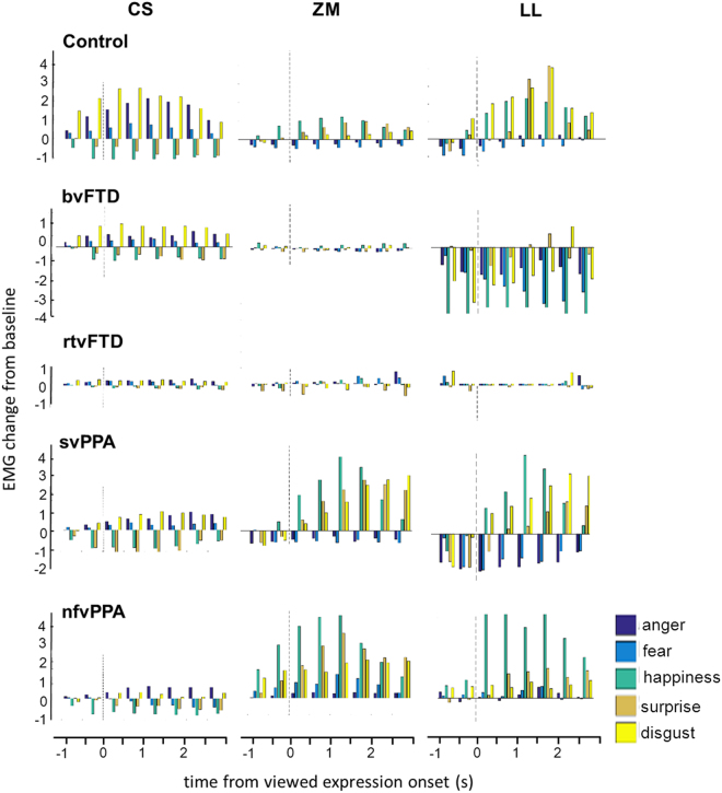
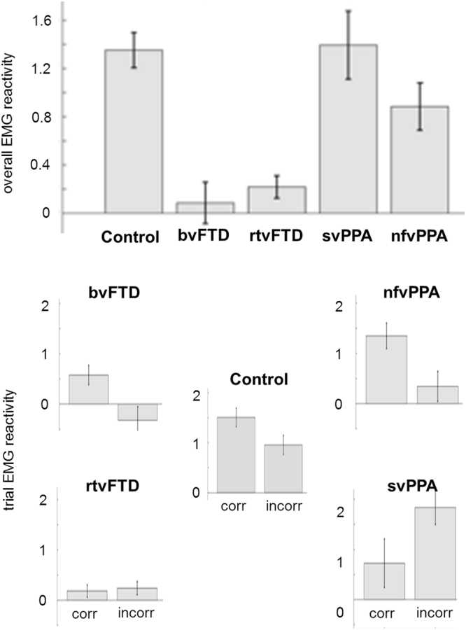
Automatic motor mimicry is essential to the normal processing of perceived emotion, and disrupted automatic imitation might underpin socio-emotional deficits in neurodegenerative diseases, particularly the frontotemporal dementias. However, the pathophysiology of emotional reactivity in these diseases has not been elucidated. We studied facial electromyographic responses during emotion identification on viewing videos of dynamic facial expressions in 37 patients representing canonical frontotemporal dementia syndromes versus 21 healthy older individuals. Neuroanatomical associations of emotional expression identification accuracy and facial muscle reactivity were assessed using voxel-based morphometry. Controls showed characteristic profiles of automatic imitation, and this response predicted correct emotion identification. Automatic imitation was reduced in the behavioural and right temporal variant groups, while the normal coupling between imitation and correct identification was lost in the right temporal and semantic variant groups. Grey matter correlates of emotion identification and imitation were delineated within a distributed network including primary visual and motor, prefrontal, insular, anterior temporal and temporo-occipital junctional areas, with common involvement of supplementary motor cortex across syndromes. Impaired emotional mimesis may be a core mechanism of disordered emotional signal understanding and reactivity in frontotemporal dementia, with implications for the development of novel physiological biomarkers of socio-emotional dysfunction in these diseases.


Conflict of interest statement

Dr. Marshall is supported by the Leonard Wolfson Experimental Neurology Centre. Mr Hardy holds a MRC UK PhD studentship. Ms Russell reports no biomedical financial interests or potential conflicts of interest. Dr. Clark was supported by The National Brain Appeal – Frontotemporal Dementia Research Fund. Ms Bond holds a MRC UK PhD studentship. Ms Dick is funded by the Alzheimer’s Society. Ms Brotherhood reports no biomedical financial interests or potential conflicts of interest. Dr. Mummery reports no biomedical financial interests or potential conflicts of interest. Dr. Schott reports no biomedical financial interests or potential conflicts of interest. Dr. Rohrer is a MRC UK Clinical Scientist. Dr. Kilner reports no biomedical financial interests or potential conflicts of interest. Prof Warren was supported by a Wellcome Trust Senior Clinical Fellowship and receives funding from the Alzheimer’s Society.

Figures

Figure 1
Figure 1
Patterns of EMG reactivity for each muscle in each participant group. For each participant group, the plots show the time course of average EMG reactivity (in microvolts) for key facial muscles while participants watched videos of emotional facial expressions. EMG reactivity, here indexed in arbitrary units as mean EMG change from baseline, is shown on the y-axis (after rectifying, high-pass filtering and removing artefacts as described in Methods). Onset of the viewed facial expression (as determined in a prior independent analysis of the video stimuli) is at time 0 (dotted line) in each panel. In healthy controls, corrugator supercilii (CS) was activated during viewing of anger, fear and disgust, but inhibited during viewing of happiness and surprise; zygomaticus major (ZM) was activated during viewing of happiness and surprise, but inhibited during viewing of anger and fear; and levator labii (LL) was inhibited during viewing of anger and fear, and maximally activated during viewing of disgust. Note that in healthy controls muscle responses consistently preceded the unambiguous onset of viewed emotional expressions. bvFTD, patient group with behavioural variant frontotemporal dementia (excluding right temporal cases); Control, healthy control group; nfvPPA, patient group with nonfluent variant primary progressive aphasia; rtvFTD, patient subgroup with right temporal variant frontotemporal dementia; svPPA, patient group with semantic variant primary progressive aphasia.
Figure 2
Figure 2
EMG reactivity in each participant group, and the relationship with identification accuracy. For each participant group, the histograms show mean overall facial muscle EMG reactivity (top) and EMG reactivity separately (below) for those trials on which viewed emotional expressions were identified correctly (corr) versus incorrectly (incorr); error bars indicate standard error of the mean (see also Table 2). bvFTD, patient group with behavioural variant frontotemporal dementia; Control, healthy control group; nfvPPA, patient group with nonfluent variant primary progressive aphasia; rtvFTD, patient subgroup with right temporal variant frontotemporal dementia; svPPA, patient group with semantic variant primary progressive aphasia.
Figure 3
Figure 3
Neuroanatomical correlates of emotion identification and EMG reactivity for each syndromic group. Statistical parametric maps (SPMs) show regional grey matter volume positively associated with overall emotion identification accuracy and facial EMG reactivity during viewing of emotional facial expressions, based on voxel-based morphometry of patients’ brain MR images (see also Table 3); T-scores are coded on the colour bar. SPMs are overlaid on sections of the normalised study-specific T1-weighted mean brain MR image; the MNI coordinate (mm) of the plane of each section is indicated (coronal and axial sections show the left hemisphere on the left). Panels code syndromic profiles of emotion identification (ID) or EMG reactivity (EMG). Note that the correlates of emotion identification and EMG reactivity in different syndromes overlapped in particular brain regions, including supplementary motor cortex and temporo-occipital junction (see Table 3). SPMs are thresholded for display purposes at p < 0.001 uncorrected over the whole brain, however local maxima of areas shown were each significant at p < 0.05 after family-wise error correction for multiple voxel-wise comparisons within pre-specified anatomical regions of interest (see Table 3). bvFTD, patient group with behavioural variant FTD; nfvPPA, patient group with nonfluent variant primary progressive aphasia; rtvFTD, patient subgroup with right temporal variant frontotemporal dementia; svPPA, patient group with semantic variant primary progressive aphasia.

References

    1. Niedenthal PM. Embodying emotion. Science. 2007;316:1002–1005. doi: 10.1126/science.1136930. - DOI - PubMed
    1. Wood A, Rychlowska M, Korb S, Niedenthal P. Fashioning the Face: Sensorimotor Simulation Contributes to Facial Expression Recognition. Trends in cognitive sciences. 2016;20:227–240. doi: 10.1016/j.tics.2015.12.010. - DOI - PubMed
    1. Dimberg U, Thunberg M. Rapid facial reactions to emotional facial expressions. Scand J Psychol. 1998;39:39–45. doi: 10.1111/1467-9450.00054. - DOI - PubMed
    1. Dimberg U, Thunberg M, Grunedal S. Facial reactions to emotional stimuli: Automatically controlled emotional responses. Cognition Emotion. 2002;16:449–471. doi: 10.1080/02699930143000356. - DOI
    1. Neumann R, Schulz SM, Lozo L, Alpers GW. Automatic facial responses to near-threshold presented facial displays of emotion: imitation or evaluation? Biological psychology. 2014;96:144–149. doi: 10.1016/j.biopsycho.2013.12.009. - DOI - PubMed

Publication types

LinkOut - more resources