. 2018 Jan 18;9(2):28.
doi: 10.1038/s41419-017-0195-0.

Preclinical evaluation of antitumor activity of the proteasome inhibitor MLN2238 (ixazomib) in hepatocellular carcinoma cells

Affiliations

Preclinical evaluation of antitumor activity of the proteasome inhibitor MLN2238 (ixazomib) in hepatocellular carcinoma cells

Giuseppa Augello et al. Cell Death Dis. .

Abstract

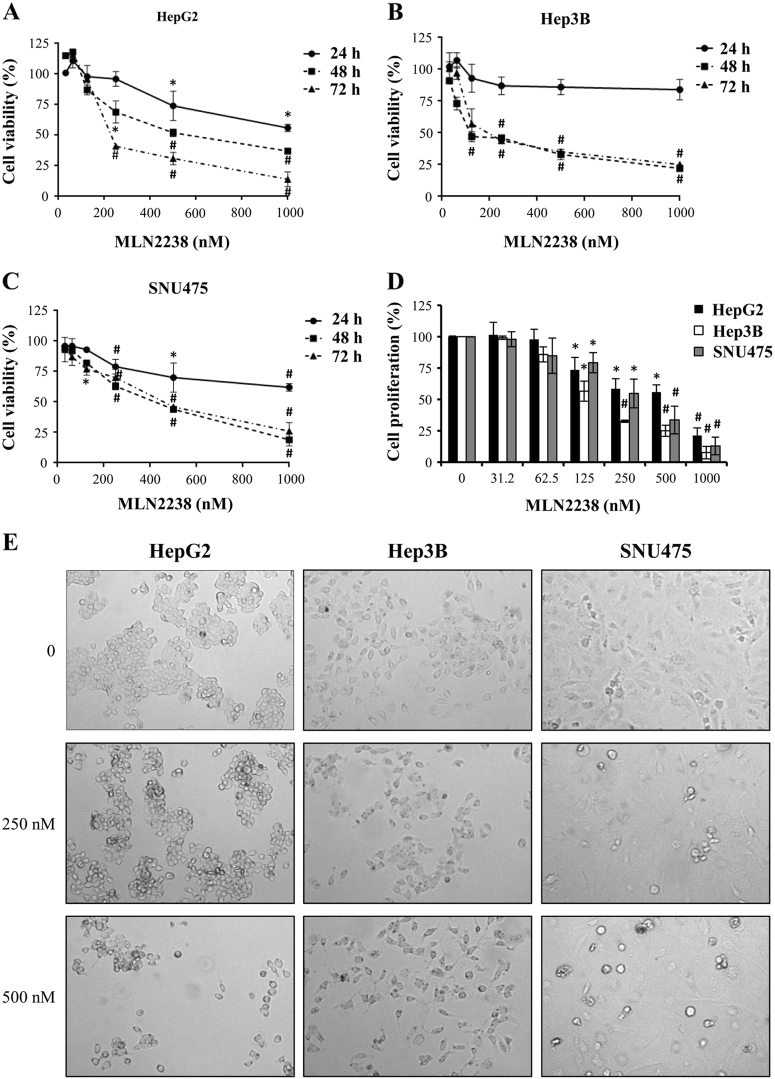
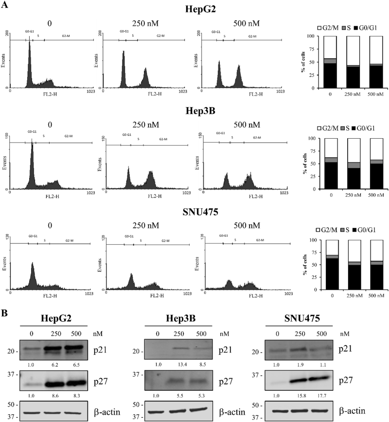
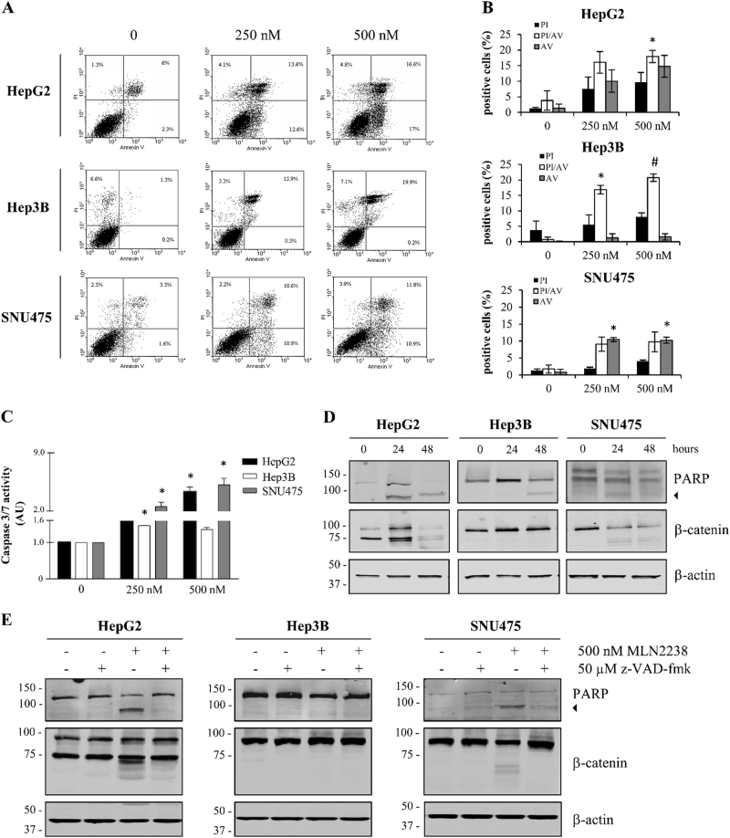
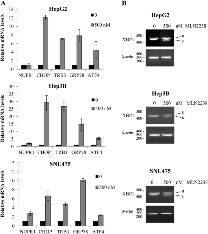
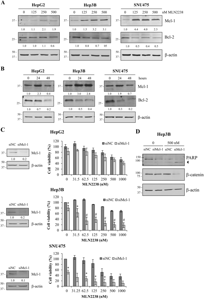
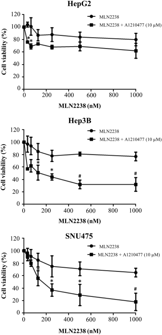
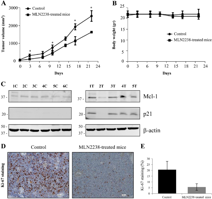
Hepatocellular carcinoma (HCC) is one of the common malignancies and is an increasingly important cause of cancer death worldwide. Surgery, chemotherapy, and radiation therapy extend the 5-year survival limit in HCC patients by only 6%. Therefore, there is a need to develop new therapeutic approaches for the treatment of this disease. The orally bioavailable proteasome inhibitor MLN2238 (ixazomib) has been demonstrated to have anticancer activity. In the present study, we investigated the preclinical therapeutic efficacy of MLN2238 in HCC cells through in vitro and in vivo models, and examined its molecular mechanisms of action. MLN2238 inhibited cell viability in human HCC cells HepG2, Hep3B, and SNU475 in a time- and dose-dependent manner. Flow cytometry analysis demonstrated that MLN2238 induced G2/M cell cycle arrest and cellular apoptosis in HCC cells. Cell cycle arrest was associated with increased expression levels of p21 and p27. MLN2238-induced apoptosis was confirmed by caspase-3/7 activation, PARP cleavage and caspase-dependent β-catenin degradation. In addition, MLN2238 activated ER stress genes in HCC cells and increased the expression of the stress-inducible gene nuclear protein-1. Furthermore, MLN2238 treatment induced upregulation of myeloid cell leukemia-1 (Mcl-1) protein, and Mcl-1 knockdown sensitized HCC cells to MLN2238 treatment, suggesting the contribution of Mcl-1 expression to MLN2238 resistance. This result was also confirmed using the novel Mcl-1 small molecule inhibitor A1210477. Association of A1210477 and MLN2238 determined synergistic antitumor effects in HCC cells. Finally, in vivo orally administered MLN2238 suppressed tumor growth of Hep3B cells in xenograft models in nude mice. In conclusion, our results offer hope for a new therapeutic opportunity in the treatment of HCC patients.


Conflict of interest statement

The authors declare that they have no conflict of interest.

Figures

Fig. 1
Fig. 1. MLN2238 treatment alters cell viability, proliferation, and morphology in HCC cells.
ac Cell viability was evaluated by MTS assay. HepG2 a, Hep3B b, and SNU475 c cells were treated with increasing MLN2238 concentrations for 24, 48, and 72 h. d Proliferation was measured by BrdU incorporation into DNA. Cells underwent treatment for 48 h with increasing MLN2238 concentrations. e Morphological observations of MLN2238-treated cells by phase contrast microscope. Photographs were taken after 24 h of treatment (× 20 magnification). *P < 0.05 MLN2238 vs. control; #P < 0.005 MLN2238 vs. control
Fig. 2
Fig. 2. MLN2238 treatment induces cell cycle arrest and upregulated p21 and p27 expression in HCC cells.
a Images representing cell cycle analysis in cells receiving 250 and 500 nM MLN2238 for 24 h. Left panels, flow cytometry was used to analyze the DNA content of cells after staining with propidium iodide. Right panels, summarized data of cell cycle distribution. Data are expressed as the percentage of cells in the G2/M, S and G0/G1 cell cycle phase, and are expressed as the means of three separate experiments. b Western blot analysis of p21 and p27 in HCC cells after treatment with 250 and 500 nM of MLN2238 for 24 h
Fig. 3
Fig. 3. MLN2238 induces apoptosis in HCC cells.
a Representative FITC-Annexin V/PI flow cytometry analysis of HepG2, Hep3B, and SNU475 cells after 24 h of incubation with 250 and 500 nM MLN2238. b Summarized data of apoptotic percentage distribution. c Caspase-3/7 activity levels in cells after treatment with 250 and 500 nM MLN2238 for 24 h. Data are given as AU normalized to control values and represent the mean ± S.D. (n = 3). d Representative western blotting of PARP and β-catenin in cell lines after treatment with 500 nM MLN2238 for 24 and 48 h. The 85 kDa form of PARP is indicated by an arrowhead. e Representative western blot analysis of PARP and β-catenin after treatment with 500 nM MLN2238 and/or 50 µM z-VAD-fmk for 24 h. The 85 kDa form of PARP is indicated by an arrowhead. *P < 0.05, MLN2238 vs. control; #P < 0.005, MLN2238 vs. control
Fig. 4
Fig. 4. MLN2238 treatment induces ER stress in HCC cells.
Effects of MLN2238 treatment with 500 nM of MLN2238 for 24 h on ER stress gene expression levels were determined by quantitative Real-Time PCR a and semiquantitative PCR b. a The relative gene expression was calculated (ratio of drug-treated samples vs. control) and corrected by the quantified level of β-actin expression. b Expression of XBP1 mRNA. u = unspliced XBP1 mRNA; s = spliced XBP1 mRNA
Fig. 5
Fig. 5. MLN2238 treatment regulates expression of anti-apoptotic proteins Mcl-1 and Bcl-2, and Mcl-1 knockdown sensitizes HCC cells to MLN2238-mediated cell death.
Dose- a and time-dependent b effects of MLN2238 treatment on Mcl-1 and Bcl-2 expression determined by western blot analysis. a Cells exposed to the specified MLN2238 concentrations for 24 h. b Cells treated with 500 nM of MLN2238 for 24 and 48 h. c Left panels, Mcl-1 expression levels after 72 h of transfection with Mcl-1 siRNA (siMcl-1) in cells compared with cells transfected with control siRNA (siNC). Right panels, viability of cells transfected with Mcl-1 siRNA (siMcl-1) after treatment with the indicated MLN2238 concentrations for 24 h. d Representative western blotting of PARP and β-catenin levels expressed in Hep3B cells transfected with Mcl-1 siRNA (siMcl-1) or control siRNA (siNC) after 24 h of treatment with 500 nM of MLN2238. The 85 kDa form of PARP is indicated by an arrowhead. *P < 0.05 MLN2238 vs. control; #P < 0.005 MLN2238 vs. control
Fig. 6
Fig. 6. Treatment with MLN2238 in combination with A1210477 synergistically inhibits cell viability in HCC cells.
Viability after treating HCC cells with different doses of MLN2238 + 10 µM A1210477 for 24 h was evaluated by MTS assay. *P < 0.05, 10 µM A1210477 + MLN2238 vs. 10 µM A1210477 alone; #P < 0.005, 10 µM A1210477 + MLN2238 vs. 10 µM A1210477 alone
Fig. 7
Fig. 7. MLN2238 inhibits tumor growth in vivo.
a Effect of MLN2238 on tumor development. A comparison was made between the tumor growth curve of MLN2238-treated mice (11 mg/kg) and that of control mice receiving only vehicle as treatment, *P < 0.05. b Analysis of body weight alterations. Animals were weighed two times/week and the data plotted in the graphs. c Representative immunoblotting of Mcl-1 and p21 expression levels in mice receiving only vehicle treatment (control: 1 C, 2 C, 3 C, 4 C, 5 C, and 6 C) and mice treated with MLN2238 (1 T, 2 T, 3 T, 4 T and 5 T). d Immunohistochemical staining of the Ki-67 proliferation marker in the control and MLN2238-treated mice (scale bar = 50 µm). e Data indicate the number of positive cells and are expressed as the means ± S.D. of five different fields in three sections of tumors from the control and the MLN2238-treated mice

References

    1. Torrecilla S, Llovet JM. New molecular therapies for hepatocellular carcinoma. Clin. Res. Hepatol. Gastroenterol. 2015;39:80–85. doi: 10.1016/j.clinre.2015.06.016. - DOI - PubMed
    1. Curran MP, McKeage K. Bortezomib: a review of its use in patients with multiple myeloma. Drugs. 2009;69:859–888. doi: 10.2165/00003495-200969070-00006. - DOI - PubMed
    1. O’Connor OA, et al. Phase II clinical experience with the novel proteasome inhibitor bortezomib in patients with indolent non-Hodgkin’s lymphoma and mantle cell lymphoma. J. Clin. Oncol. 2005;23:676–684. doi: 10.1200/JCO.2005.02.050. - DOI - PubMed
    1. Lee AH, Iwakoshi NN, Anderson KC, Glimcher LH. Proteasome inhibitors disrupt the unfolded protein response in myeloma cells. Proc. Natl. Acad. Sci. USA. 2003;100:9946–9951. doi: 10.1073/pnas.1334037100. - DOI - PMC - PubMed
    1. Jiang HY, Wek RC. Phosphorylation of the alpha-subunit of the eukaryotic initiation factor-2 (eIF2alpha) reduces protein synthesis and enhances apoptosis in response to proteasome inhibition. J. Biol. Chem. 2005;280:14189–14202. doi: 10.1074/jbc.M413660200. - DOI - PubMed

Publication types

MeSH terms