. 2018 Jan 18;8(1):1161.
doi: 10.1038/s41598-018-19516-6.

Umbilical cord extracts improve osteoporotic abnormalities of bone marrow-derived mesenchymal stem cells and promote their therapeutic effects on ovariectomised rats

Affiliations

Umbilical cord extracts improve osteoporotic abnormalities of bone marrow-derived mesenchymal stem cells and promote their therapeutic effects on ovariectomised rats

Akira Saito et al. Sci Rep. .

Erratum in

Abstract

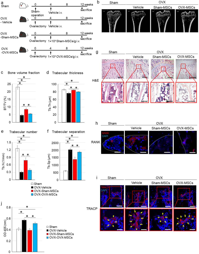
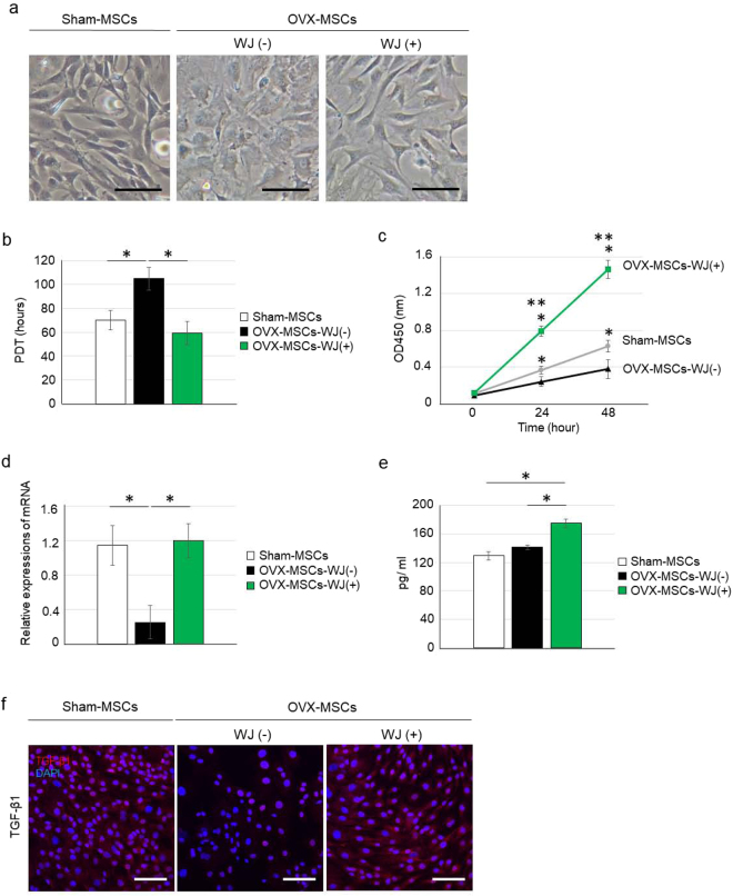
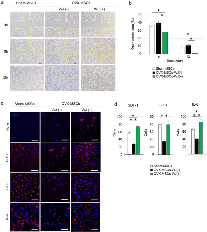
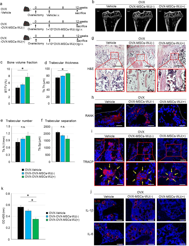
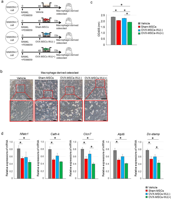
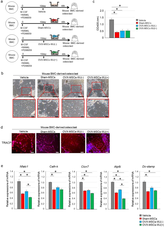
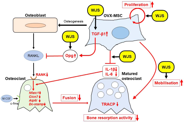
Bone marrow-derived mesenchymal stem cells (BM-MSCs) are the most valuable source of autologous cells for transplantation and tissue regeneration to treat osteoporosis. Although BM-MSCs are the primary cells responsible for maintaining bone metabolism and homeostasis, their regenerative ability may be attenuated in postmenopausal osteoporosis patients. Therefore, we first examined potential abnormalities of BM-MSCs in an oestrogen-deficient rat model constructed by ovariectomy (OVX-MSCs). Cell proliferation, mobilisation, and regulation of osteoclasts were downregulated in OVX-MSCs. Moreover, therapeutic effects of OVX-MSCs were decreased in OVX rats. Accordingly, we developed a new activator for BM-MSCs using human umbilical cord extracts, Wharton's jelly extract supernatant (WJS), which improved cell proliferation, mobilisation and suppressive effects on activated osteoclasts in OVX-MSCs. Bone volume, RANK and TRACP expression of osteoclasts, as well as proinflammatory cytokine expression in bone tissues, were ameliorated by OVX-MSCs activated with WJS (OVX-MSCs-WJ) in OVX rats. Fusion and bone resorption activity of osteoclasts were suppressed in macrophage-induced and primary mouse bone marrow cell-induced osteoclasts via suppression of osteoclast-specific genes, such as Nfatc1, Clcn7, Atp6i and Dc-stamp, by co-culture with OVX-MSCs-WJ in vitro. In this study, we developed a new activator, WJS, which improved the functional abnormalities and therapeutic effects of BM-MSCs on postmenopausal osteoporosis.


Conflict of interest statement

The authors declare that they have no competing interests.

Figures

Figure 1
Figure 1
Therapeutic effect of Sham-MSCs and OVX-MSCs in OVX rats. (a) Experimental protocol for Sham and OVX rats treated with Vehicle, Sham-MSCs, or OVX-MSCs. (b) Representative micro-CT images of tibias. (cf) Quantitative changes in trabecular parameters, including trabecular bone volume, expressed as c: percentage of total tissue volume (BV/TV), d: trabecular thickness (Tb.Th), (e) Trabecular number (Tb.N), and (f) Trabecular separation (Tb.Sp). *P < 0.05. Data are expressed as mean ± SE of 4–5 animals. (g) Histological findings of the tibia in H&E-stained sections. Bar: 500 μm in upper panels, 100 μm in lower panels. (h) Immunofluorescence staining of the tibia with an anti-RANK antibody (red). DAPI was used for counterstaining nuclei (blue). Bar: 25 µm. (i) Immunofluorescence staining of the tibia with an anti-TRACP antibody (red). DAPI was used for counterstaining nuclei (blue). Bar: upper 50 μm, lower 25 µm. (j) Serum TRACP levels at the time of sacrifice of rats. *P < 0.05. Data are expressed as mean ± SE of 4–5 animals.
Figure 2
Figure 2
Abnormalities and activating effects of WJS for cell morphology, proliferation, secretion, and protein expression of OVX-MSCs. (a) Phase contrast observations of Sham-MSCs (left panel), OVX-MSCs-WJ(-) (middle panel) and OVX-MSCs-WJ(+) (right panel). Images were obtained at 48 h after activation with WJS. Bar: 100 μm. (b) Population doubling time of Sham-MSCs, OVX-MSCs-WJ(-), and OVX-MSCs-WJ(+) at passage 2. *P < 0.05. Data are expressed as mean ± SE of 5–6 BM-MSC cultures. (c) MTT proliferation assay of Sham-MSCs, OVX-MSCs-WJ(-), and OVX-MSCs-WJ(+). *P < 0.05. Data are expressed as mean ± SE of 5–6 BM-MSC cultures. (d) Relative mRNA expression of Sham-MSCs, OVX-MSCs-WJ(-), and OVX-MSCs-WJ(+). *P < 0.05. Data are expressed as mean ± SE of 4 BM-MSCs. Opg, osteoprotegerin. (e) OPG levels in the culture supernatant of Sham-MSCs, OVX-MSCs-WJ(-), and OVX-MSCs-WJ(+). *P < 0.05. Data are expressed as mean ± SE of 4 BM-MSC cultures. (f) Immunofluorescence staining of Sham-MSCs (left panel), OVX-MSCs-WJ(-) (middle panel), and OVX-MSCs-WJ(+) (right panel) with an anti-TFG-β antibody (red). DAPI was used for counterstaining nuclei (blue). Bar: 100 μm.
Figure 3
Figure 3
Activating effects of WJS on migration capacity of OVX-MSCs. (a) Wound assay of Sham-MSCs, OVX-MSCs-WJ(-), and OVX-MSCs-WJ(+). *P < 0.05. Data are expressed as mean ± SE of 4 BM-MSC cultures. The open wound area was measured at 6 h and 12 h after performing the cross-scratch. Yellow lines indicate the boundary of cell migration. (b) The ratio of open wound area at 6 h and 12 h compared with 0 h. Six cross-scratch points in each BM-MSC culture were measured. *P < 0.05. Data are expressed as mean ± SE of 4 BM-MSC cultures. (c) Chemotaxis assay of Sham-MSCs, OVX-MSCs-WJ(-), and OVX-MSCs-WJ(+). BM-MSCs induced to migrate to the opposite side of the membrane by SDF-1, IL-1β, and IL-6 as observed by confocal microscope. DAPI was used for counterstaining nuclei (blue). Bar: 100 μm. SDF-1, stromal cell-derived factor 1; IL-1β, interleukin-1 beta; IL-6, interleukin-6. (d) Numbers of BM-MSCs migrated to the opposite side of the membrane were counted in three fields of view for each BM-MSC type. *P < 0.05. Data are expressed as mean ± SE of 3 BM-MSC cultures.
Figure 4
Figure 4
Therapeutic effect of OVX-MSCs activated with WJS in OVX rats. (a) Experimental protocol for Vehicle, OVX-MSC-WJ(-), and OVX-MSC-WJ(+) therapies in OVX rats. (b) Representative micro-CT images of tibias. (cf) Quantitative changes in trabecular parameters, including trabecular bone volume, expressed as c: percentage of total tissue volume (BV/TV), d: trabecular thickness (Tb.Th), (e) Trabecular number (Tb.N), and (f) Trabecular separation (Tb.Sp). *P < 0.05. Data are expressed as mean ± SE of 4–5 animals. (g) Histological findings of the tibia in H&E-stained sections at 8 weeks after administration of Vehicle, OVX-MSC-WJ(-), or OVX-MSC-WJ(+) therapies in OVX rats. Bar: 500 μm in upper panel, 100 μm in lower panel. (h) Immunofluorescence staining of the tibia with an anti-RANK antibody (red). DAPI was used for counterstaining nuclei (blue). Bar: 25 µm. (i) Immunofluorescence staining of the tibia with an anti-TRACP antibody (red). DAPI was used for counterstaining nuclei (blue). Bar: upper 50 μm, lower 25 µm. (j) Immunofluorescence staining of the tibia with an anti-IL-1β antibody (red) (upper) and with an anti-IL-6 antibody (red) (lower). DAPI was used for counterstaining nuclei (blue). Bar: 100 μm. (k) Serum TRACP levels at 8 weeks after administration of Vehicle, OVX-MSC-WJ(-), and OVX-MSC-WJ(+) therapies in OVX rats. *P < 0.05. Data are expressed as mean ± SE of 4–5 animals.
Figure 5
Figure 5
Regulatory effect of OVX-MSCs activated with WJS on RAW264.7 cell-derived osteoclasts. (a) Experimental protocol for co-culture of RAW264.7 cell-derived osteoclasts with Vehicle, Sham-MSCs, OVX-MSCs-WJ(-), or OVX-MSCs-WJ(+). (b) Phase contrast observations of matured osteoclasts co-cultured without MSCs (left panel) or with Sham-MSCs (middle left panel), OVX-MSCs-WJ(-) (middle right panel), or OVX-MSCs-WJ(+) (right panel) using a Transwell. Images were obtained 72 h after adding RANKL and PD98059 to the culture medium, and 24 h after starting co-culture. Bar: 500 μm in upper panel, 100 μm in lower panel. (c) TRACP levels in the supernatant of RAW264.7 cell-derived osteoclasts co-cultured without MSCs and with Sham-MSCs, OVX-MSCs-WJ(-), or OVX-MSCs-WJ(+). Data are expressed as mean ± SE of osteoclasts from 3 experiments. *P < 0.05. (d) Relative mRNA expression in RAW264.7 cell-derived osteoclasts. Values represent mean ± SE of osteoclasts co-cultured without MSCs or with Sham-MSCs, OVX-MSCs-WJ(-), or OVX-MSCs-WJ(+) from 3 experiments. *P < 0.05. Nfatc1, nuclear factor of activated T cells; Cath-k, cathepsin K; Clc7, chloride channel-7; Atp6i, ATPase, H+ transporting, (vacuolar proton pump) member I; Dc-stamp, dendritic cell-specific transmembrane protein.
Figure 6
Figure 6
Regulatory effect of OVX-MSCs activated with WJS on mouse bone marrow cell-derived osteoclasts. (a) Experimental protocol for co-culture of mouse BMC-derived osteoclasts with Vehicle, Sham-MSCs, OVX-MSCs-WJ(-), or OVX-MSCs-WJ(+). (b) Phase contrast observations of matured osteoclasts co-cultured without MSCs (left panel) or with Sham-MSCs (middle left panel), OVX-MSCs-WJ(-) (middle right panel), or OVX-MSCs-WJ(+) (right panel) using a Transwell. Images were obtained 10 days after adding RANKL and PD98059 to the culture medium, and 48 h after starting co-culture. Bar: 500 μm in upper panel, 100 μm in lower panel. (c) TRACP levels in the supernatant of mouse BMC-derived osteoclasts co-cultured without MSCs and with Sham-MSCs, OVX-MSCs-WJ(-), or OVX-MSCs-WJ(+). Data are expressed as mean ± SE of osteoclasts from 4–5 experiments. *P < 0.05. (d) Immunofluorescence staining of mouse BMC-derived osteoclasts with an anti-TRACP antibody (red). DAPI was used for counterstaining nuclei (blue). Bar: 100 μm. (e) Relative mRNA expression in mouse BMC-derived osteoclasts. Values represent mean ± SE of osteoclasts co-cultured without MSCs and with Sham-MSCs, OVX-MSCs-WJ(-), or OVX-MSCs-WJ(+) from 4–5 experiments. *P < 0.05. Nfatc1, nuclear factor of activated T cells; Cath-k, cathepsin K; Clc7, chloride channel-7; Atp6i, ATPase, H+ transporting, (vacuolar proton pump) member I; Dc-stamp, dendritic cell-specific transmembrane protein.
Figure 7
Figure 7
Presumed activation mechanisms of WJS on OVX-MSCs and resulting therapeutic effects for osteoporosis. The site of action of WJS on OVX-MSCs and the resulting therapeutic mechanism for osteoporosis.

Similar articles

Cited by

References

    1. Pacifici R. Estrogen deficiency, T cells and bone loss. Cellular immunology. 2008;252:68–80. doi: 10.1016/j.cellimm.2007.06.008. - DOI - PubMed
    1. Martin-Millan M, et al. The estrogen receptor-alpha in osteoclasts mediates the protective effects of estrogens on cancellous but not cortical bone. Mol Endocrinol. 2010;24:323–334. doi: 10.1210/me.2009-0354. - DOI - PMC - PubMed
    1. Raisz LG. Pathogenesis of osteoporosis: concepts, conflicts, and prospects. J Clin Invest. 2005;115:3318–3325. doi: 10.1172/JCI27071. - DOI - PMC - PubMed
    1. Pittenger MF, Martin BJ. Mesenchymal stem cells and their potential as cardiac therapeutics. Circ Res. 2004;95:9–20. doi: 10.1161/01.RES.0000135902.99383.6f. - DOI - PubMed
    1. Bernardo ME, Fibbe WE. Safety and efficacy of mesenchymal stromal cell therapy in autoimmune disorders. Annals of the New York Academy of Sciences. 2012;1266:107–117. doi: 10.1111/j.1749-6632.2012.06667.x. - DOI - PubMed

MeSH terms