. 2018 Jan 18;9(2):25.
doi: 10.1038/s41419-017-0176-3.

Human colorectal cancer-derived mesenchymal stem cells promote colorectal cancer progression through IL-6/JAK2/STAT3 signaling

Affiliations

Human colorectal cancer-derived mesenchymal stem cells promote colorectal cancer progression through IL-6/JAK2/STAT3 signaling

Xiaochao Zhang et al. Cell Death Dis. .

Abstract

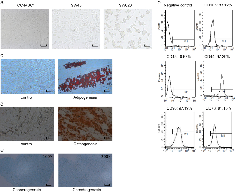
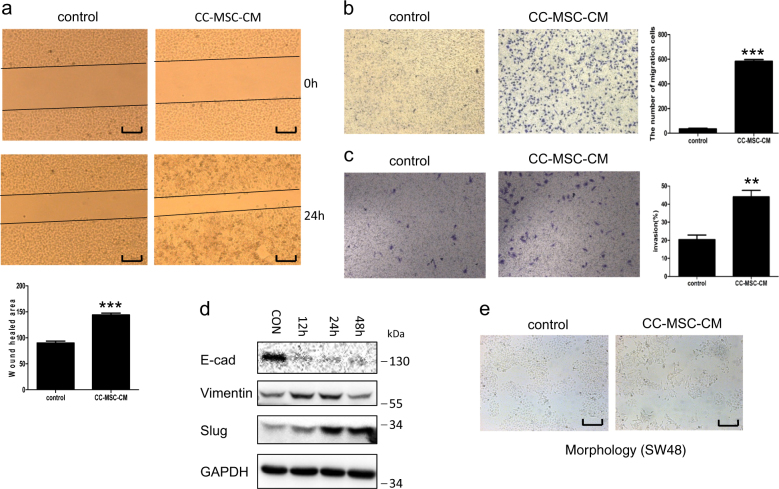
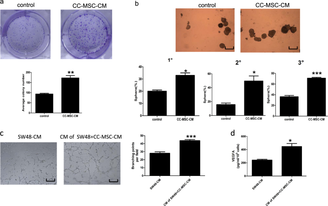
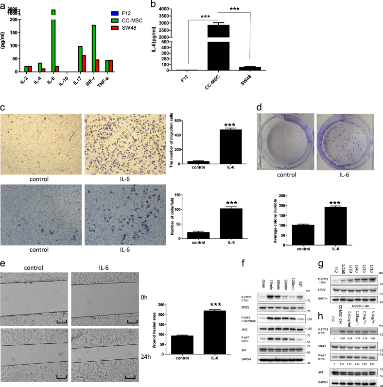
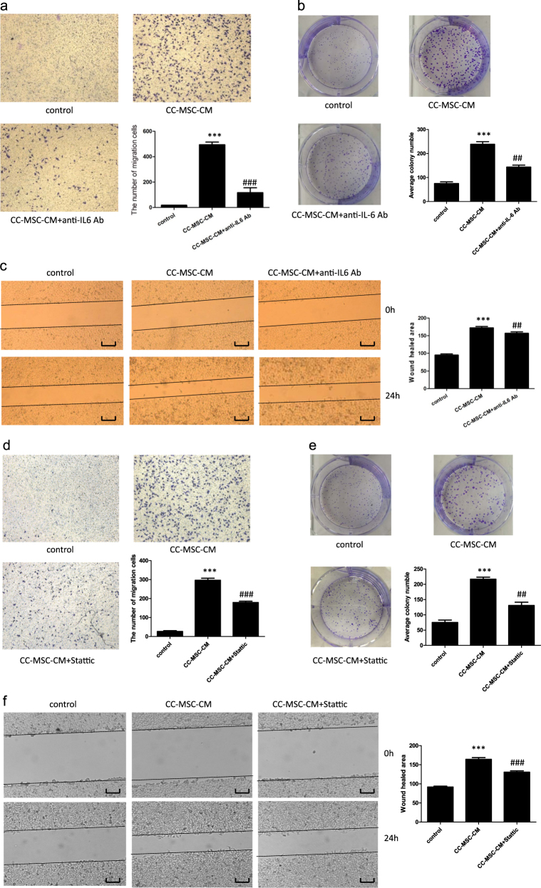
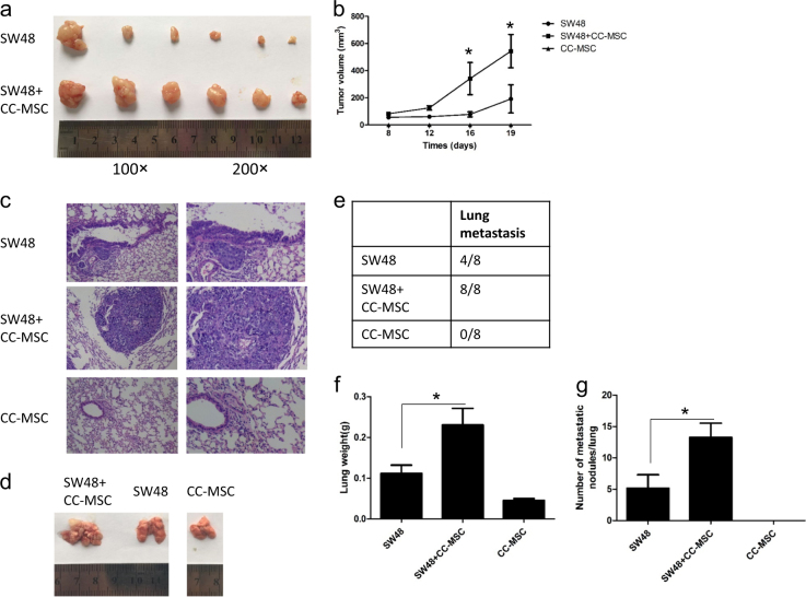
Mesenchymal stem cells (MSCs) have been reported to localize in colorectal carcinomas, and participate in the formation of the tumor microenvironment. They have recently been isolated from colorectal cancer tissues, and are implicated in the growth, invasion, and metastasis of cancer cells. However, the roles and detailed mechanisms associated with human colorectal cancer-derived MSCs (CC-MSCs) have not been fully addressed. In this study, we found that CC-MSCs increased the migration and invasion of colorectal cancer cells and promoted the tumorigenesis of colorectal cancer through epithelial-to-mesenchymal transition (EMT) in vitro. We also found that CC-MSCs enhanced the growth and metastasis of colorectal cancer in vivo. Mechanistically, we determined that interleukin-6 (IL-6) was the most highly expressed cytokine in the CC-MSC conditioned medium, and promoted the progression of colorectal cancer cells through IL-6/JAK2/STAT3 signaling, which activated PI3K/AKT signaling. We used anti-IL-6 antibody to target IL-6. Collectively, these results reveal that the IL-6 secreted by CC-MSCs enhances the progression of colorectal cancer cells through IL-6/JAK2/STAT3 signaling, and could provide a novel therapeutic or preventive target.


Conflict of interest statement

The authors declare that they have no conflict of interest.

Figures

Fig. 1
Fig. 1. The characterizations of human colorectal cancer-derived mesenchymal stem cells (CC-MSCs).
a Morphology of CC-MSCs, SW620 colorectal cancer cells, and SW48 colorectal cancer cells (magnification, ×100; scale bar: 250 μm). The CC-MSCs were fibroblastic in shape, but the SW620 and SW48 cancer cells were round. b Flow cytometric analysis of CC-MSCs. The CC-MSCs were positive for CD105, CD90, CD73, and CD44, and negative for CD45. c CC-MSCs cultivated in control medium or adipogenic differentiation medium were stained with Oil Red O. The red lipid droplets indicate adipogenic differentiation (magnification, ×100; scale bar: 250 μm). d CC-MSCs cultivated in control medium or osteogenic differentiation medium were stained with alizarin red (magnification, ×100; scale bar: 250 μm). e CC-MSCs cultivated in chondrogenic differentiation medium were stained with alcian blue (left panel, magnification, ×100; scale bar: 250 μm; right panel, magnification, ×200; scale bar: 125 μm)
Fig. 2
Fig. 2. Human colorectal cancer-derived mesenchymal stem cells promote the migration and invasion of SW48 colorectal cancer in vitro, and mediate epithelial-to-mesenchymal transition (EMT) in colorectal cancer.
a Wound-healing closure assays of SW48 colorectal cancer cells were performed in the presence or absence of CC-MSC conditioned medium (CC-MSC-CM) (magnification, ×50; scale bar: 500 μm). b Chemotaxis assays of SW48 cells with CC-MSCs-CM in the lower chamber (×100). c Invasion assays of SW48 colorectal cancer cells were also carried out in the presence or absence of CC-MSCs-CM (×100). The addition of the CC-MSC-CM enhanced both the migration and invasion of the SW48 cells. *P < 0.05 compared with mock treatment. d SW48 cells were cultivated with CC-MSC-CM for different times, and several markers associated with the EMT process, such as E-cadherin, vimentin, and SLUG, were detected by western blotting analysis. F12 served as the control. e SW48 cells in the presence or absence of CC-MSC-CM were photographed using a phase contrast microscope. A mesenchymal phenotype in SW48 cells was viewed in the presence of CC-MSC-CM and compared with the control group (magnification, ×50; scale bar: 500 μm)
Fig. 3
Fig. 3. Human colon cancer-derived mesenchymal stem cells promote the generation of cancer stem cells and enhance angiogenesis in vitro.
a SW48 cells treated with CC-MSC-CM were assayed for clonogenicity in adherent cultures. Separated clones were counted and plotted. b Effect of CC-MSC-CM on serial sphere-formation assays of SW620. Tumor spheres were generated in suspension culture for 2 weeks using Ultra-Low Attachment plates (magnification, ×100; scale bar: 250 μm). *P < 0.05 compared with mock treatment. c Human umbilical vein endothelial cells (HUVECs) were cultivated in CM from control SW48 cells and SW48 cells pretreated with CC-MSC-CM for 24 h. The cells’ tube formation ability was quantified by counting the branching points per field (magnification, ×100; scale bar: 250 μm). ***P < 0.001. d ELISA was used to examine the VEGFA level in the CM obtained in Fig. 3c. All experiments were carried out in triplicate. *P < 0.05
Fig. 4
Fig. 4. IL-6 secreted by CC-MSCs enhances the proliferation, migration and invasion of colorectal cancer cells through IL-6/JAK2/STAT3 signaling.
a The levels of various factors in the cell-free culture supernatants were measured using Bio-Plex cytokine arrays. b Quantitative analysis of IL-6 levels using enzyme-linked immunosorbent assay (ELISA). The conditioned media from the cultured CC-MSCs and SW48 cells were collected to detect the levels of IL-6, and representative results from one of the three independent experiments are presented. c Transwell migration (top) and invasion (bottom) assay of SW48 cells with or without 10 ng/mL recombinant IL-6 treatment (×100). d Colony-formation assay of SW48 cells treated with or without 10 ng/mL recombinant IL-6. e Wound-healing assay of SW48 cells in the presence or absence of recombinant IL-6 (magnification, ×50; scale bar: 500 μm). ***P < 0.001. f SW48 cells were cultured with CC-MSC-CM for different times. Protein expression was determined by western blotting and representative results from one of the three independent experiments are presented. g SW48 cells were cultured with different dilution ratios of CC-MSC-CM for 30 min. Protein expression was determined by western blotting and representative results from one of the three independent experiments are presented. h SW48 cells were cultured for 30 min in F12 or CC-MSC-CM, which had been pre-incubated with different concentrations of anti-IL-6 antibody for 2 h, and representative results from one of the three independent experiments are presented
Fig. 5
Fig. 5. Tumor-promoting effect of CC-MSCs on SW48 cells is reduced by the addition of anti-IL-6 antibody and the inhibitor of STAT3.
a Transwell migration assay of SW48 cells exposed to CC-MSC-CM with or without anti-IL-6 antibody treatment (×100). b Colony-formation assay of SW48 cells treated with CC-MSC-CM in the presence or absence of anti-IL-6 antibody. c Wound-healing assay of SW48 cells exposed to CC-MSC-CM was performed in the presence or absence of anti-IL-6 antibody (magnification, ×50; scale bar: 500 μm). d Transwell migration assay of SW48 cells exposed to CC-MSC-CM with or without the STAT3 inhibitor (Stattic) treatment (×100). e Colony-formation assay of SW48 cells treated with CC-MSC-CM in the presence or absence of the STAT3 inhibitor (Stattic). f Wound-healing assay of SW48 cells exposed to CC-MSC-CM was performed in the presence or absence of the STAT3 inhibitor (Stattic) (magnification, 50X; scale bar: 500 μm). *P < 0.05, **P < 0.01, ***P < 0.001: compared with the control group; #P < 0.05, ##P < 0.01, ###P < 0.001: compared with the CC-MSC-CM-treated group
Fig. 6
Fig. 6. Human colorectal cancer-derived mesenchymal stem cells enhance the growth and metastasis of colorectal cancer in vivo.
a Subcutaneous tumors from SW48 cells injected into the flank of nude mice with or without CC-MSCs (1:1). b Volume of subcutaneous tumors in mice with SW48 cells with or without CC-MSC transplantation, or CC-MSCs alone (n = 6). c Representative hematoxylin and eosin staining and image d of lung tissue sections from BALB/c (nu/nu) mice from each group killed at 8 weeks. e Incidence of lung metastasis in the different groups of nude mice (n = 8). f Weight of lung tissue in each group. g Number of lung metastatic foci in each group (n = 8). *P < 0.05 compared with the SW48 alone group

Similar articles

Cited by

References

    1. Chamberlain G, Fox J, Ashton B. Concise review: mesenchymal stem cells: their phenotype, differentiation capacity, immunological features, and potential for homing. Stem Cells. 2007;25:2739–2749. doi: 10.1634/stemcells.2007-0197. - DOI - PubMed
    1. Tse WT, Pendleton JD, Beyer WM, Egalka MC, Guinan EC. Suppression of allogeneic T-cell proliferation by human marrow stromal cells: implications in transplantation. Transplantation. 2003;75:389–397. doi: 10.1097/01.TP.0000045055.63901.A9. - DOI - PubMed
    1. Zuk PA, et al. Multilineage cells from human adipose tissue: implications for cell-based therapies. Tissue Eng. 2001;7:211–228. doi: 10.1089/107632701300062859. - DOI - PubMed
    1. Salem HK, Thiemermann C. Mesenchymal stromal cells: current understanding and clinical status. Stem Cells. 2010;28:585–596. - PMC - PubMed
    1. Lin TM, et al. Isolation and identification of mesenchymal stem cells from human lipoma tissue. Biochem. Biophys. Res. Commun. 2007;361:883–889. doi: 10.1016/j.bbrc.2007.07.116. - DOI - PubMed

Publication types

MeSH terms