. 2018 Jan 18;8(1):1022.
doi: 10.1038/s41598-018-19314-0.

Myelination of Purkinje axons is critical for resilient synaptic transmission in the deep cerebellar nucleus

Affiliations

Myelination of Purkinje axons is critical for resilient synaptic transmission in the deep cerebellar nucleus

Tara Barron et al. Sci Rep. .

Abstract

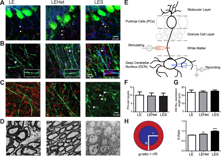
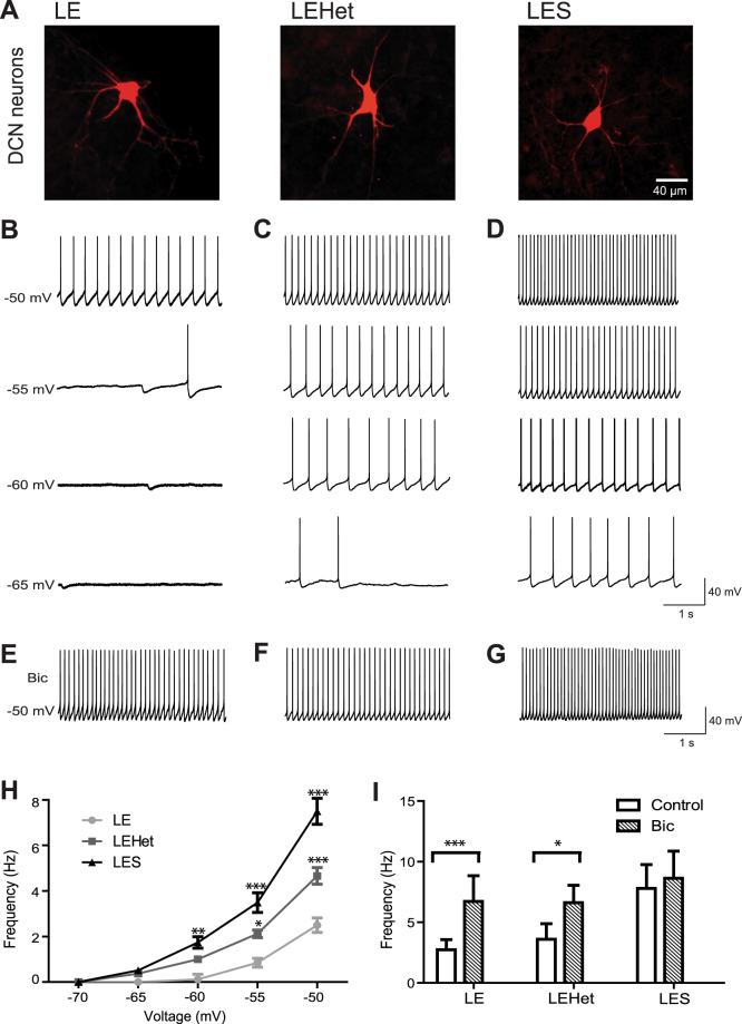
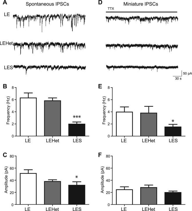
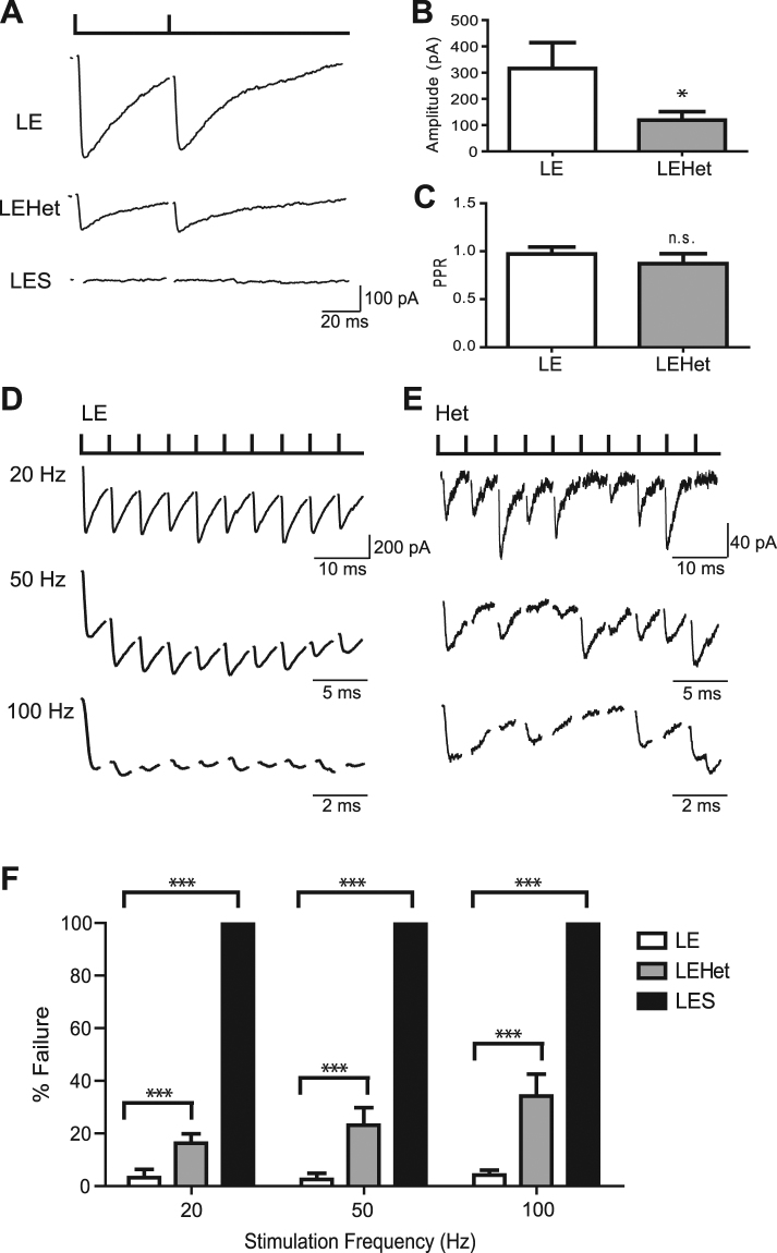
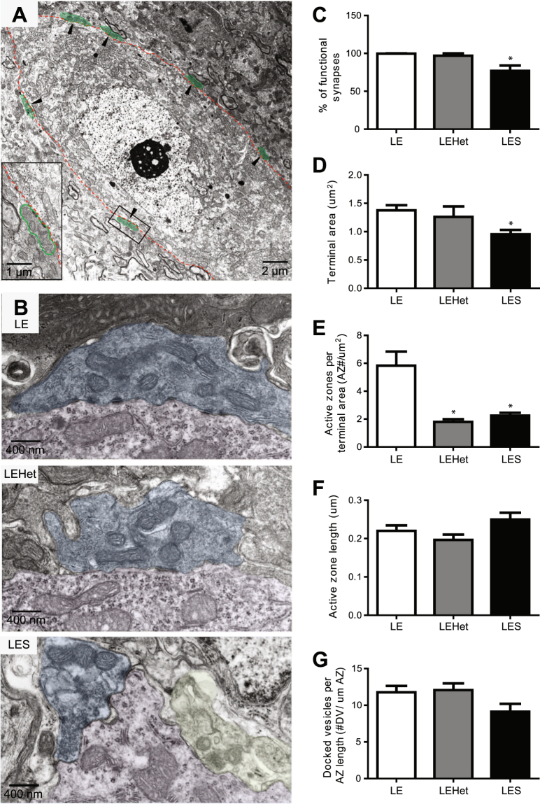
The roles of myelin in maintaining axonal integrity and action potential (AP) propagation are well established, but its role in synapse maintenance and neurotransmission remains largely understudied. Here, we investigated how Purkinje axon myelination regulates synaptic transmission in the Purkinje to deep cerebellar nuclei (DCN) synapses using the Long Evans Shaker (LES) rat, which lacks compact myelin and thus displays severe locomotion deficits. DCN neurons fired spontaneous action potentials (APs), whose frequencies were dependent on the extent of myelin. In the LES cerebellum with severe myelin deficiency, DCN neurons were hyper-excitable, exhibiting spontaneous AP firing at a much higher frequency compared to those from wild type (LE) and heterozygote (LEHet) rats. The hyper-excitability in LES DCN neurons resulted from reduced inhibitory GABAergic inputs from Purkinje cells to DCN neurons. Corresponding with functional alterations including failures of AP propagation, electron microscopic analysis revealed anatomically fewer active zones at the presynaptic terminals of Purkinje cells in both LEHet and LES rats. Taken together, these studies suggest that proper axonal myelination critically regulates presynaptic terminal structure and function and directly impacts synaptic transmission in the Purkinje cell-DCN cell synapse in the cerebellum.


Conflict of interest statement

The authors declare that they have no competing interests.

Figures

Figure 1
Figure 1
Structural changes of the Purkinje axon in the LES cerebellum (A). Calbindin (CB)-expressing Purkinje cells (green) in LE, LEHet, and LES cerebella. Caspr (blue) and NavPan (red) expression along Purkinje axons (arrowheads). (B) Purkinje axons expressing CB (green) in the white matter of the cerebellum of LE, LEHet, and LES. Arrowheads mark nodal and paranodal structures, expressing Caspr (blue) and NavPan (red), enlarged in inset. Inset width is 18 μm. While the nodal structure of Purkinje axons in LE and LEHet remain intact, Nav and Caspr expression in LES Purkinje axons is diffuse. (C) CB-expressing Purkinje axons (green) and myelin basic protein (MBP) expression (red) in the white matter of the cerebellum of LE, LEHet, and LES. MBP expression is absent in the LES cerebellum, where spheroid swellings were observed in Purkinje axons (arrowheads). (D) EM images of myelination around Purkinje axons in LE, LEHet, and LES. Myelin was thinner in LEHet than LE, and nearly absent in LES. (E) Diagram of a Purkinje cell with dendrites that extend into the molecular layer and an axon projecting through the granule cell layer and white matter that synapses in the DCN. Recording electrode (blue) represents the recording site in Fig. 2 through 4. Stimulating electrode (red) indicates the stimulating site in Fig. 4. (F) Number of Purkinje cells per 304.5 μm × 304.5 μm square was not different between LE, LEHet, and LES animals. (G) There were no significant differences between the length of NavPan staining along Purkinje AIS between LE, LEHet, and LES animals. (H) Quantification of the g-ratio is calculated by dividing the radius of the axon (blue) by the total radius of the axon and myelin (red). The g-ratio quantified from electron microscopy (EM) images was significantly higher in LEHet and LES compared to LE.
Figure 2
Figure 2
LES and LEHet DCN neurons display altered tonic inhibition and spontaneous AP firing. (A) Representative deep cerebellar nuclei (DCN) cells in LE, LEHet, and LES, which were filled with Alexa 568 during whole-cell recordings. (B–D) Whole-cell current clamp recordings of spontaneous APs at membrane potentials of −50, −55, −60, and −65 mV in DCN neurons of LE (B), LEHet (C), and LES (D) rats, demonstrating increased firing frequency in LES, and to a lesser extent, LEHet DCN neurons. (E–G) Spontaneous action potential firing in DCN neurons of LE (E), LEHet (F), and LES (G) rats at −50 mV in the presence of 10 μM bicuculline. (H) Summary of spontaneous firing frequency at various membrane potentials from −70 mV to −50 mV in LE, LEHet, and LES DCN cells. (I) The effect of bicuculline on spontaneous firing frequency (at −50 mV) in LE, LEHet, and LES DCN cells. *, **, and *** indicate p < 0.05, p < 0.01, and p < 0.001, respectively.
Figure 3
Figure 3
LES animals show reduced spontaneous inhibitory input into DCN cells. (A) Recordings of sIPSCs in DCN cells from LE, LEHet, and LES animals in the presence of CNQX (25 μM). (B,C) Summary of sIPSC frequency (B) and amplitude (C) in LE, LEHet, and LES DCN cells. (D) Recordings of mIPSCs in the presence of TTX (1 μM) in LE, LEHet, and LES DCN cells. (E,F) Summary of mIPSC frequency (E) and amplitude (F) in LE, LEHet, and LES DCN cells. *, **, and *** indicate p < 0.05, p < 0.01, and p < 0.001, respectively.
Figure 4
Figure 4
LES and LEHet show reduced inhibitory transmission at the Purkinje cell-DCN cell synapse. (A) Recordings of eIPSCs in DCN neurons evoked by stimulating Purkinje axon with bipolar stimulator placed in the cerebellar white matter in LE, LEHet, and LES rats. Paired pulse stimulation with an interval of 50 ms. Note no identifiable current in LES DCN cells in response to Purkinje axon stimulation. (B) Summary of eIPSC amplitude in LE and LEHet DCN neurons. (C) No significant difference between the paired pulse ratio (PPR) in eIPSCs from LEHet and LE DCN neurons. (D) and (E) Whole cell voltage clamp recordings of eIPSCs in DCN neurons of LE (D) and LEHet (E) rats after stimulation at 20, 50, and 100 Hz. (F) Summary of failure rate in LE, LEHet, and LES DCN cells at 20 Hz, 50 Hz, and 100 Hz. LES cells showed 100% failure rate. *, **, and *** indicate p < 0.05, p < 0.01, and p < 0.001, respectively.
Figure 5
Figure 5
Purkinje cell-DCN neuron synapse structure disruptions in LES and LEHet animals. (A) EM image from the DCN of an LE rat. Arrowheads indicate examples of Purkinje cell terminals (colored green). (B) Electron microscopy images of Purkinje cell-DCN neuron synapses including all active zones in LE, LEHet, and LES rats. Yellow colored terminal indicates a degenerating terminal. (C–G) Summary of the number of functional (non-degenerating) Purkinje terminals per DCN cell (C), Purkinje terminal area (D), number of active zones per terminal (E), length of active zones (F) and number of docked vesicles (DV) per active zone (G) in LE, LEHet and LES rats. *, **, and *** indicate p < 0.05, p < 0.01, and p < 0.001, respectively.

References

    1. Baumann N, Pham-Dinh D. Biology of Oligodendrocyte and Myelin in the Mammalian Central Nervous System. Physiol Rev. 2001;81(2):871–927. doi: 10.1152/physrev.2001.81.2.871. - DOI - PubMed
    1. Haines JD, Inglese M, Casaccia P. Axonal Damage in Multiple Sclerosis. Mt Sinai J Med. 2011;78(2):231–243. doi: 10.1002/msj.20246. - DOI - PMC - PubMed
    1. Herbert AL, Monk KR. Advances in myelinating glial cell development. Curr Opin Neurobiol. 2017;42:53–60. doi: 10.1016/j.conb.2016.11.003. - DOI - PMC - PubMed
    1. Smith KJ, McDonald WI. The pathophysiology of multiple sclerosis: the mechanisms underlying the production of symptoms and the natural history of the disease. Philos Trans R Soc Lond B Biol Sci. 1999;354:1649–1673. doi: 10.1098/rstb.1999.0510. - DOI - PMC - PubMed
    1. Wolswijk G. Oligodendrocyte survival, loss and birth in lesions of chronic-stage multiple sclerosis. Brain. 2000;123(1):105–115. doi: 10.1093/brain/123.1.105. - DOI - PubMed

Publication types