. 2018 Jan 18;8(1):1117.
doi: 10.1038/s41598-018-19463-2.

Intradermal delivery of STAT3 siRNA to treat melanoma via dissolving microneedles

Affiliations

Intradermal delivery of STAT3 siRNA to treat melanoma via dissolving microneedles

Jingtong Pan et al. Sci Rep. .

Abstract

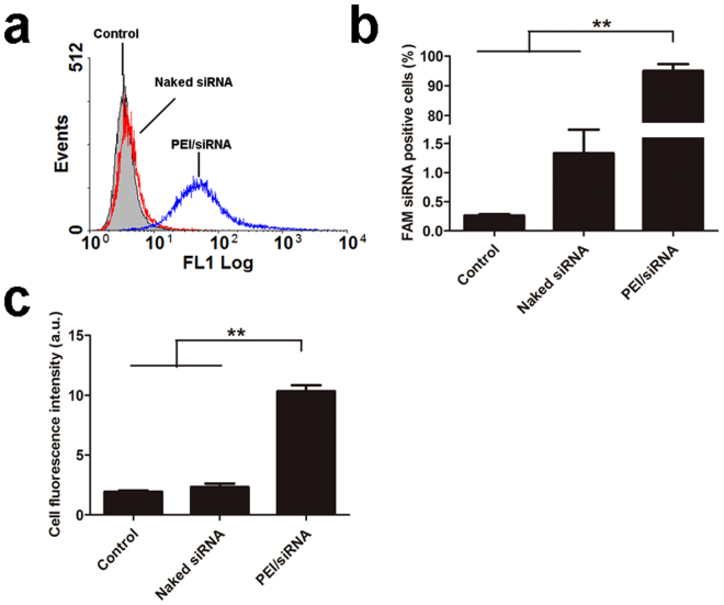
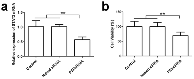
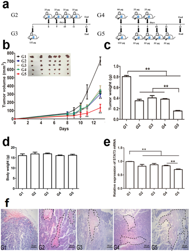
Hyperactivity of signal transducer and activity of transcription 3 (STAT3) plays a crucial role in melanoma invasion and metastasis. Gene therapy applying siRNA targeting STAT3 is a potential therapeutic strategy for melanoma. In this article, we first fabricated safe and novel dissolving microneedles (MNs) for topical application of STAT3 siRNA to enhance the skin penetration of siRNA and used polyethylenimine (PEI, 25 kDa) as carrier to improve cellular uptake of siRNA. The results showed that MNs can effectively penetrate skin and rapidly dissolve in the skin. In vitro B16F10 cell experiments presented that STAT3 siRNA PEI complex can enhance cellular uptake and transfection of siRNA, correspondingly enhance gene silencing efficiency and inhibit tumor cells growth. In vivo experiments indicated that topical application of STAT3 siRNA PEI complex delivered by dissolving MNs into skin can effectively suppress the development of melanoma through silencing STAT3 gene, and the inhibition effect is dose-dependent. STAT3 siRNA delivery via dissolving MNs is a promising approach for skin melanoma treatment with targeting inhibition efficacy and minimal adverse effects.


Conflict of interest statement

The authors declare that they have no competing interests.

Figures

Figure 1
Figure 1
Images of dissolving microneedles. (a) The dissolving microneedle arrays loaded with Rhodamine B were photographed by digital camera. (b) The dissolving microneedle arrays were observed by scanning electron microscope. (c) The image of microneedle loaded with PEI/FAM-siRNA photographed by confocal laser scanning microscope.
Figure 2
Figure 2
Mechanical strength of dissolving microneedles. (a) Fracture force determination of PEI/siRNA-loaded dissolving microneedles (n = 5). (b) Number of pinholes produced by different forces (10 N, 20 N, and 30 N) pressed on PEI/siRNA-loaded dissolving microneedles. Each value represented mean ± standard derivation (n = 3), **P < 0.01. (c) Optical coherence tomography (OCT) presented the depth of PEI/siRNA loaded microneedle puncturing into rat skin.
Figure 3
Figure 3
Micrograph of the residual PEI/FAM-siRNA loaded dissolving microneedles observed with confocal laser scanning microscope. The microneedles were pierced into the rat skin for maintaining 1 min, 3 min, or 5 min, and then removed.
Figure 4
Figure 4
CLSM images of B16 cells uptake of PEI/FAM-siRNA complex. (a) Control; (b) Naked FAM-siRNA; (c) PEI/FAM-siRNA complex. Scale bar: 10 μm.
Figure 5
Figure 5
Cellular uptake of FAM-siRNA complex by B16 cells. (a) The image of characteristic flow cytometry histogram; (b) Percentage of FAM-siRNA positive B16 cells; (c) Average fluorescence intensity of B16 cells after 4 h incubation. Each value represented mean ± standard derivation (n = 3), **P < 0.01.
Figure 6
Figure 6
(a) In vitro STAT3 gene silencing. (b) Percentage of B16 cells viability was analyzed in comparison with the untreated control using MTT assay. Each value represented mean ± standard derivation (n = 3), **P < 0.01.
Figure 7
Figure 7
In vivo effect of PEI/STAT3 siRNA loaded with dissolving microneedles on melanoma. (a) Schematics of different administration protocols. (b) The average tumor volume versus time. The tumor volume of G2, G3, G4, or G5 was keeping significant smaller than that of G1 from the time point of day 9 to the termination of day 13 (*P < 0.05 at time point of day 9, and 10, and **P < 0.01 at time point of day 11 and 13), and at time of day 8, the tumor volume of G3 or G5 significant smaller than that of G1 (P < 0.05). However, there was no significant difference between G2, G3, and G4 from the time point of day 9 to the termination of day 13. The tumor volume of G5 was further significantly smaller than that of G2, G3, or G4 (P < 0.01) (mean ± SD, n = 6; n = 6), (c) Tumor weight was compared at the end of experiment (mean ± SD, n = 6, **P < 0.01). (d) Body weight was determined at the end of experiment (mean ± SD, n = 6, **P < 0.01). (e) In vivo STAT3 gene silencing efficiency determined with RT-PCR (n = 3). (f) HE staining of representative tumor tissue from different groups. The tumor necrosis was outlined with black dotted lines.

References

    1. Ruan R, et al. Topical and Targeted Delivery of siRNAs to Melanoma Cells Using a Fusion Peptide Carrier. Scientific reports. 2016;6:29159. doi: 10.1038/srep29159. - DOI - PMC - PubMed
    1. Jerant, A. F., Johnson, J. T., Sheridan, C. D. & Caffrey, T. J. Early detection and treatment of skin cancer. Am Fam Physician62, 357–368, 375–376, 381–382 (2000). - PubMed
    1. Jemal A, Devesa SS, Hartge P, Tucker MA. Recent trends in cutaneous melanoma incidence among whites in the United States. J Natl Cancer Inst. 2001;93:678–683. doi: 10.1093/jnci/93.9.678. - DOI - PubMed
    1. Kim CJ, Dessureault S, Gabrilovich D, Reintgen DS, Slingluff CL. Immunotherapy for melanoma. Cancer control. 2002;9:22–30. doi: 10.1177/107327480200900104. - DOI - PubMed
    1. O’Day SJ, Kim CJ, Reintgen DS. Metastatic melanoma: chemotherapy to biochemotherapy. Cancer control. 2002;9:31–38. doi: 10.1177/107327480200900105. - DOI - PubMed

Publication types

MeSH terms