. 2017:2017:5658681.
doi: 10.1155/2017/5658681. Epub 2017 Oct 24.

Dangguijihwang-tang and Dangguijakyak-san Prevent Menopausal Symptoms and Dangguijihwang-tang Prevents Articular Cartilage Deterioration in Ovariectomized Obese Rats with Monoiodoacetate-Induced Osteoarthritis

Affiliations

Dangguijihwang-tang and Dangguijakyak-san Prevent Menopausal Symptoms and Dangguijihwang-tang Prevents Articular Cartilage Deterioration in Ovariectomized Obese Rats with Monoiodoacetate-Induced Osteoarthritis

Hye Won Lee et al. Evid Based Complement Alternat Med. 2017.

Abstract

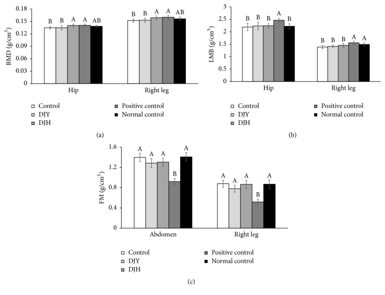
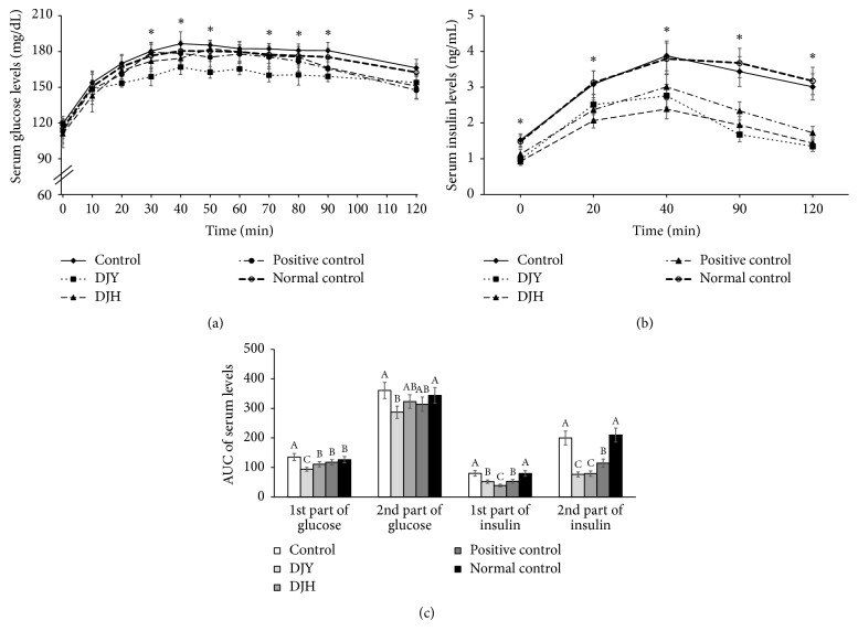
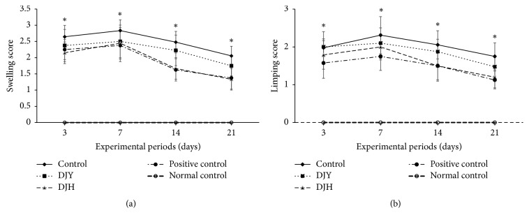
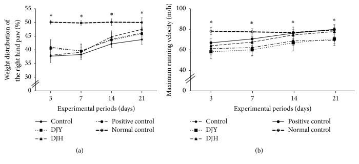
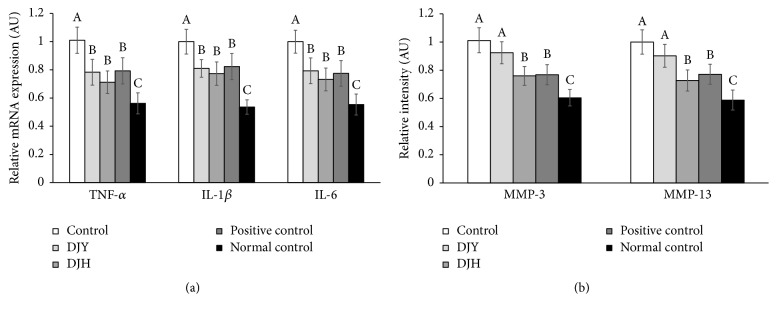
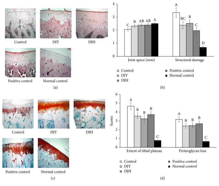
We investigated whether dangguijakyak-san (DJY) and dangguijihwang-tang (DJH), oriental medicines traditionally used for inflammatory diseases, could prevent and/or delay the progression of postmenopausal symptoms and osteoarthritis in osteoarthritis-induced estrogen-deficient rats. Treated ovariectomized (OVX) rats consumed either 1% DJY or 1% DJH in the diets. Positive-control rats were given 30 μg/kg bw 17β-estradiol and control rats were given 1% fat as were the normal-control rats. All rats received high-fat diets for 8 weeks. At the 9th week, OVX rats received articular injections of monoiodoacetate (MIA) or saline (normal control) into the right knee. At 3 weeks after MIA injection, DJY reduced visceral-fat mass and improved glucose metabolism by reducing insulin resistance, whereas DJH increased BMD and decreased insulin resistance. DJH improved weight distribution in the right knee and maximum running velocity on a treadmill at days 14 and 21 as much as those of the positive control. TNF-α, IL-1β, and IL-6 levels in articular cartilage were much higher in the control than the positive control, whereas both DJY and DJH reduced the levels to those of the positive control. The histological analysis assessed articular cartilage damage near the tidemark and proteoglycan loss in the control versus the positive control; DJY and DJH prevented this damage and proteoglycan loss. In conclusion, DJY may provide an effective treatment for improving glucose tolerance, and DJH may be appropriate for preventing osteoarthritis.


Figures

Figure 1
Figure 1
HPLC chromatogram. (a) References for dangguijakyak-san: 1, albiflorin; 2, paeoniflorin; 3, nodakenin; 4, z-ligustilide; 5, decursin. (b) Dangguijakyak-san water extract. (c) References for dangguijihwang-tang: 1,5-hydroxymethyl-2-furaldehyde; 2, nodakenin; 3, decursin. (d) Dangguijihwang-tang water extract.
Figure 2
Figure 2
Body composition at the end of experiments. Ovariectomized (OVX) rats were provided with a 45% fat diet containing (1) 1% lyophilized DJY powder + MIA injection (DJY), (2) 1% lyophilized DJH powder + MIA injection (DJH), (3) 30 μg/kg body weight 17β-estradiol + 1% dextrose + MIA injection (positive control), (4) 1% dextrose + MIA injection (placebo, control), or (5) 1% dextrose with no MIA injection (normal control). After 4 weeks of the assigned diets, an articular injection of MIA into the right knee was made in all OVX groups except the normal-control group and the assigned diets were provided for an additional 3 weeks. At the 7th week body composition was measured by DEXA. (a) Bone mineral density (BMD). (b) Lean body mass (LBM) at the hip and right leg. (c) Fat mass (FM) at the abdomen and right leg. Each bar and error bar represents the mean ± SD from 10 rats per group. A,BDifferent letters indicate significant differences in the treatment groups of OVX rats by Tukey's test at p < 0.05.
Figure 3
Figure 3
Changes of serum glucose and insulin levels during oral glucose tolerance test at the end of experiment. Ovariectomized (OVX) rats were provided with a 45% fat diet containing (1) 1% lyophilized DJY powder + MIA injection (DJY), (2) 1% lyophilized DJH powder + MIA injection (DJH), (3) 30 μg/kg body weight 17β-estradiol + 1% dextrose + MIA injection (positive control), (4) 1% dextrose + MIA injection (placebo, control), or (5) 1% dextrose with no MIA injection (normal control). After 4 weeks of the assigned diets, an articular injection of MIA into the right knee was made in all OVX groups except the normal-control group and the assigned diets were provided for an additional 3 weeks. At 7th week rats were orally given 2 g glucose/kg body weight and serum glucose and insulin levels were measured. (a) Changes of serum glucose levels. (b) Changes of serum insulin levels. (c) Area under the curve of serum glucose and insulin levels. Each bar and error bar represents the mean ± SD from 10 rats per group. A,B,CDifferent letters indicate significant differences in the treatment groups of OVX rats at each time point identified by Tukey's test at p < 0.05.
Figure 4
Figure 4
Gross observation of osteoarthritis symptoms at 3, 7, 14, and 21 days after monoiodoacetate (MIA) injection. Ovariectomized (OVX) rats were provided with a 45% fat diet containing (1) 1% lyophilized DJY powder + MIA injection (DJY), (2) 1% lyophilized DJH powder + MIA injection (DJH), (3) 30 μg/kg body weight 17β-estradiol + 1% dextrose + MIA injection (positive control), (4) 1% dextrose + MIA injection (placebo, control), or (5) 1% dextrose with no MIA injection (normal control). After 4 weeks of the assigned diets, an articular injection of MIA into the right knee was made in all OVX groups except the normal-control group and the assigned diets were provided for an additional 3 weeks. (a) The scores of the swelling in the right knee. (b) The scores of limping in the right knee. Each data point and error bar represents the mean ± SD from 10 rats per group. A,B,C,DDifferent letters indicate significant differences in the treatment groups of OVX rats at each time point identified by Tukey's test at p < 0.05.
Figure 5
Figure 5
Pain-related behaviors after osteoarthritis induction at 3, 7, 14, and 21 days after monoiodoacetate (MIA) injection. Ovariectomized (OVX) rats were provided with a 45% fat diet containing (1) 1% lyophilized DJY powder + MIA injection (DJY), (2) 1% lyophilized DJH powder + MIA injection (DJH), (3) 30 μg/kg body weight 17β-estradiol + 1% dextrose + MIA injection (positive control), (4) 1% dextrose + MIA injection (placebo, control), or (5) 1% dextrose with no MIA injection (normal control). After 4 weeks of the assigned diets, an articular injection of MIA into the right knee was made in all OVX groups except the normal-control group and the assigned diets were provided for an additional 3 weeks. The pain indicators were measured. (a) Differences in weight distribution of the right hind paw (%) measured by an incapacitance tester. (b) Maximum velocity run on a treadmill; each data point and error bar represents the mean ± SD from 10 rats per group. A,B,CDifferent letters indicate significant differences in the treatment groups of OVX rats at each time point identified by Tukey's test at p < 0.05.
Figure 6
Figure 6
The mRNA expression of proinflammatory cytokines and matrix metalloproteinases (MMPs) in the articular cartilage. Ovariectomized (OVX) rats were provided with a 45% fat diet containing (1) 1% lyophilized DJY powder + MIA injection (DJY), (2) 1% lyophilized DJH powder + MIA injection (DJH), (3) 30 μg/kg body weight 17β-estradiol + 1% dextrose + MIA injection (positive control), (4) 1% dextrose + MIA injection (placebo, control), or (5) 1% dextrose with no MIA injection (normal control). After 4 weeks of the assigned diets, an articular injection of MIA into the right knee was made in all OVX groups except the normal-control group and the assigned diets were provided for an additional 3 weeks. The mRNA expression of genes related to collagen degradation and proinflammatory cytokines was measured in the articular cartilage by real-time PCR. (a) mRNA expression of MMP-3 and MMP-13. (b) Proinflammatory cytokines (TNF-α, IL-1β, and IL-6). Each bar and error bar represents the mean ± SD from 4 rats per group. A,B,CDifferent letters indicate significant differences in the treatment groups of OVX rats at each time point identified by Tukey's test at p < 0.05.
Figure 7
Figure 7
Histopathological features of osteoarthritic lesions in the knee joints of rats at 21 days after intra-articular injection of monoiodoacetate (MIA). Ovariectomized (OVX) rats were provided with a 45% fat diet containing (1) 1% lyophilized DJY powder + MIA injection (DJY), (2) 1% lyophilized DJH powder + MIA injection (DJH), (3) 30 μg/kg body weight 17β-estradiol + 1% dextrose + MIA injection (positive control), (4) 1% dextrose + MIA injection (placebo, control), or (5) 1% dextrose with no MIA injection (normal control). After 4 weeks of the assigned diets, an articular injection of MIA into the right knee was made in all OVX groups except the normal-control group and the assigned diets were provided for an additional 3 weeks. (a) Images of H-E staining in the right knee (×10). (b) Quantification of the joint space and structural damage. (c) Safranin O-fast green staining in the right knee (×10). (d) Quantification of cartilage damage. Each bar and error bar represents the mean ± SD from 4 rats per group. A,B,C,D,EDifferent letters indicate significant differences in the treatment groups of OVX rats at each time point identified by Tukey's test at p < 0.05.

Similar articles

Cited by

References

    1. Jia M., Dahlman-Wright K., Gustafsson J.-Å. Estrogen receptor alpha and beta in health and disease. Best Practice & Research Clinical Endocrinology & Metabolism. 2015;29(4):557–568. doi: 10.1016/j.beem.2015.04.008. - DOI - PubMed
    1. Mauvais-Jarvis F. Estrogen and androgen receptors: Regulators of fuel homeostasis and emerging targets for diabetes and obesity. Trends in Endocrinology & Metabolism. 2011;22(1):24–33. doi: 10.1016/j.tem.2010.10.002. - DOI - PMC - PubMed
    1. Mauvais-Jarvis F., Clegg D. J., Hevener A. L. The role of estrogens in control of energy balance and glucose homeostasis. Endocrine Reviews. 2013;34(3):309–338. doi: 10.1210/er.2012-1055. - DOI - PMC - PubMed
    1. Abu-Taha M., Rius C., Hermenegildo C., et al. Menopause and ovariectomy cause a low grade of systemic inflammation that may be prevented by chronic treatment with low doses of estrogen or losartan. The Journal of Immunology. 2009;183(2):1393–1402. doi: 10.4049/jimmunol.0803157. - DOI - PubMed
    1. Au A., Feher A., McPhee L., Jessa A., Oh S., Einstein G. Estrogens, inflammation and cognition. Frontiers in Neuroendocrinology. 2016;40:87–100. doi: 10.1016/j.yfrne.2016.01.002. - DOI - PubMed

LinkOut - more resources