Post-Traumatic Peripheral Giant Osteoma in the Frontal Bone
- PMID: 29349054
- PMCID: PMC5759665
- DOI: 10.7181/acfs.2017.18.4.273
Post-Traumatic Peripheral Giant Osteoma in the Frontal Bone
Abstract
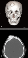
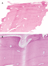
Osteomas are benign, slow-growing tumors that most frequently occur in the craniomaxillofacial region. These tumors are mostly asymptomatic and are generally found incidentally. A giant osteoma is generally considered to be greater than 30 mm in diameter or 110 g in weight. A 35-year-old female presented to us with complaints of a firm mass that showed continuous growth on the forehead following trauma. A hairline incision was made to expose the osteoma. Biopsy of the tumor confirmed a osteoma. There were no complications after surgery. Postoperative computed tomography revealed that the tumor was completely removed. Because a peripheral giant osteoma of the frontal bone with a history of trauma is a rare finding, thorough history-taking, physical examination, and preoperative imaging tests are needed for patients with a history of trauma to rule out a giant osteoma.
Keywords: Forehead; Frontal bone; Osteoma.
Conflict of interest statement
CONFLICT OF INTEREST: No potential conflict of interest relevant to this article was reported.
Figures


Similar articles
-
Endoscopic Approach for Resection of Frontal Forehead Osteoma: Technical Case Report Instruction.World Neurosurg. 2023 Jul;175:11. doi: 10.1016/j.wneu.2023.03.135. Epub 2023 Apr 6. World Neurosurg. 2023. PMID: 37028484
-
Giant Frontal Sinus Osteomas: Demographic, Clinical Presentation, and Management of 10 Cases.Am J Rhinol Allergy. 2019 Jan;33(1):36-43. doi: 10.1177/1945892418804911. Epub 2018 Oct 11. Am J Rhinol Allergy. 2019. PMID: 30306798
-
Endoscopic resection of forehead osteomas: Technical notes.Ann Chir Plast Esthet. 2020 Feb;65(1):91-99. doi: 10.1016/j.anplas.2019.04.004. Epub 2019 May 14. Ann Chir Plast Esthet. 2020. PMID: 31101396
-
Peripheral Solitary Osteoma of the Zygomatic Arch: A Case Report and Literature Review.Open Dent J. 2017 Feb 28;11:120-125. doi: 10.2174/1874210601711010120. eCollection 2017. Open Dent J. 2017. PMID: 28357005 Free PMC article. Review.
-
Mandibular traumatic peripheral osteoma: a case report.Oral Surg Oral Med Oral Pathol Oral Radiol Endod. 2011 Dec;112(6):e44-8. doi: 10.1016/j.tripleo.2011.05.006. Epub 2011 Sep 8. Oral Surg Oral Med Oral Pathol Oral Radiol Endod. 2011. PMID: 21862366 Review.
Cited by
-
Huge osteoma in the frontoparietal bone caused by trauma from 18 years ago: a case report.J Med Case Rep. 2024 Feb 9;18(1):48. doi: 10.1186/s13256-024-04373-x. J Med Case Rep. 2024. PMID: 38331951 Free PMC article.
-
Giant Post-Traumatic Frontoethmoid Osteoma: Diagnostic, Therapeutic and Reconstructive Approach.Turk Arch Otorhinolaryngol. 2020 Mar;58(1):61-64. doi: 10.5152/tao.2020.4858. Epub 2020 Mar 1. Turk Arch Otorhinolaryngol. 2020. PMID: 32313898 Free PMC article.
References
-
- Pohranychna K. A rare clinical case of the isolated primary frontal bone osteoma. Exp Oncol. 2016;38:204–206. - PubMed
-
- Bodner L, Gatot A, Sion-Vardy N, Fliss DM. Peripheral osteoma of the mandibular ascending ramus. J Oral Maxillofac Surg. 1998;56:1446–1449. - PubMed
-
- Regezi JA, Sciubba JJ. Oral pathology: clinical pathologic correlations. Philadelphia: W.B. Saunders; 1999.
-
- White SC, Pharoah MJ. Oral radiology: principles and interpretation. St. Louis: Mosby/Elsevier; 2009.
LinkOut - more resources
Full Text Sources
Other Literature Sources

