Review
. 2018 Jun;23(4):635-644.
doi: 10.1007/s00775-018-1534-z. Epub 2018 Jan 19.

Contribution of Mössbauer spectroscopy to the investigation of Fe/S biogenesis

Affiliations
Review

Contribution of Mössbauer spectroscopy to the investigation of Fe/S biogenesis

Ricardo Garcia-Serres et al. J Biol Inorg Chem. 2018 Jun.

Erratum in

Abstract

Fe/S cluster biogenesis involves a complex machinery comprising several mitochondrial and cytosolic proteins. Fe/S cluster biosynthesis is closely intertwined with iron trafficking in the cell. Defects in Fe/S cluster elaboration result in severe diseases such as Friedreich ataxia. Deciphering this machinery is a challenge for the scientific community. Because iron is a key player, 57Fe-Mössbauer spectroscopy is especially appropriate for the characterization of Fe species and monitoring the iron distribution. This minireview intends to illustrate how Mössbauer spectroscopy contributes to unravel steps in Fe/S cluster biogenesis. Studies were performed on isolated proteins that may be present in multiple protein complexes. Since a few decades, Mössbauer spectroscopy was also performed on whole cells or on isolated compartments such as mitochondria and vacuoles, affording an overview of the iron trafficking. This minireview aims at presenting selected applications of 57Fe-Mössbauer spectroscopy to Fe/S cluster biogenesis.

Keywords: Fe/S biogenesis; Iron trafficking; Iron–sulfur cluster; Mössbauer spectroscopy.


Figures

Fig. 1
Fig. 1
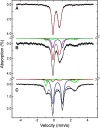
Characterization by Mössbauer spectroscopy of oxidized (top) and dithionite-reduced (bottom) human mitoNEET recorded at 4.2 K in a magnetic field of 600 G applied parallel to the direction of the γ-rays. The solid and dashed blue lines represent the contributions of [2Fe-2S]2+ clusters, and the solid and dashed red lines represent the contributions of [2Fe-2S]+ clusters. From [15]
Fig. 2
Fig. 2
Ferredoxin-mediated reductive coupling of [2Fe-2S]2+ clusters on IscU monitored by Mössbauer spectroscopy (4.2 K; 50 mT applied field parallel to γ radiation). 2 × [2Fe-2S]2+ IscU as prepared (a), after reduction with 1.06 reducing equivalents of Fdx [2Fe-2S]2+ cluster (b), and after reduction with 1.28 reducing equivalents of dithionite per IscU [2Fe-2S]2+ cluster (c). From [18]
Fig. 3
Fig. 3
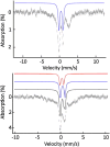
4.2 K, 60 mT parallel applied field Mössbauer spectra of the reconstituted quaternary (ISCU/NFS1/ISD11/FXN) complex (top) with 3.5 Fe/Cplx or reconstituted ternary (ISCU/NFS1/ISD11) complex (bottom) with 2.4 Fe/Cplx. The blue lines represent contributions from [4Fe-4S]2+ clusters and the red lines represent contributions from [2Fe-2S]2+ clusters. Adapted from [47]
Fig. 4
Fig. 4
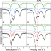
Mössbauer spectra recorded at 5.5 K using a 60 mT external magnetic field applied parallel to the γ-beam. The panels a and b reproduce spectra of whole control cells and panels c and d those of ISCA1 or ISCA2-overexpressing cells. Experimental spectra are shown with hatched marks and simulations are overlaid as solid black lines. Five components were used for simulation: HS FeII (light and dark green), [4Fe-4S]2+ clusters and LS ferrous hemes (light blue), FeIII NP (dark blue) and [2Fe-2S]2+ (red). From [55]
Fig. 5
Fig. 5
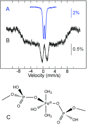
Top: Mössbauer spectra of ∆yfh1 yeast mitochondria. Spectrum a was recorded at 78 K in zero field and spectrum b at 4.2 K in a 7 T magnetic field applied parallel to the γ-beam. Experimental spectra are shown with hatched marks and simulations are overlaid as solid lines. Simulation of spectrum b was achieved assuming a distribution of the hyperfine field. From [59]. Bottom (c): Coordination of the ferric ion in NP according to EXAFS and electron microscopy results [60]
Fig. 6
Fig. 6
Theoretical Mössbauer spectra at 6 K with a 50 mT external magnetic field applied parallel to the γ-beam of the five main components identified in spectra recorded on S. cerevisiae cells, isolated mitochondria or isolated vacuoles. They are scaled to the same area and calculated according to published parameters [62, 66]

References

    1. Beinert H, Holm RH, Münck E. Iron–sulfur clusters: nature’s modular, multipurpose structures. Science. 1997;277:653–659. doi: 10.1126/science.277.5326.653. - DOI - PubMed
    1. Bill E. Iron–sulfur clusters—new features in enzymes and synthetic models. Hyperfine Interact. 2012;205:139–147. doi: 10.1007/s10751-011-0411-8. - DOI
    1. Pandelia M-E, Lanz ND, Booker SJ, Krebs C. Mössbauer spectroscopy of Fe/S proteins. Biochim Biophys Acta. 2015;1853:1395–1405. doi: 10.1016/j.bbamcr.2014.12.005. - DOI - PubMed
    1. Münck E, Stubna A. 2.21—Mössbauer spectroscopy: bioinorganic A2—McCleverty, Jon A. In: Meyer TJ, editor. Comprehensive coordination chemistry II. Oxford: Pergamon; 2003. pp. 279–286.
    1. Gütlich P, Bill E, Trautwein AX. Mössbauer spectroscopy and transition metal chemistry: fundamentals and applications. Berlin Heidelberg: Springer; 2010.

Publication types

MeSH terms

LinkOut - more resources