. 2018 Sep;12(3):561-573.
doi: 10.1007/s12079-018-0449-3. Epub 2018 Jan 19.

Syringic acid, a phenolic acid, promotes osteoblast differentiation by stimulation of Runx2 expression and targeting of Smad7 by miR-21 in mouse mesenchymal stem cells

Affiliations

Syringic acid, a phenolic acid, promotes osteoblast differentiation by stimulation of Runx2 expression and targeting of Smad7 by miR-21 in mouse mesenchymal stem cells

B Arumugam et al. J Cell Commun Signal. 2018 Sep.

Abstract

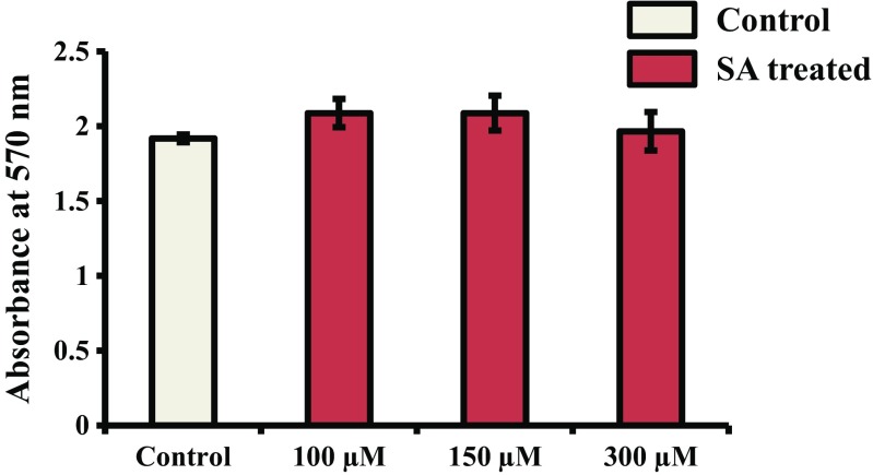
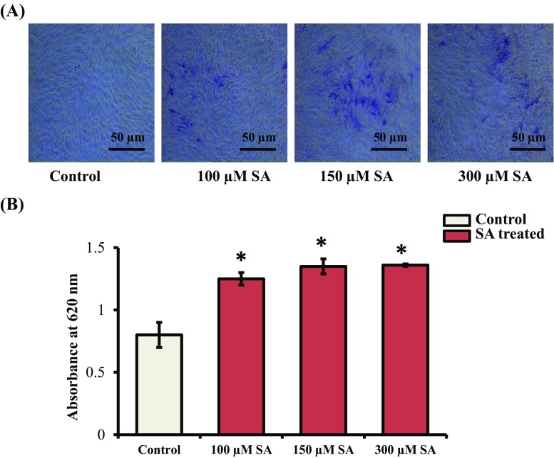
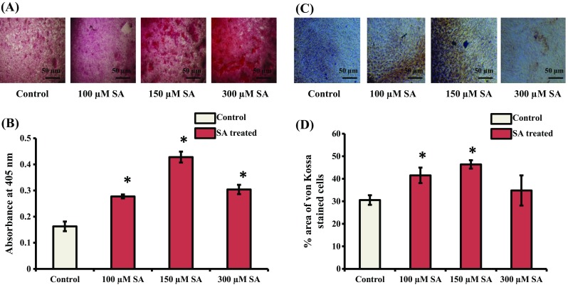
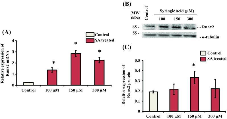
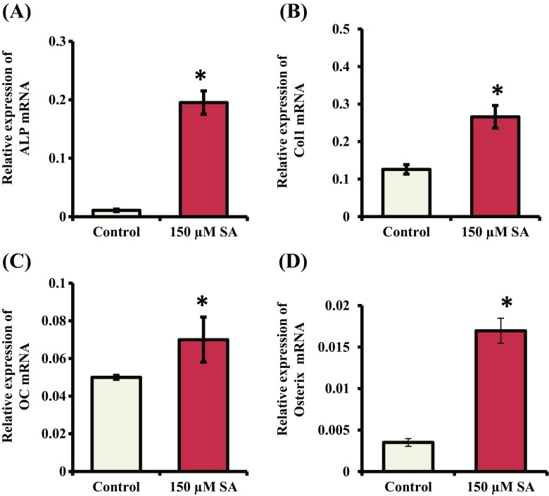
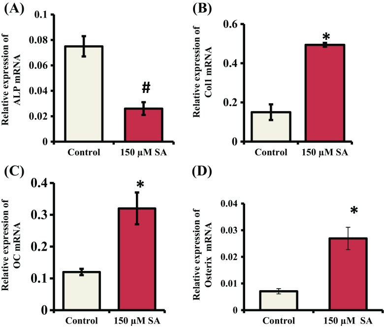
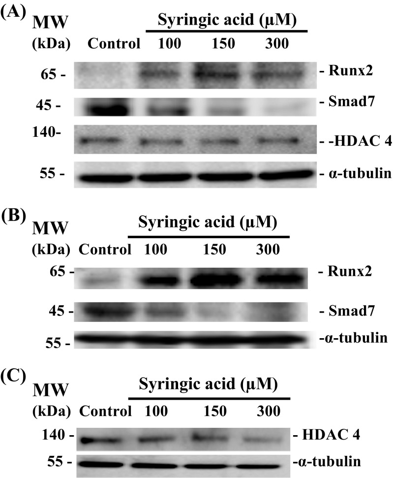
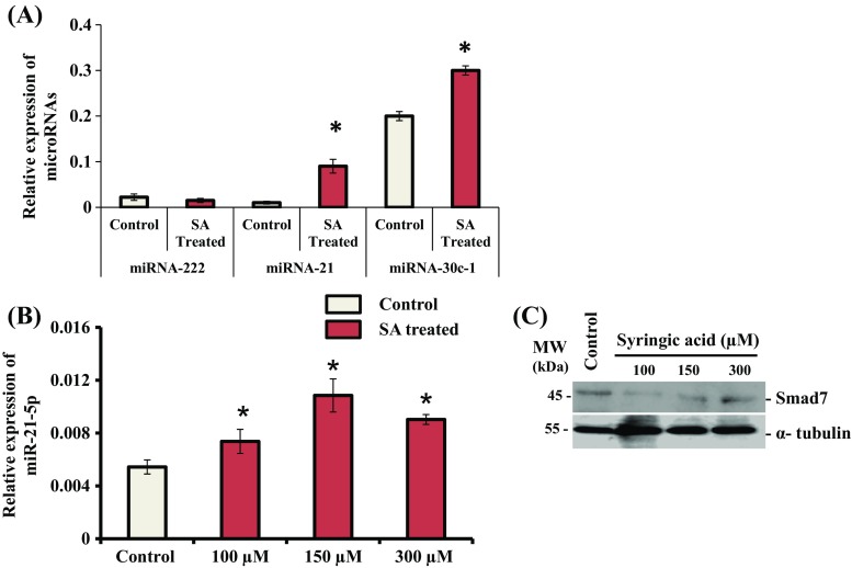
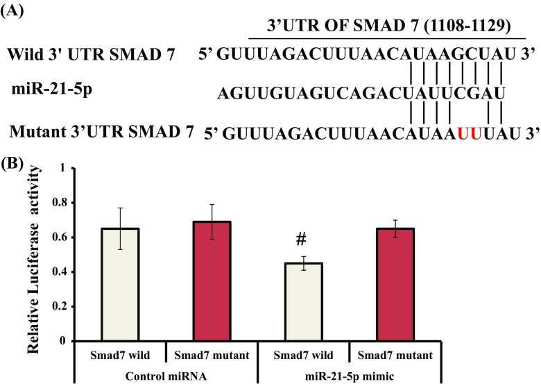
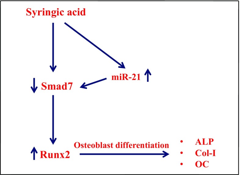
Syringic acid (SA), a phenolic acid, has been used in Chinese and Indian medicine for treating diabetes but its role in osteogenesis has not yet been investigated. In the present study, at the molecular and cellular levels, we evaluated the effects of SA on osteoblast differentiation. At the cellular level, there was increased alkaline phosphatase (ALP) activity and calcium deposition by SA treatment in mouse mesenchymal stem cells (mMSCs). At the molecular level, SA treatment of these cells stimulated expression of Runx2, a bone transcription factor, and of osteoblast differentiation marker genes such as ALP, type I collagen, and osteocalcin. It is known that Smad7 is an antagonist of TGF-β/Smad signaling and is a negative regulator of Runx2. microRNAs (miRNAs) play a key role in the regulation of osteogenesis genes at the post-transcriptional level and studies have reported that Smad7 is one of the target genes of miR-21. We found that there was down regulation of Smad7 and up regulation of miR-21 in SA-treated mMSCs. We further identified that the 3'-untranslated region (UTR) of Smad7 was directly targeted by miR-21 in these cells. Thus, our results suggested that SA promotes osteoblast differentiation via increased expression of Runx2 by miR-21-mediated down regulation of Smad7. Hence, SA may have potential in orthopedic applications.

Keywords: Bone; Runx2; Smad7; Syringic acid; miR-21.


Figures

Fig. 1
Fig. 1
Cytotoxicity assessment of SA. mMSCs were treated with SA at different concentrations (100, 150, and 300 μM) for 48 h. The MTT assay was then performed as described in the methods section
Fig. 2
Fig. 2
Effect of SA on ALP activity. (A) mMSCs were treated with SA at different concentrations (100, 150, and 300 μM) in DMEM media for 7 d. The cells were then stained with BCIP-NBT reagent. Images were obtained using an inverted microscope. (B) Quantification of ALP activity done by the calorimetric method at 620 nm. *indicates a significant increase compared to control (p < 0.05)
Fig. 3
Fig. 3
Effect of SA on Alizarin red and von Kossa staining of cells. (A) mMSCs were treated with SA at different concentrations (100, 150, and 300 μM) in DMEM media for 14 d. The cells were then subjected to Alizarin red staining. (B) Quantification analysis of cells stained using Alizarin red. (C) mMSCs were treated with SA at different concentrations (100, 150, and 300 μM) in DMEM media containing osteogenic supplements for 14 d. The cells were then subjected to von Kossa staining. (D) Calorimetric estimation of von Kossa stained cells. *indicates a significant increase compared to control (p < 0.05)
Fig. 4
Fig. 4
Effect of SA on the expression of Runx2. mMSCs were treated with SA at different concentrations (100, 150 and 300 μM) in DMEM media for 24 h. (A) Total RNA was isolated, and cDNA was synthesized and subjected to RT-qPCR analysis using Runx2 gene primers. RPL13A was used as an internal control for normalization. *indicates a significant increase compared to control (p < 0.05). (B) Whole cell lysates were prepared and subjected to western blot analysis using Runx2 antibody. α-tubulin was used as an internal control for normalization. (C) Relative quantification analysis of biological triplicate samples. *indicates a significant increase compared to control (p < 0.05)
Fig. 5
Fig. 5
Effect of SA on the expression of osteoblast differentiation marker genes and Osterix. mMSCs were treated with SA at 150 μM in DMEM media for 7 d. Total RNA was isolated and subjected to RT-qPCR analysis using primers for (A) ALP, (B) Col-I, (C) OC and (D) Osterix genes. RPL13A was used as an internal control for normalization. *indicates a significant increase compared to control (p < 0.05)
Fig. 6
Fig. 6
Effect of SA on the expression of osteoblast differentiation marker genes and Osterix. mMSCs were treated with SA at 150 μM in DMEM media for 14 d. Total RNA was isolated and subjected to RT-qPCR analysis using the primers for (A) ALP, (B) Col-I, (C) OC and (D) Osterix genes. RPL13A was used as an internal control for normalization. *indicates a significant increase compared to control (p < 0.05)
Fig. 7
Fig. 7
Effect of SA on the expression of Smad7 protein. mMSCs were treated with SA at different concentrations (100, 150, and 300 μM) in DMEM media for (A) 7 d and (B) and (C) 14 d. Whole cell lysates were prepared and subjected to western blot analysis using antibodies against Smad7, HDAC4, Runx2 and α-tubulin. α-tubulin was used as an internal control for normalization
Fig. 8
Fig. 8
Effect of SA on the expression of miRNAs. (A) mMSCs were treated with SA at 150 uM in DMEM media for 14 d. Total RNA was isolated, and cDNA was synthesized and subjected to qPCR analysis using the primers for mir-222, mir-21 and mir-30-c-1. (B) mMSCs were treated with SA at different concentrations (100, 150 and 300 uM) in DMEM media for 24 h. Total RNA was isolated, and cDNA was synthesized and subjected to qPCR analysis using the primers for mir-21. RPL13A was as used as an internal control for normalization. *indicates a significant increase compared to control (p < 0.05). (C) mMSCs were treated with SA at different concentrations (100, 150, and 300 μM) in DMEM media for 24 h. Whole cell lysates were prepared and subjected to western blot analysis using Smad7 and α-tubulin antibodies. α-tubulin was used as an internal control for normalization
Fig. 9
Fig. 9
(A) A region of wild or mutant Smad7 3’ UTR binding site (1108–1129) with the seed sequence of miR-21-5p. (B) miR-21 directly targets Smad7. mMSCs were transiently transfected with the pmirGLO construct containing the wild-type or mutant Smad7 3′-UTR along with control miRNA or miR-21-5p. After 24 h, cell lysates were prepared and relative luciferase activity was calculated after normalization of firefly luciferase activity with Renilla luciferase activity. # indicates significant decrease compared to control miRNA (p < 0.05)
Fig. 10
Fig. 10
Schematic diagram of SA action on mMSCs in osteoblast differentiation. SA treatment upregulated the expression of miR-21, and the increased miR-21 targeted Smad7 expression, resulting in increased Runx2 expression, thus leading to expression of osteoblast differentiation marker genes such as ALP, Col-I, and OC in mMSCs

References

    1. Al Mamun MA, Hosen MJ, Islam K, Khatun A, Alam MM, Al-Bari MAA (2015) Tridax procumbens flavonoids promote osteoblast differentiation and bone formation. Biol Res 48(65) - PMC - PubMed
    1. Al-Obaidi MM, Al-Bayaty FH, Al Batran R, Hussaini J, Khor GH (2014) Impact of ellagic acid in bone formation after tooth extraction: an experimental study on diabetic rats. Sci World J: 10.1155/2014/908098 - PMC - PubMed
    1. Arumugam B, Vairamani M, Partridge NC, Selvamurugan N (2017) Characterization of Runx2 phosphorylation sites required for TGF-β1-mediated stimulation of matrix Metalloproteinase-13 expression in osteoblastic cells. J Cell Physiol 10.1002/jcp.25964 - PubMed
    1. Auh QS, Park KR, Yun HM, Lim HC, Kim GH, Lee DS, Kim YC, Oh H, Kim EC. Sulfuretin promotes osteoblastic differentiation in primary cultured osteoblasts and in vivo bone healing. Oncotarget. 2016;7(48):78320. - PMC - PubMed
    1. Baselga-Escudero L, Blade C, Ribas-Latre R, Casanova E, Susarez M, Torres JL, Salvado MJ, Arola L, Arola-Arnal A. Resveratrol and EGCG bind directly and distinctively to miR-33a and miR-122 and modulate divergently their levels in hepatic cells. Nucleic Acids Res. 2014;42:882–892. - PMC - PubMed