Review
. 2018 Feb 15;26(4):e68-e76.
doi: 10.5435/JAAOS-D-16-00600.

Angiography and Embolization in the Management of Bleeding Pelvic Fractures

Affiliations
Review

Angiography and Embolization in the Management of Bleeding Pelvic Fractures

Rahul Vaidya et al. J Am Acad Orthop Surg. .

Abstract

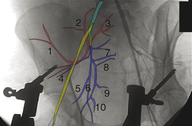
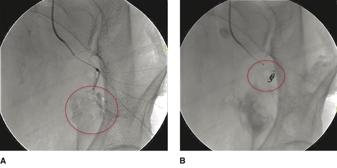
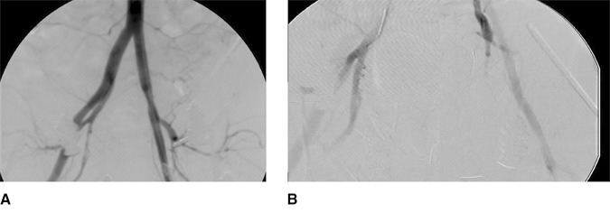
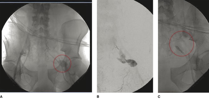
The use, timing, and priority of angioembolization in the management of bleeding pelvic fractures remain ambiguous. The most common vessels for angioembolization are, in decreasing order, the internal iliac artery and its branches, the superior gluteal artery, the obturator artery, and the internal pudendal artery. Technical success rates for this treatment option range from 74% to 100%. The fracture patterns most commonly requiring angioembolization are the Young and Burgess lateral compression and anterior-posterior compression types and Tile type C. Mortality rates after angioembolization of 16% to 50% have been reported, but deaths are usually related to concomitant injuries. The sensitivity and specificity of contrast-enhanced CT in detecting the need for angioembolization range from 60% to 90% and 92% to 100%, respectively. Angioembolization can be effective in the management of bleeding pelvic fractures, but as with any treatment, the risks of complications must be considered. Availability of angioembolization and institutional expertise/preference for the alternative strategy of pelvic packing influence its use.
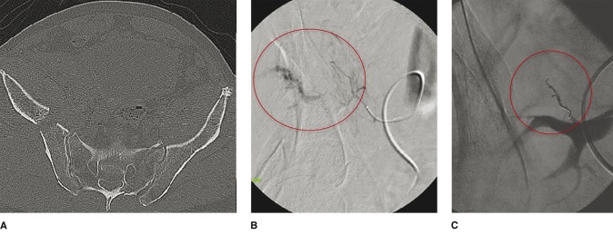

Figures

Figure 1
Figure 1
Diagram depicting the pelvic arterial system, overlaid on a pelvic angiogram. The common iliac artery is shown in aqua; the external iliac artery, in yellow; the internal iliac artery, in green; the posterior branch of the internal iliac artery, in red; and the anterior branch of the internal iliac artery, in blue. Smaller vessels include the superior gluteal artery (1), iliolumbar artery (2), lateral sacral artery (3), inferior gluteal artery (4), umbilical artery (5), obturator artery (6), internal pudendal artery (7), medial rectal artery (8), uterine artery or ductus deferens (9), and superior vesical artery (10).
Figure 2
Figure 2
A, Angiogram showing posterior branch/superior gluteal artery extravasation (circled) in a 47-year-old woman who sustained a Young and Burgess lateral compression type III pelvic injury in a motor vehicle accident. B, Angiogram showing successful embolization (circled).
Figure 3
Figure 3
A, Pelvic angiogram showing extravasation resulting from a Young and Burgess anterior-posterior compression type III pelvic injury in a 41-year-old man who was crushed by a forklift. B, The patient underwent laparotomy, external fixation with a femoral distractor, pelvic packing, and angiography with gel foam embolization of both internal iliac artery arteries.
Figure 4
Figure 4
A and B, Angiograms showing extravasation (circled in panel A) from the anterior division of the internal iliac artery in a 41-year-old patient who sustained a Young and Burgess anterior-posterior compression type III pelvic injury as a result of a motor vehicle accident. The patient was treated initially with a binder. C, Angiogram showing successful embolization with a coil (circled).
Figure 5
Figure 5
Axial pelvic CT (A) and aortopelvic angiogram (B) showing bleeding off the posterior division of the right internal iliac artery in an 81-year-old man who sustained a hemodynamically unstable Young and Burgess lateral compression type III pelvic injury in a fall from a roof. The patient was treated initially with a pelvic binder. C, Angiogram showing successful coil embolization (circled).

References

    1. Durkin A, Sagi HC, Durham R, Flint L: Contemporary management of pelvic fractures. Am J Surg 2006;192(2):211-223. - PubMed
    1. Costantini TW, Bosarge PL, Fortlage D, Bansal V, Coimbra R: Arterial embolization for pelvic fractures after blunt trauma: Are we all talk? Am J Surg 2010;200(6):752-758. - PubMed
    1. Jeroukhimov I, Ashkenazi I, Kessel B, et al. : Selection of patients with severe pelvic fracture for early angiography remains controversial. Scand J Trauma Resusc Emerg Med 2009;17:62. - PMC - PubMed
    1. Magnussen RA, Tressler MA, Obremskey WT, Kregor PJ: Predicting blood loss in isolated pelvic and acetabular high-energy trauma. J Orthop Trauma 2007;21(9):603-607. - PubMed
    1. El-Haj M, Bloom A, Mosheiff R, Liebergall M, Weil YA: Outcome of angiographic embolisation for unstable pelvic ring injuries: Factors predicting success. Injury 2013;44(12):1750-1755. - PubMed