. 2018 Jan 19;13(1):e0191318.
doi: 10.1371/journal.pone.0191318. eCollection 2018.

Inhibition of interleukin-6 decreases atrogene expression and ameliorates tail suspension-induced skeletal muscle atrophy

Affiliations

Inhibition of interleukin-6 decreases atrogene expression and ameliorates tail suspension-induced skeletal muscle atrophy

Mitsutaka Yakabe et al. PLoS One. .

Abstract

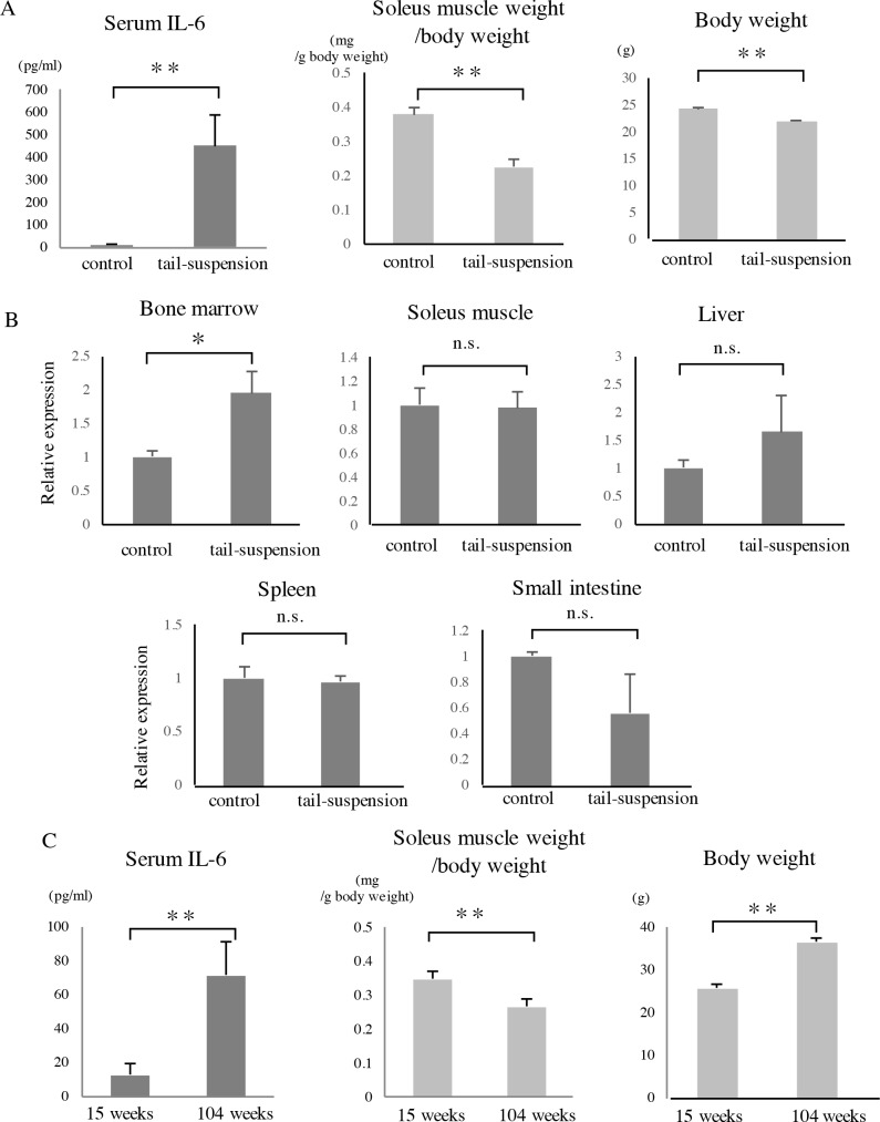
Background: Interleukin-6 (IL-6) is an inflammatory cytokine. Whether systemic IL-6 affects atrogene expression and disuse-induced skeletal muscle atrophy is unclear.

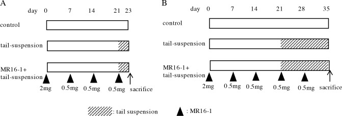
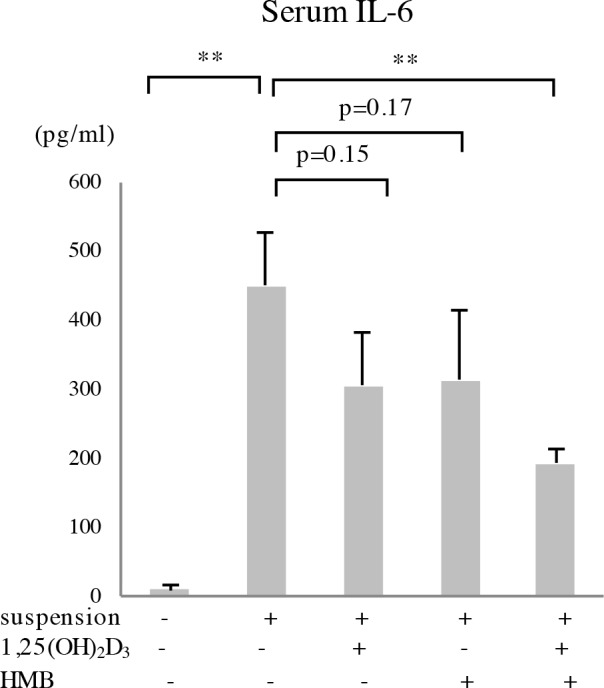
Methods: Tail-suspended mice were used as a disuse-induced muscle atrophy model. We administered anti-mouse IL-6 receptor antibody, beta-hydroxy-beta-methylbutyrate (HMB) and vitamin D to the mice and examined the effects on atrogene expression and muscle atrophy.

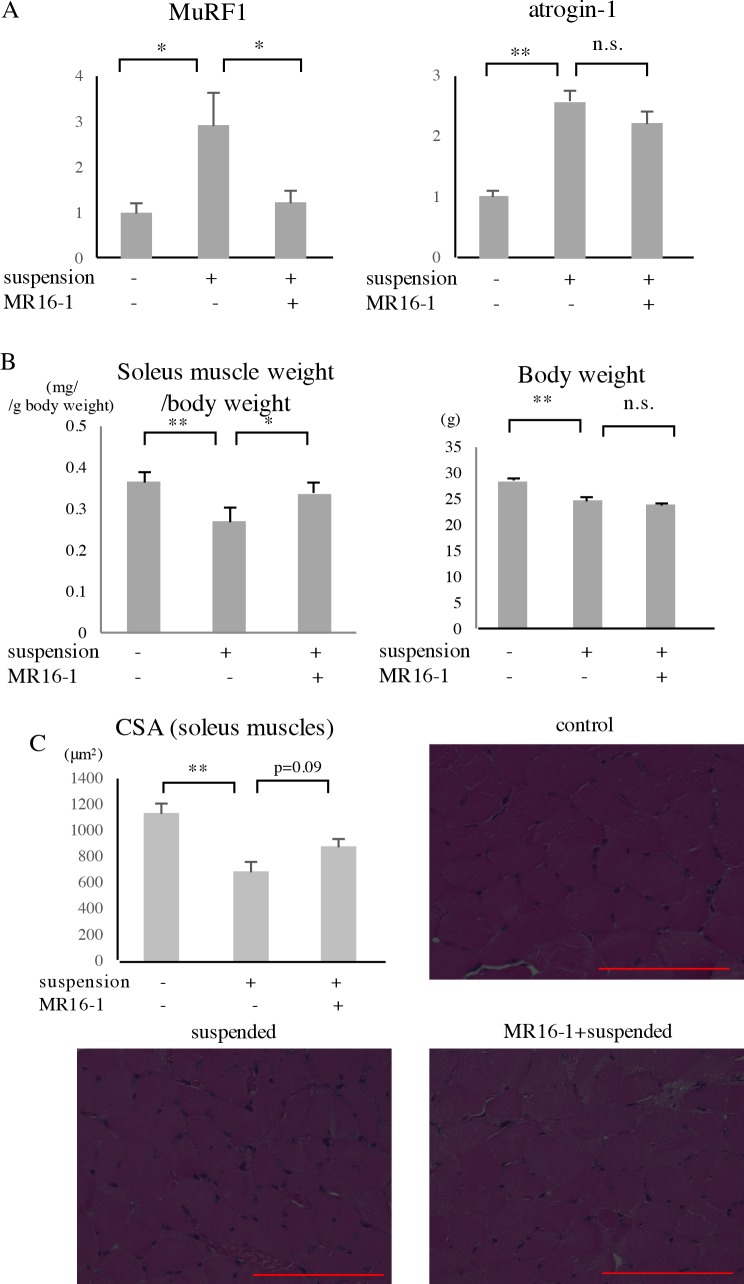
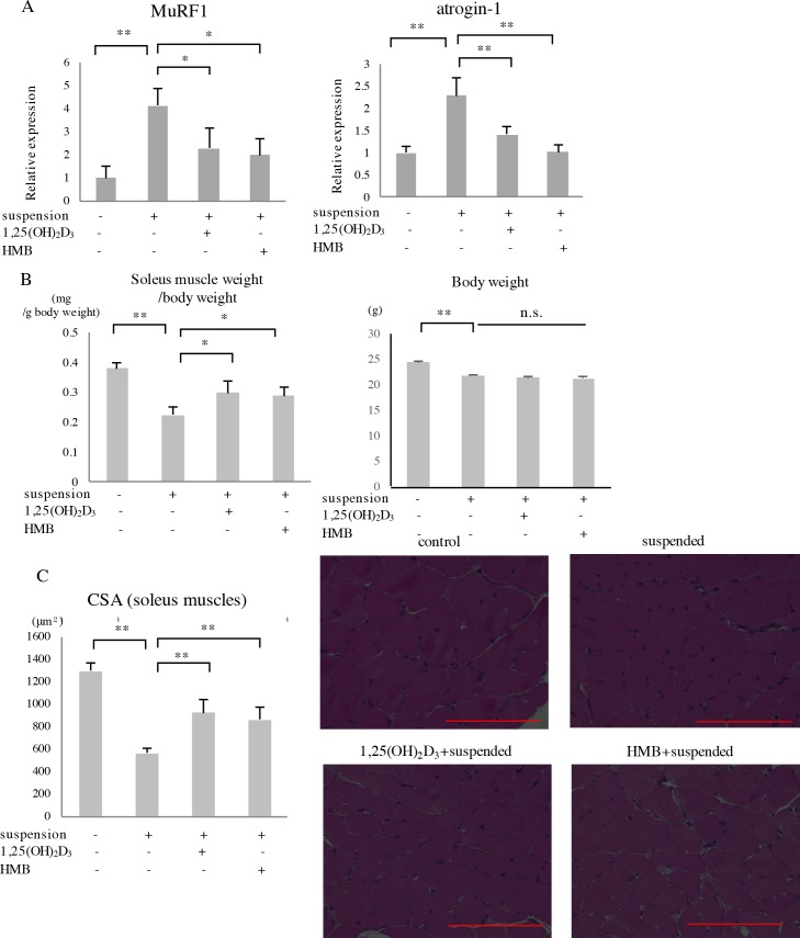
Results: Serum IL-6 levels were elevated in the mice. Inhibition of IL-6 receptor suppressed muscle RING finger 1 (MuRF1) expression and prevented muscle atrophy. HMB and vitamin D inhibited the serum IL-6 surge, downregulated the expression of MuRF1 and atrogin-1 in the soleus muscle, and ameliorated atrophy in the mice.

Conclusion: Systemic IL-6 affects MuRF1 expression and disuse-induced muscle atrophy.


Conflict of interest statement

Competing Interests: The authors have declared that no competing interests exist.

Figures

Fig 1
Fig 1. MR16-1 administration protocol.
Eleven-week-old C57BL/6J male mice were administered MR16-1 via intraperitoneal injection and subjected to tail suspension. The mice were subjected to two days of tail suspension to examine atrogene expression in the soleus muscles (A, n = 7–8), and to two weeks of tail suspension to measure soleus muscle weight and CSA (B, n = 5). The arrowheads indicate MR16-1 administration and its amount.
Fig 2
Fig 2. Characteristics of tail-suspended mice and aged mice.
(A) Twelve-week-old C57BL/6J male mice were subjected to two weeks of tail suspension. Mice were sacrificed, and then serum samples and soleus muscles were collected. Serum IL-6 level was determined using ELISA. Soleus muscle weight/body weight ratio and the body weight of mice are shown (n = 4). (B) Twelve-week-old mice were subjected to seven days of tail suspension, and then RNA was extracted from the following tissues: whole bone marrow from femurs (n = 4), soleus muscles (n = 3), livers (n = 6), spleens (n = 3), and small intestines (n = 3). Quantitative real-time PCR was performed, and the relative IL-6 expression in each tissue is shown. (C) C57BL/6J male mice aged 15 weeks and 104 weeks were sacrificed, and then serum samples and soleus muscles were collected (n = 4). *p<0.05, **p<0.01, n.s.: not significant.
Fig 3
Fig 3. Effects of MR16-1 on atrogene expression and muscle atrophy.
Eleven-week-old C57BL/6J male mice were administered with MR16-1 via intraperitoneal injection and subjected to tail suspension according to the protocol in Fig 1. (A) After two days of tail suspension, soleus muscles were collected, and then the expression of MuRF1 and atrogin-1 was measured by real-time PCR. (n = 7–8). (B, C) After two weeks of tail suspension, mice were sacrificed, and soleus muscles were collected. Soleus muscle weight/body weight (B) and CSA of the soleus muscles (C) are shown (n = 5). In Fig 3C, representative microscopic images of the soleus muscles (HE staining) are shown. CSA of each fiber was calculated using ImageJ, and the values were averaged for each muscle. Red bars correspond to 100μm. *p<0.05, **p<0.01, n.s.: not significant.
Fig 4
Fig 4. Effects of 1,25(OH)2D3 and HMB on tail suspension-induced atrogene expression and muscle atrophy.
(A) Twelve-week-old C57BL/6J male mice were administered 1,25(OH)2D3 or HMB by gavage and subjected to three days of tail suspension. MuRF1 and atrogin-1 expression levels were measured by real-time PCR. (B) Twelve-week-old mice were administered 1,25(OH)2D3 or HMB and subjected to two weeks of tail suspension. Soleus muscle weight/body weight ratio is shown. (C) Values of soleus muscle CSAs measured using ImageJ are shown. Representative images of soleus muscles (HE staining) are shown. Red bars correspond to 100μm. Each n = 4. *p<0.05, **p<0.01, n.s.: not significant.
Fig 5
Fig 5. Effects of 1,25(OH)2D3 and HMB on the tail suspension-induced serum IL-6 surge.
Twelve-week-old C57BL/6J male mice were administered 1,25(OH)2D3, HMB or both by gavage for seven days and then subjected to two weeks of tail suspension. Serum IL-6 level was determined by ELISA. n = 4, **p<0.01.

Similar articles

Cited by

References

    1. Foletta VC, White LJ, Larsen AE, Leger B, Russell AP. The role and regulation of MAFbx/atrogin-1 and MuRF1 in skeletal muscle atrophy. Pflugers Arch. 2011;461:325–35. doi: 10.1007/s00424-010-0919-9 - DOI - PubMed
    1. Cruz-Jentoft AJ, Baeyens JP, Bauer JM, Boirie Y, Cederholm T, Landi F, et al. Sarcopenia: European consensus on definition and diagnosis: Report of the European Working Group on Sarcopenia in Older People. Age Ageing. 2010;39:412–23. doi: 10.1093/ageing/afq034 - DOI - PMC - PubMed
    1. Bodine SC, Latres E, Baumhueter S, Lai VK, Nunez L, Clarke BA, et al. Identification of ubiquitin ligases required for skeletal muscle atrophy. Science. 2001;294:1704–8. doi: 10.1126/science.1065874 - DOI - PubMed
    1. Gomes MD, Lecker SH, Jagoe RT, Navon A, Goldberg AL. Atrogin-1, a muscle-specific F-box protein highly expressed during muscle atrophy. Proc Natl Acad Sci U S A. 2001;98:14440–5. doi: 10.1073/pnas.251541198 - DOI - PMC - PubMed
    1. Sandri M, Sandri C, Gilbert A, Skurk C, Calabria E, Picard A, et al. Foxo transcription factors induce the atrophy-related ubiquitin ligase atrogin-1 and cause skeletal muscle atrophy. Cell. 2004;117:399–412. - PMC - PubMed

LinkOut - more resources