. 2018 Apr 1;314(4):G471-G482.
doi: 10.1152/ajpgi.00153.2017. Epub 2018 Jan 11.

Proliferation of hepatic stellate cells, mediated by YAP and TAZ, contributes to liver repair and regeneration after liver ischemia-reperfusion injury

Affiliations

Proliferation of hepatic stellate cells, mediated by YAP and TAZ, contributes to liver repair and regeneration after liver ischemia-reperfusion injury

Takanori Konishi et al. Am J Physiol Gastrointest Liver Physiol. .

Abstract

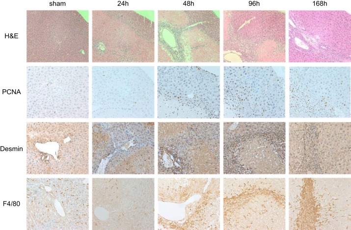
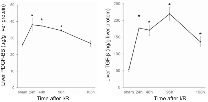
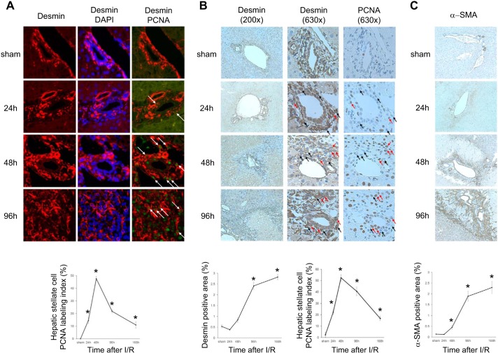
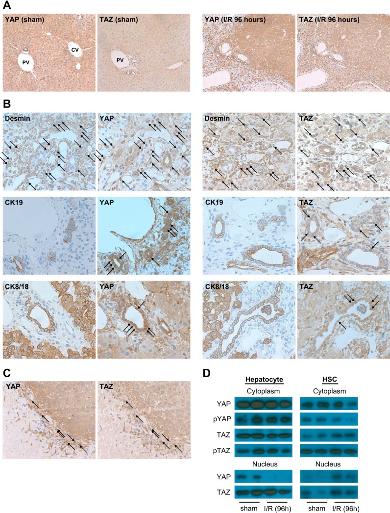
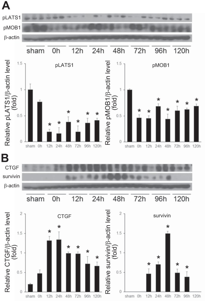
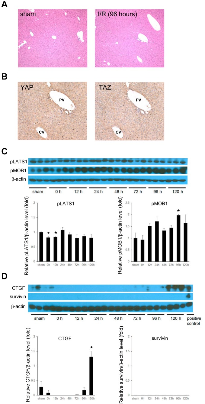
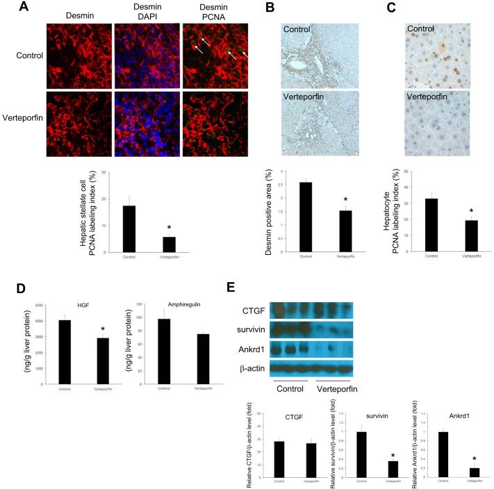
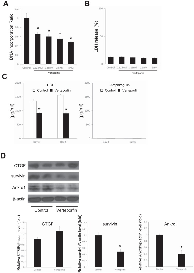
Yes-associated protein (YAP) and transcriptional coactivator with PDZ-binding motif (TAZ) are key regulators of cell proliferation and organ size; however, their physiological contribution after liver injury has not been fully understood. In this study, we sought to determine the role of YAP and TAZ during liver recovery after ischemia-reperfusion (I/R). A murine model of partial (70%) I/R was used to induce liver injury and study the reparative and regenerative response. After liver injury, there was marked activation and proliferation of hepatic stellate cells. The Hippo pathway components, large tumor suppressor 1 (LATS1) and its adapter protein, Mps one binder 1 (MOB1), were inactivated during liver repair, and YAP and TAZ were activated selectively in hepatic stellate cells. Concurrently, the expression of connective tissue growth factor and survivin, both of which are YAP and TAZ target genes, were upregulated. Hepatic stellate cell expansion and concomitant activation of YAP and TAZ occurred only in the injured liver and were not observed in the nonischemic liver. Treatment of mice with verteporfin, an inhibitor of YAP and TAZ, decreased hepatic stellate cell proliferation, survivin, and cardiac ankyrin repeat protein expression. These changes were associated with a significant decrease in hepatocyte proliferation. The data suggest that liver repair and regeneration after I/R injury are dependent on hepatic stellate cell proliferation, which is mediated by YAP and TAZ. NEW & NOTEWORTHY This study is the first to assess the proliferation of hepatic stellate cells (HSCs) after ischemia-reperfusion (I/R) injury and their role in the reparative and regenerative process. Here we show that the Hippo pathway is inactivated after I/R and that Yes-associated protein/transcriptional coactivator with PDZ-binding motif (YAP/TAZ) activation is detected in HSC. HSC proliferation and expansion are prominent during liver recovery after I/R injury. Inhibition of YAP/TAZ activation with verteporfin reduces HSC proliferation and target gene expression and attenuates hepatocyte proliferation.

Keywords: hepatic stellate cell proliferation; liver injury; liver regeneration; survivin.


Figures

Fig. 1.
Fig. 1.
Temporal course of liver repair and regeneration after ischemia-reperfusion (I/R) injury. Hematoxylin and eosin (H and E) staining shows massive liver injury after 24 h of reperfusion with repair and restoration of nearly normal liver architecture 168 h after reperfusion. Hepatocyte proliferation was determined by immunohistochemical staining for proliferating cell nuclear antigen (PCNA) and shows robust proliferation of hepatocytes 48 and 96 h after reperfusion that tapers off at 168 h of reperfusion. Hepatic stellate cells (HSCs) and macrophages were detected by desmin and F4/80 staining, respectively. Proliferating hepatocytes were located prominently in the parenchyma adjacent to HSCs and macrophages. Original magnification is ×100 for H and E and ×200 for PCNA, desmin, and F4/80.
Fig. 2.
Fig. 2.
Expression of liver platelet-derived growth factor-BB (PDGF-BB) and transforming growth factor-β (TGF-β) after ischemia-reperfusion (I/R) injury. Liver PDGF-BB and TGF-β protein levels were measured by ELISA. PDGF-BB and TGF-β levels increased during I/R liver injury and remained elevated during liver repair and regeneration. Data are means ± SE with n = 3–7 per group. *P < 0.05 compared with sham mice.
Fig. 3.
Fig. 3.
Proliferation and activation of hepatic stellate cells (HSCs) after ischemia-reperfusion (I/R). HSC proliferation was assessed by 2 independent techniques (A and B) over a time course of I/R. Both demonstrate the same temporal pattern of HSC proliferation after I/R. A: HSC proliferation was determined by dual immunofluorescence staining for desmin (red) and proliferating cell nuclear antigen (PCNA, green). DAPI was used for nuclear staining. HSCs positive for PCNA are marked by arrows. Quantitative analysis of PCNA labeling was calculated as a percentage of PCNA-positive HSCs. Original magnification is ×630. B: HSC proliferation was determined by immunohistochemistry for desmin and PCNA in serial sections. Desmin-labeled area was measured as a percentage of total area. Original magnification is ×200. Representative HSCs are marked by arrows (red: PCNA positive, black: PCNA negative). Quantitative analysis of PCNA labeling was calculated as a percentage of PCNA-positive HSCs. Original magnification is ×630. C: activated HSCs were assessed by immunohistochemical staining for α-smooth muscle actin (α-SMA). Stellate cell activation increased during after I/R. α-SMA-labeled area was measured as a percentage of total area. Original magnification is ×200. For all graphs, data are means ± SE with n = 3–4. *P < 0.05 compared with sham mice.
Fig. 4.
Fig. 4.
Cell-specific Yes-associated protein/transcriptional coactivator with PDZ-binding motif (YAP/TAZ) activation in postischemic liver after ischemia-reperfusion (I/R). A: YAP and TAZ nuclear expression in sham liver and postischemic liver 96 h after I/R. Original magnification is ×200. PV, portal vein; CV, central vein. B: nuclear YAP and TAZ expression in hepatic stellate cells (HSCs), biliary epithelial cells, and reactive ductular cells was determined 96 h after I/R by staining of serial sections for YAP or TAZ with desmin, CK19, or CK8/18, respectively. Representative HSCs, biliary epithelial cells, and reactive ductular cells positive for YAP or TAZ are marked by arrows. Original magnification is ×400. C: concomitant activation of YAP and TAZ occurs in HSCs. Serial liver sections obtained 96 h after I/R were stained for YAP and TAZ. Representative HSCs positive for YAP and TAZ are marked by arrows. Original magnification is ×400. D: nuclear YAP and TAZ expression increases in HSCs, but not hepatocytes, after I/R. Cytoplasmic and nuclear fractions from hepatocytes and HSCs isolated 96 h after I/R were analyzed by Western blot and stained with YAP, phosphorylated YAP (Ser127), TAZ, and phosphorylated TAZ (Ser89).
Fig. 5.
Fig. 5.
Hippo pathway and Yes-associated protein/transcriptional coactivator with PDZ-binding motif (YAP/TAZ) target protein expression in postischemic liver after ischemia-reperfusion (I/R). A: I/R leads to reduced phosphorylation of the Hippo pathway proteins, large tumor suppressor 1 (LATS1) and Mps one binder 1 (MOB1). B: I/R leads to increased expression of the YAP/TAZ target genes, CTGF and survivin. Lysates of postischemic liver tissue were analyzed by Western blot. Results were quantitated by image analysis. Data are means ± SE with n = 3. *P < 0.05 compared with sham mice.
Fig. 6.
Fig. 6.
Yes-associated protein/transcriptional coactivator with PDZ-binding motif (YAP/TAZ) and Hippo pathway regulation in nonischemic liver after ischemia-reperfusion (I/R). A: liver histopathology (H and E staining) in nonischemic liver after I/R showed no changes in liver architecture and no expansion of nonparenchymal cells. Original magnification is ×100. B: YAP and TAZ expression (immunohistochemistry) in nonischemic liver lobes after 96 h of reperfusion was minimal in hepatocytes. Original magnification is ×200. PV, portal vein; CV, central vein. C: no change in activation of Hippo pathway during I/R injury. Lysates of the nonischemic liver tissue were analyzed by Western blot and stained with phosphorylated large tumor suppressor 1 (LATS) and phosphorylated Mps one binder 1 (MOB1). D: expression of the YAP/TAZ target genes, CTGF and survivin, were assessed in lysates of nonischemic liver by Western blot. Results were quantitated by image analysis. Data are means ± SE with n = 3. *P < 0.05 compared with sham mice.
Fig. 7.
Fig. 7.
Yes-associated protein (YAP) and transcriptional coactivator with PDZ-binding motif (TAZ) contribute to hepatic stellate cell (HSC) proliferation after ischemia-reperfusion (I/R). Mice were injected intraperitoneally with 10% dimethylsulfoxide solution (control) or 100 mg/kg verteporfin after 24 h of reperfusion and every 24 h thereafter. After 96 h of reperfusion, postischemic liver was analyzed. A: verteporfin treatment decreased HSC proliferation in ischemic liver after I/R. HSC proliferation was determined by dual immunofluorescence staining for desmin (red) and proliferating cell nuclear antigen (PCNA, green). DAPI was used for nuclear staining. Representative HSCs positive for PCNA are marked by arrows. Quantitative analysis of PCNA labeling was calculated as a percentage of PCNA-positive HSCs. Original magnification is ×630. B: verteporfin treatment decreased the number of HSCs in postischemic liver after I/R. Desmin-labeled cells were measured as a percentage of total area. Original magnification is ×200. C: verteporfin treatment decreased hepatocyte proliferation in postischemic liver after I/R. Hepatocyte proliferation was determined by immunohistochemical staining for PCNA. Original magnification is ×200. D: verteporfin treatment decreased growth factors in postischemic liver after I/R. Liver hepatocyte growth factor (HGF) and amphiregulin levels were analyzed by ELISA. E: verteporfin treatment decreased the expression of survivin and Ankrd1, but not connective tissue growth factor (CTGF). The lysates of ischemic liver were analyzed by Western blot. Results were quantitated by image analysis. Data are means ± SE with n = 3–4. *P < 0.05 compared with control group.
Fig. 8.
Fig. 8.
Inhibition of Yes-associated protein/transcriptional coactivator with PDZ-binding motif (YAP/TAZ) by verteporfin decreases hepatic stellate cell (HSC) proliferation and target gene expression in vitro. HSCs were isolated from mouse liver and treated with verteporfin or control culture medium for 5 days. A: verteporfin treatment decreased HSC proliferation as measured by DNA incorporation (n = 8). B: verteporfin treatment did not induce cytotoxicity (lactate dehydrogenase, LDH, release) of HSCs (n = 6). C: effect of verteporfin treatment (5 nM) on the release of hepatocyte growth factor (HGF) and amphiregulin by HSCs (n = 3). HGF and amphiregulin were measured by ELISA. Open bars, control; solid bars, verteporfin. D: effect of verteporfin treatment (5 nM) on expression of the YAP/TAZ target genes, CTGF, survivin, and Ankrd1, measured by Western blot. Results were quantitated by image analysis (n = 3). *P < 0.05 compared with control group.

References

    1. Altieri DC. Molecular circuits of apoptosis regulation and cell division control: the survivin paradigm. J Cell Biochem 92: 656–663, 2004. doi: 10.1002/jcb.20140. - DOI - PubMed
    1. Baba HA, Wohlschlaeger J, Schmitz KJ, Nadalin S, Lang H, Benesch A, Gu Y, Biglarnia AR, Sotiropoulos GC, Takeda A, Takeda N, von Wnuck Lipinski K, Levkau B. Survivin is upregulated during liver regeneration in rats and humans and is associated with hepatocyte proliferation. Liver Int 29: 585–592, 2009. doi: 10.1111/j.1478-3231.2008.01911.x. - DOI - PubMed
    1. Bai H, Zhang N, Xu Y, Chen Q, Khan M, Potter JJ, Nayar SK, Cornish T, Alpini G, Bronk S, Pan D, Anders RA. Yes-associated protein regulates the hepatic response after bile duct ligation. Hepatology 56: 1097–1107, 2012. doi: 10.1002/hep.25769. - DOI - PMC - PubMed
    1. Baier PK, Baumgartner U, Hempel S, Wolff-Vorbeck G, von Dobschuetz E, Hopt UT. Kupffer cells infiltrate liver tissue early after ischemia-reperfusion and partial hepatectomy. Eur Surg Res 37: 290–297, 2005. doi: 10.1159/000089239. - DOI - PubMed
    1. Barone S, Okaya T, Rudich S, Petrovic S, Tenrani K, Wang Z, Zahedi K, Casero RA, Lentsch AB, Soleimani M. Distinct and sequential upregulation of genes regulating cell growth and cell cycle progression during hepatic ischemia-reperfusion injury. Am J Physiol Cell Physiol 289: C826–C835, 2005. doi: 10.1152/ajpcell.00629.2004. - DOI - PubMed

Publication types

MeSH terms

LinkOut - more resources