. 2018 Apr 1;314(4):L569-L582.
doi: 10.1152/ajplung.00565.2016. Epub 2017 Dec 20.

Activated human T lymphocytes inhibit TGFβ-induced fibroblast to myofibroblast differentiation via prostaglandins D2 and E2

Affiliations

Activated human T lymphocytes inhibit TGFβ-induced fibroblast to myofibroblast differentiation via prostaglandins D2 and E2

Shannon H Lacy et al. Am J Physiol Lung Cell Mol Physiol. .

Abstract

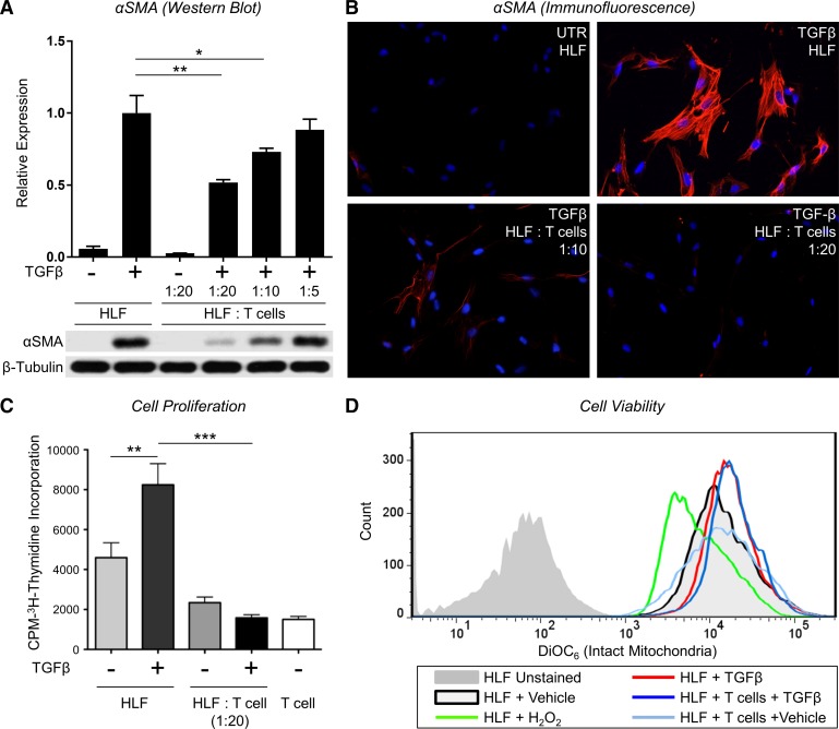
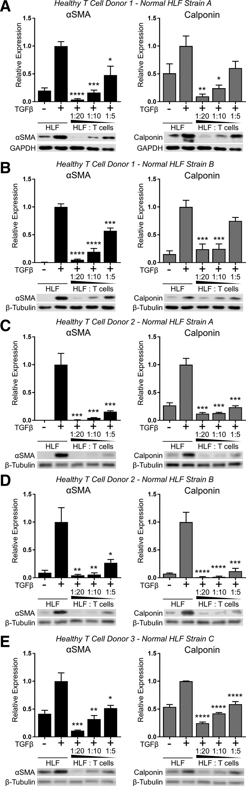
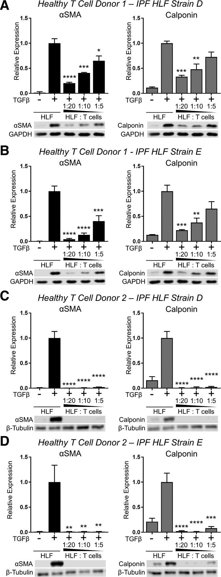
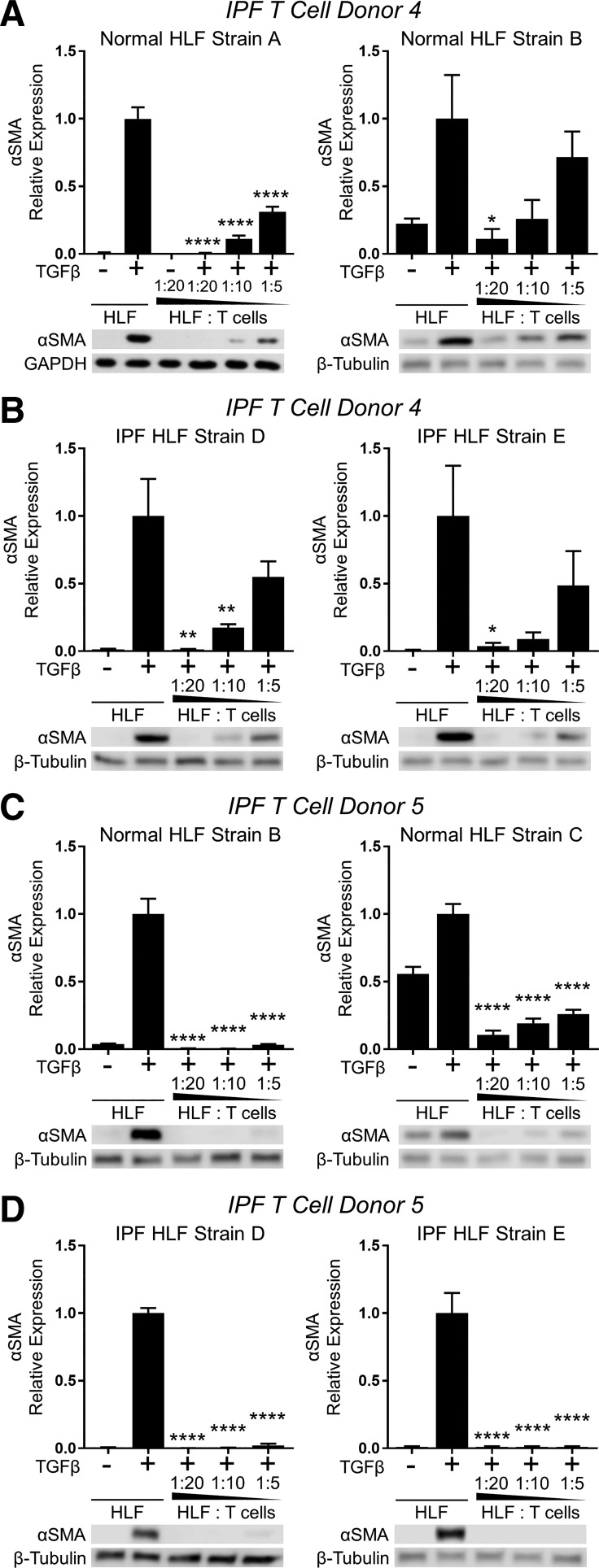
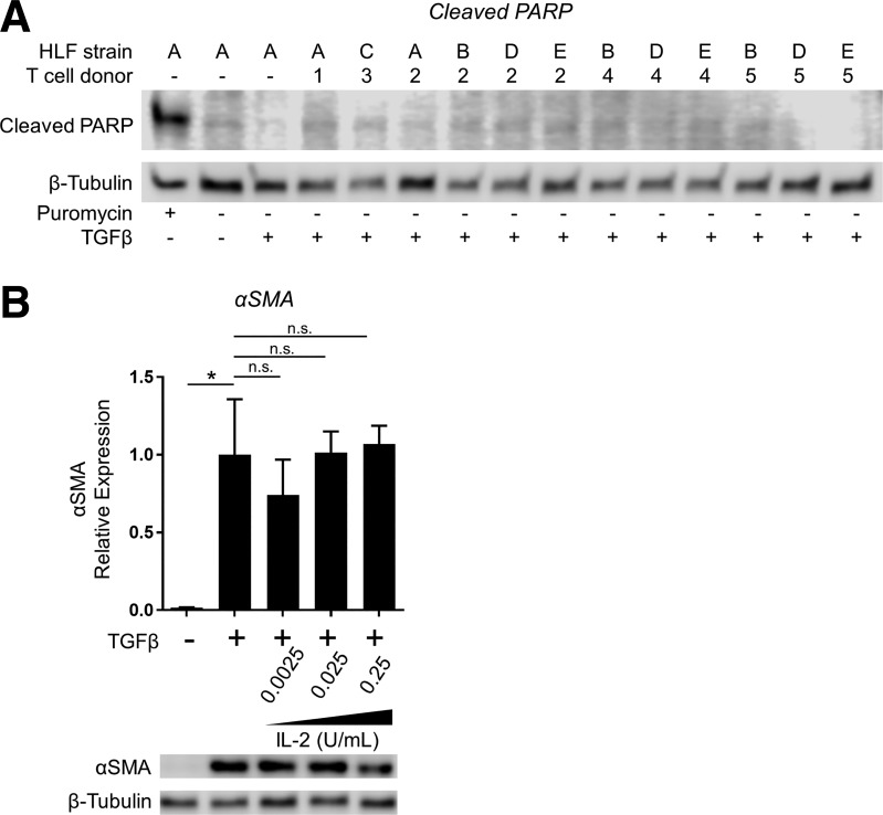
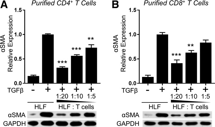
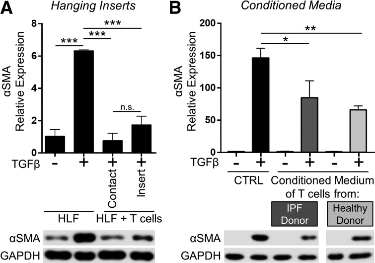
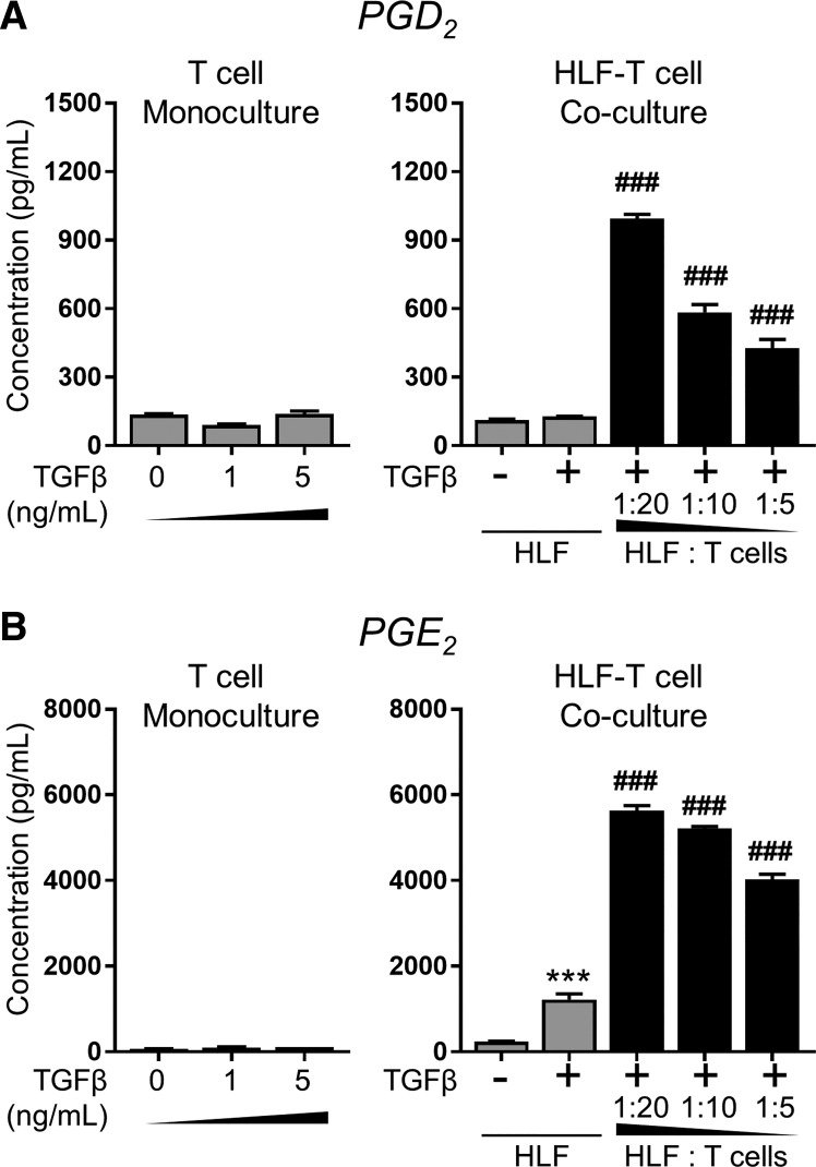
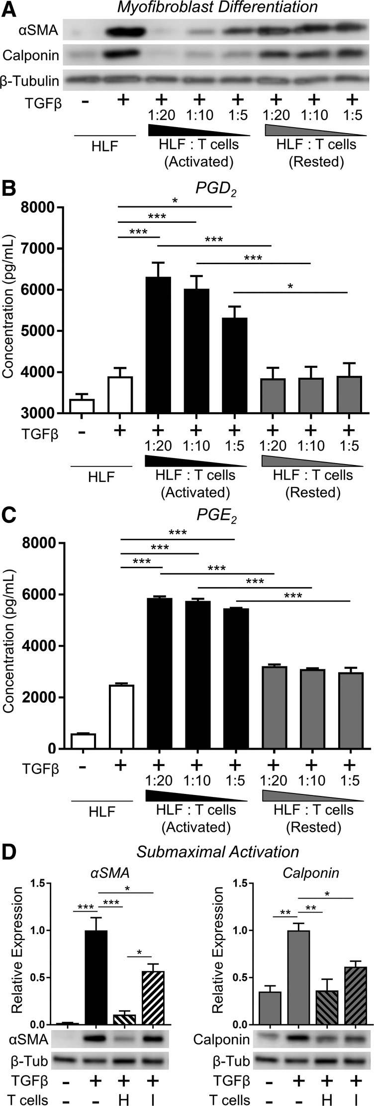
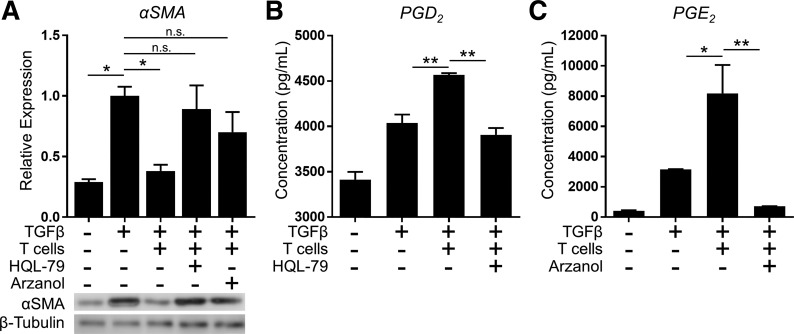
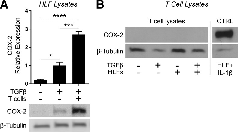
In pulmonary fibrosis (PF), fibroblasts and myofibroblasts proliferate and deposit excessive extracellular matrix in the interstitium, impairing normal lung function. Because most forms of PF have a poor prognosis and limited treatment options, PF represents an urgent unmet need for novel, effective therapeutics. Although the role of immune cells in lung fibrosis is unclear, recent studies suggest that T lymphocyte (T cell) activation may be impaired in PF patients. Furthermore, we have previously shown that activated T cells can produce prostaglandins with anti-scarring potential. Here, we test the hypothesis that activated T cells directly inhibit myofibroblast differentiation using a coculture system. Coculture with activated primary blood-derived T cells, from both healthy human donors and PF patients, inhibited transforming growth factor β-induced myofibroblast differentiation in primary human lung fibroblasts isolated from either normal or PF lung tissue. Coculture supernatants contained anti-fibrotic prostaglandins D2 and E2, and the inhibitory effect of coculture on myofibroblast differentiation was largely reversed when prostaglandin production was abrogated either by resting the T cells before coculture or via specific pharmacological inhibitors. Moreover, coculture conditions induced COX-2 in HLFs but not in T cells, suggesting that T cells deliver an activating signal to HLFs, which in turn produce anti-fibrotic prostaglandins. We show for the first time that coculture with activated primary human T lymphocytes strongly inhibits myofibroblast differentiation, revealing a novel cell-to-cell communication network with therapeutic implications for fibrotic lung diseases.


Figures

Fig. 1.
Fig. 1.
Jurkat T lymphocytes inhibit transforming growth factor-β (TGFβ)-induced α-smooth muscle actin (α-SMA) expression and proliferation in human lung fibroblasts without inducing cell death. A: human lung fibroblasts (HLFs) were treated with 5 ng/ml TGFβ in the presence or absence of irradiated Jurkat T cells at indicated ratios. After 72 h, T cells were removed by repeated PBS washes, and α-SMA protein expression in the HLFs was analyzed by Western blot and densitometric analysis. Densitometry of n = 3 replicates per cell strain normalized to TGFβ alone. Data shown are means ± SE. *P < 0.05 and **P < 0.01 by ANOVA. B: HLFs were grown in chamber slides and treated with 5 ng/ml TGFβ in the presence or absence of irradiated Jurkat T cells at indicated ratios. After 72 h, T cells were removed by repeated PBS washes, and HLFs were stained with an antibody to α-SMA (red) and DAPI (blue). C: HLFs were treated with 5 ng/ml TGFβ in the presence or absence of irradiated Jurkat T cells for 48 h. Cells were allowed to incorporate [3H]thymidine for another 18 h; n = 6/group. Data shown are means ± SE. **P < 0.01 and ***P < 0.001 by ANOVA. D: HLFs were treated with 5 ng/ml TGFβ in the presence or absence irradiated Jurkat T cells for 72 h. During the last 15 min, T cells were removed by washing, and HLFs were labeled with 40 nM DiOC6. HLFs with reduced mitochondrial membrane potential were detected by FACS Canto II cytometer using Blue E detector (488 laser + 530/30 filter).
Fig. 2.
Fig. 2.
Primary T lymphocytes from healthy donors inhibit TGFβ-induced myofibroblast differentiation in lung fibroblasts isolated from nonfibrotic lung tissue. Three strains of HLFs isolated from normal lung tissue of donors without idiopathic pulmonary fibrosis (IPF; normal HLF strains A, B, and C) were cultured with activated peripheral blood mononuclear cell (PBMC)-derived primary T cells from 3 healthy donors (donors 1, 2, and 3) at indicated ratios and treated with 5 ng/ml TGFβ for 72 h. Expression of α-SMA and calponin was analyzed by Western blot and densitometric analysis. AE: different combinations of T cell and HLF donors as labeled at top. Data shown are means ± SE of n = 3 replicates/condition, protein expression relative to loading control, normalized to TGFβ alone. *P < 0.05, **P < 0.01, ***P < 0.001, and ****P < 0.0001 by ANOVA compared with HLF + TGFβ control.
Fig. 3.
Fig. 3.
Primary T lymphocytes from healthy donors inhibit TGFβ-induced myofibroblast differentiation in lung fibroblasts isolated from IPF lung tissue. Two strains of HLFs isolated from the fibrotic lung tissue of donors with IPF (IPF HLF strains D and E) were cultured with activated PBMC-derived primary T cells from 2 healthy donors (donors 1 and 2) at indicated ratios and treated with 5 ng/ml TGFβ for 72 h. Expression of α-SMA and calponin were analyzed by Western blot and densitometric analysis. AD: different combinations of T cell and HLF donors as labeled at top. Data shown are means ± SE of n = 3 replicates/condition, protein expression relative to loading control, normalized to TGFβ alone. *P < 0.05, **P < 0.01, ***P < 0.001, and ****P < 0.0001 by ANOVA compared with HLF + TGFβ control.
Fig. 4.
Fig. 4.
Primary T lymphocytes from IPF patients inhibit TGFβ-induced myofibroblast differentiation in both normal and fibrotic human lung fibroblasts. PBMC-derived primary T cells were purified, expanded, and activated from 2 IPF patient donors (IPF donors 4 and 5). T cells from each donor were used in coculture with 2 normal HLF strains (normal HLF strains A and B or B and C) and 2 IPF HLF strains (IPF HLF strains D and E) at indicated ratios in the presence of 5 ng/ml TGFβ for 72 h. Expression of α-SMA was analyzed by Western blot and densitometric analysis. AD: different combinations of T cell and HLF donors as labeled at top. Data shown are means ± SE of n = 3 replicates/condition, protein expression relative to loading control, normalized to TGFβ alone. *P < 0.05, **P < 0.01, and ****P < 0.0001 by ANOVA compared with HLF + TGFβ control.
Fig. 5.
Fig. 5.
Inhibition of myofibroblast differentiation in coculture with primary T cells is not attributable to IL-2 spillover or HLF cell death. A: HLFs from normal lung tissue (strains A, B, and C) and IPF lung tissue (strains D and E) were cocultured with PBMC-derived primary T cells from healthy donors (donors 1, 2, and 3) and IPF patient donors (donors 4 and 5) at 1:10 HLF/T cell ratio in the presence of 5 ng/ml TGFβ for 72 h. Expression of cleaved PARP was analyzed by Western blot. As a positive control (lane 1), HLFs were treated with puromycin (5 ug/ml) for 24 h to induce apoptosis. B: HLFs were treated with 5 ng/ml TGFβ in the absence or presence of IL-2 at the doses indicated. Expression of α-SMA was analyzed by Western blot with densitometry. Data shown are means ± SE of n = 3 replicates/condition, protein expression relative to loading control, normalized to TGFβ alone. *P < 0.05 by ANOVA. NS, not significant.
Fig. 6.
Fig. 6.
Purified CD4+ and CD8+ primary T lymphocytes inhibit myofibroblast differentiation. CD4+ (A) and CD8+ (B) T cells were purified from the expanded, enriched T cell population using negative selection. HLFs (normal HLF strain A) were cultured with the purified T cell subsets and treated with 5 ng/ml TGFβ for 72 h. Expression of α-SMA was analyzed by Western blot and densitometric analysis. Data shown are means ± SE of n = 3 replicates/condition, protein expression relative to loading control, normalized to TGFβ alone. **P < 0.01 and ***P < 0.001 by ANOVA compared with HLF + TGFβ control.
Fig. 7.
Fig. 7.
Primary T-lymphocyte-mediated inhibition of myofibroblast differentiation is contact independent. A: HLFs were cocultured with primary T cells (1:20 ratio) either in contact or separated by a semipermeable hanging insert and treated with 5 ng/ml TGFβ for 72 h. B: conditioned media from activated normal T cells and IPF T cells were collected. HLFs were treated with 5 ng/ml TGF-β in the presence of T cell-conditioned medium for 72 h. α-SMA protein expression was analyzed by Western blot and densitometric analysis. Data shown are means ± SE of n = 3 replicates/condition, protein expression relative to loading control, normalized to untreated control. *P < 0.05, **P < 0.01, and ***P < 0.001 by ANOVA. NS, not significant. Note that the indicated samples were resolved on the same gel, and intervening irrelevant lanes are not shown.
Fig. 8.
Fig. 8.
Antifibrotic prostaglandins are present in the conditioned medium of HLF primary T cell cocultures, but not monocultures. For T cell monocultures, PBMC-derived primary T cells from healthy donor 1 were plated at the same density as that used in 1:20 HLF-T cell cocultures and treated with TGFβ at the indicated doses for 48 h. For HLF monocultures and HLF-T cell cocultures, HLFs (normal strain A) were cultured either alone (gray) or with activated PBMC-derived primary T cells from healthy donor 1 at indicated ratios and treated with 5 ng/ml TGFβ for 72 h. Supernatants were collected and analyzed for prostaglandins D2 (PGD2; A) and E2 (PGE2; B) content by enzyme immunoassay (EIA). Data shown are means ± SE of n = 3 biological replicates/condition. ***P < 0.001 by ANOVA, compared with HLF − TGFβ, or HLF alone, or HLF without TGFβ; ###P < 0.001 by ANOVA compared with HLF + TGFβ.
Fig. 9.
Fig. 9.
Prostaglandin production and antifibrotic function are abrogated in rested and submaximally activated primary human T cells. A–C: HLFs were cocultured with PBMC-derived primary T cells that were activated with CD3/CD28 beads and IL-2 (activated) or removed from activating media and rested for 18 h before coculture (rested). Cocultures were established at the indicated HLF/T cell ratios and treated with 5 ng/ml TGFβ for 72 h. Expression of α-SMA and calponin were analyzed by Western blot (A), and supernatants were collected and analyzed for PGD2 (B) and PGE2 (C) content by EIA. D: PBMC-derived primary T cells from healthy donor 1 (H) and IPF patient donor 4 (I) were cultured in “submaximal activation” conditions by reducing IL-2 and CD3/CD28 beads to one-tenth that of normal activation concentrations (i.e., 2.5 U/ml IL-2 and bead/T cell ratio of 1:10). After 24 h, cocultures were established at a 1:10 ratio of HLFs (normal strain A) to submaximally activated T cells, and cocultures were treated with TGFβ (5 ng/ml); after 72 h, T cells were removed and HLF lysates prepared for Western blot and densitometric analysis of markers of myofibroblast differentiation. Data shown are means ± SE of n = 3 biological replicates per condition. *P < 0.05, **P < 0.01, and ***P < 0.001 by ANOVA.
Fig. 10.
Fig. 10.
Inhibition of myofibroblast differentiation by T cells is dependent on hematopoietic PGD synthase (hPGDS) and microsomal PGE synthase-1 (mPGES-1). HLFs were cocultured with activated PBMC-derived primary T cells at a HLF/T cell ratio of 1:10 and treated with 5 ng/ml TGFβ for 72 h. Pharmacological inhibitors of hPGDS (HQL-79, 25 µM) or mPGES-1 (Arzanol, 2.5 μM) were added to the separate monocultures for 18 h before coculture and daily throughout the 3-day coculture experiment. A: expression of α-SMA was analyzed by Western blot. B and C: supernatants were collected and analyzed for PGD2 (B) and PGE2 (C) content by EIA. Data shown are means ± SE of n = 3 biological replicates/condition. *P < 0.05 and **P < 0.01 by ANOVA. NS, not significant.
Fig. 11.
Fig. 11.
Coculture conditions cause cyclooxygenase-2 (COX-2) expression in human lung fibroblasts but not T cells. HLFs were cocultured with Jurkat T cells (1:20 ratio of HLFs to T cells) separated by a porous hanging insert and treated with 5 ng/ml TGFβ for 24 h. Expression of COX-2 in both populations was analyzed by Western blot. A: data shown are means ± SE of n = 3 biological replicates/condition, protein expression relative to loading control, normalized to TGFβ alone. *P < 0.05, ***P < 0.001, and ****P < 0.0001 by ANOVA. B: all samples, including positive control (lysate from HLFs treated with 1 ng/ml IL-1β), were resolved on the same gel, and irrelevant intervening lanes are not shown.

Similar articles

Cited by

References

    1. Adegunsoye A, Hrusch CL, Bonham CA, Jaffery MR, Blaine KM, Sullivan M, Churpek MM, Strek ME, Noth I, Sperling AI. Skewed Lung CCR4 to CCR6 CD4+T Cell Ratio in Idiopathic Pulmonary Fibrosis Is Associated with Pulmonary Function. Front Immunol 7: 516, 2016. doi:10.3389/fimmu.2016.00516. - DOI - PMC - PubMed
    1. Baglole CJ, Reddy SY, Pollock SJ, Feldon SE, Sime PJ, Smith TJ, Phipps RP. Isolation and phenotypic characterization of lung fibroblasts. Methods Mol Med 117: 115–127, 2005. doi:10.1385/1-59259-940-0:115. - DOI - PubMed
    1. Birjandi SZ, Palchevskiy V, Xue YY, Nunez S, Kern R, Weigt SS, Lynch JP III, Chatila TA, Belperio JA. CD4(+)CD25(hi)Foxp3(+) cells exacerbate bleomycin-induced pulmonary fibrosis. Am J Pathol 186: 2008–2020, 2016. doi:10.1016/j.ajpath.2016.03.020. - DOI - PMC - PubMed
    1. Broekelmann TJ, Limper AH, Colby TV, McDonald JA. Transforming growth factor beta 1 is present at sites of extracellular matrix gene expression in human pulmonary fibrosis. Proc Natl Acad Sci USA 88: 6642–6646, 1991. doi:10.1073/pnas.88.15.6642. - DOI - PMC - PubMed
    1. Burgess HA, Daugherty LE, Thatcher TH, Lakatos HF, Ray DM, Redonnet M, Phipps RP, Sime PJ. PPARgamma agonists inhibit TGF-beta induced pulmonary myofibroblast differentiation and collagen production: implications for therapy of lung fibrosis. Am J Physiol Lung Cell Mol Physiol 288: L1146–L1153, 2005. doi:10.1152/ajplung.00383.2004. - DOI - PubMed

Publication types

MeSH terms

LinkOut - more resources