. 2018 May 1;314(5):H1053-H1060.
doi: 10.1152/ajpheart.00472.2017. Epub 2017 Dec 22.

Telomerase reverse transcriptase protects against angiotensin II-induced microvascular endothelial dysfunction

Affiliations

Telomerase reverse transcriptase protects against angiotensin II-induced microvascular endothelial dysfunction

Karima Ait-Aissa et al. Am J Physiol Heart Circ Physiol. .

Abstract

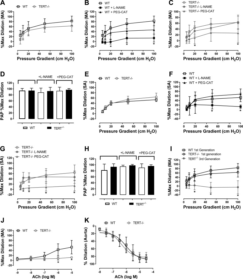
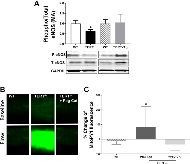
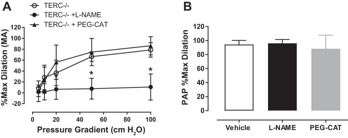
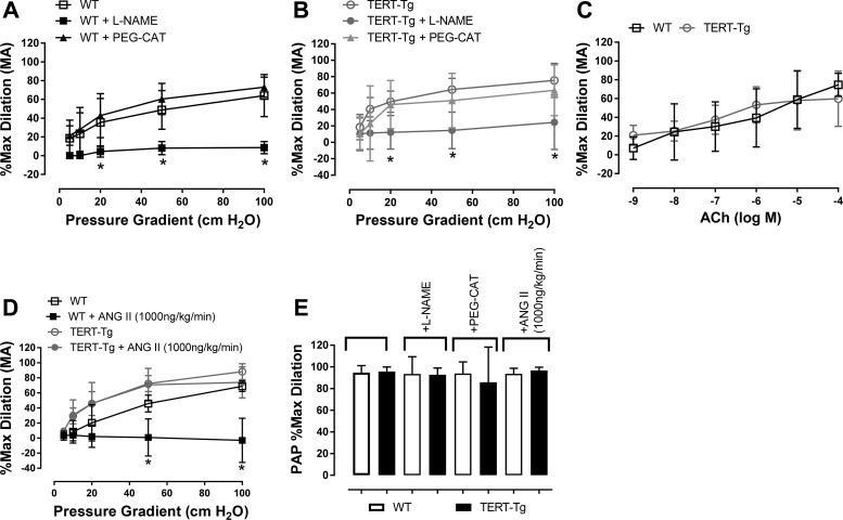
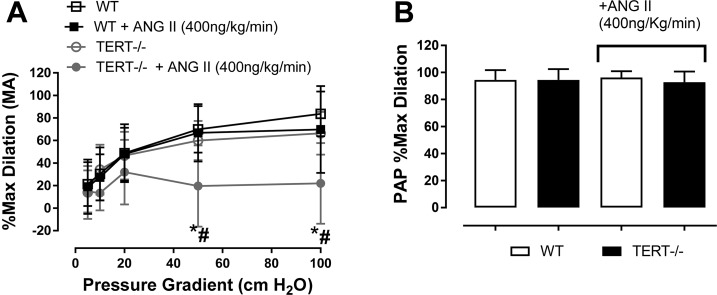
A rise in reactive oxygen species (ROS) may contribute to cardiovascular disease by reducing nitric oxide (NO) levels, leading to loss of NO's vasodilator and anti-inflammatory effects. Although primarily studied in larger conduit arteries, excess ROS release and a corresponding loss of NO also occur in smaller resistance arteries of the microcirculation, but the underlying mechanisms and therapeutic targets have not been fully characterized. We examined whether either of the two subunits of telomerase, telomerase reverse transcriptase (TERT) or telomerase RNA component (TERC), affect microvascular ROS production and peak vasodilation at baseline and in response to in vivo administration to angiotensin II (ANG II). We report that genetic loss of TERT [maximal dilation: 52.0 ± 6.1% with vehicle, 60.4 ± 12.9% with Nω-nitro-l-arginine methyl ester (l-NAME), and 32.2 ± 12.2% with polyethylene glycol-catalase (PEG-Cat) ( P < 0.05), means ± SD, n = 9-19] but not TERC [maximal dilation: 79 ± 5% with vehicle, 10.7 ± 9.8% with l-NAME ( P < 0.05), and 86.4 ± 8.4% with PEG-Cat, n = 4-7] promotes flow-induced ROS formation. Moreover, TERT knockout exacerbates the microvascular dysfunction resulting from in vivo ANG II treatment, whereas TERT overexpression is protective [maximal dilation: 88.22 ± 4.6% with vehicle vs. 74.0 ± 7.3% with ANG II (1,000 ng·kg-1·min-1) ( P = not significant), n = 4]. Therefore, loss of TERT but not TERC may be a key contributor to the elevated microvascular ROS levels and reduced peak dilation observed in several cardiovascular disease pathologies. NEW & NOTEWORTHY This study identifies telomerase reverse transcriptase (TERT) but not telomerase RNA component as a key factor regulating endothelium-dependent dilation in the microcirculation. Loss of TERT activity leads to microvascular dysfunction but not conduit vessel dysfunction in first-generation mice. In contrast, TERT is protective in the microcirculation in the presence of prolonged vascular stress. Understanding the mechanism of how TERT protects against vascular stress represents a novel target for the treatment of vascular disorders.

Keywords: angiotensin II; flow-mediated dilation; microcirculation; telomerase; telomerase reverse transcriptase.


Figures

Fig. 1.
Fig. 1.
Effect of telomerase deficiency on microvascular dilation to flow and acetylcholine (ACh) in first-generation mice. A: magnitude of flow-mediated dilation (FMD) in wild-type (WT) and telomerase reverse transcriptase knockout (TERT KO; TERT−/−) mice (n = 18 mice). The mechanism of FMD in WT (B) and TERT KO (C) mice was investigated after incubation with Nω-nitro-l-arginine methyl ester (l-NAME; nitric oxide synthase inhibitor) or polyethylene glycol-catalase (PEG-Cat; H2O2 scavenger) in isolated mesenteric arteries (MAs) (n = 7 mice). *P < 0.05 vs. control at specific pressure gradients. D: dilation to papaverine (Pap) in MAs treated with l-NAME and PEG-Cat. EG: magnitude and mechanism of FMD in septal arteries (SAs) of WT (n = 7 mice) and TERT KO mice (n = 5 mice). *P < 0.05 at specific pressure gradients. H: dilation to Pap in SAs treated with l-NAME and PEG-Cat. I: magnitude of FMD in microvessels of first- vs. third-generation TERT KO mice. FMD was preserved in WT mice, reduced slightly in first-generation TERT KO (TERT−/−) mice (n = 18 mice), and severely impaired in third-generation TERT−/− mice (n = 10 mice). *P < 0.05 at specific pressure gradients. J and K: ACh-mediated dilation in MAs and aortas of WT (n = 8 mice) and TERT KO mice (n = 6 mice) at specific pressure gradients. *P < 0.05 at specific pressure gradients. Values are means ± SD. *P < 0.05 via two-way repeated-measures ANOVA with a post hoc Tukey test. Max, maximal.
Fig. 2.
Fig. 2.
Absence of telomerase reverse transcriptase (TERT) decreases endothelial nitric oxide synthase (eNOS) protein levels and increases mitochondrial reactive oxygen species. A: protein levels of phosphorylated eNOS (P-eNOS) and total eNOS (T-eNOS) were evaluated via Western blot analysis. T-eNOS and P-eNOS levels in mesenteric arteries (MAs) from wild-type (WT), TERT knockout (KO), and TERT transgenic (Tg) mice are shown. n = 6 mice/group. *P < 0.05. B and C: change in Mito Peroxy Yellow 1 (mitoPY1) fluorescence in WT and TERT KO mice in response to elevations in flow and in the presence or absence of polyethylene glycol-catalase (PEG-Cat). Values are means ± SD; n = 5 mice/group. *P < 0.05 vs. WT mice by t-test and one-way ANOVA with a post hoc Tukey test, respectively.
Fig. 3.
Fig. 3.
Effect of telomerase RNA component (TERC) deletion on microvascular dilation to flow. A: mechanism of flow-mediated dilation in mesenteric arteries (MAs) of TERC knockout (KO) mice. n = 7 mice/group. *P < 0.05 at specific pressure gradients. B: dilation to papaverine (Pap) in MAs of TERC KO mice. Values are means ± SD. P = not significant vs. vehicle via two-way repeated-measures ANOVA with a post hoc Tukey test. l-NAME, Nω-nitro-l-arginine methyl ester; max, maximal; PEG-Cat, polyethylene glycol-catalase.
Fig. 4.
Fig. 4.
Effect of telomerase reverse transcriptase overexpression (TERT Tg) on baseline flow-mediated dilation (FMD) and angiotensin II (ANG II)-induced endothelial dysfunction. The mediator of FMD was determined by incubation with Nω-nitro-l-arginine methyl ester (l-NAME; nitric oxide synthase inhibitor) or polyethylene glycol-catalase (PEG-Cat; H2O2 scavenger) in isolated mesenteric arteries (MAs) from wild-type (WT; A) mice (n = 15 mice) and TERT Tg (B) mice (n = 12 mice). C: acetylcholine (ACh)-induced dilation in WT and TERT Tg mice (n = 5 mice/group). D and E: FMD was assessed in MAs from WT and TERT Tg mice after treatment with 1,000 ng·kg−1·min−1 ANG II (osmotic minipump, n = 4 mice/group). E: dilation to papaverine (Pap) in all treatment conditions. Dilation to ACh and Pap was unchanged in conduit arteries (aorta) in TERT Tg mice at specific pressure gradients. Values are means ± SD. *P < 0.05 via two-way repeated-measures ANOVA with a post hoc Tukey test. Max, maximal.
Fig. 5.
Fig. 5.
Effect of telomerase reverse transcriptase (TERT) deletion on the angiotensin II (ANG II)-induced endothelial dysfunction. A: flow-mediated dilation was assessed in isolated mesenteric arteries (MAs) from wild-type (WT) and telomerase reverse transcriptase knockout (TERT−/−) mice after treatment with 400 ng·kg−1·min−1 ANG II (osmotic minipump). n = 6 mice/group. *P < 0.05 at specific pressure gradients; #P < XXXXXX. B: dilation to papaverine (Pap) in both treatment conditions. Values are means ± SD; n = 6. P = not significant via two-way repeated-measures ANOVA with a post hoc Tukey test. Max, maximal.

References

    1. Ait-Aissa K, Ebben JD, Kadlec AO, Beyer AM. Friend or foe? Telomerase as a pharmacological target in cancer and cardiovascular disease. Pharmacol Res 111: 422–433, 2016. doi: 10.1016/j.phrs.2016.07.003. - DOI - PMC - PubMed
    1. Beyer AM, Freed JK, Durand MJ, Riedel M, Ait-Aissa K, Green P, Hockenberry JC, Morgan RG, Donato AJ, Peleg R, Gasparri M, Rokkas CK, Santos JH, Priel E, Gutterman DD. Critical role for telomerase in the mechanism of flow-mediated dilation in the human microcirculation. Circ Res 118: 856–866, 2016. doi: 10.1161/CIRCRESAHA.115.307918. - DOI - PMC - PubMed
    1. Beyer AM, Guo DF, Rahmouni K. Prolonged treatment with angiotensin 1–7 improves endothelial function in diet-induced obesity. J Hypertens 31: 730–738, 2013. doi: 10.1097/HJH.0b013e32835ecbe5. - DOI - PMC - PubMed
    1. Cai H, Harrison DG. Endothelial dysfunction in cardiovascular diseases: the role of oxidant stress. Circ Res 87: 840–844, 2000. doi: 10.1161/01.RES.87.10.840. - DOI - PubMed
    1. Cecchi F, Olivotto I, Gistri R, Lorenzoni R, Chiriatti G, Camici PG. Coronary microvascular dysfunction and prognosis in hypertrophic cardiomyopathy. N Engl J Med 349: 1027–1035, 2003. doi: 10.1056/NEJMoa025050. - DOI - PubMed

Publication types

MeSH terms

LinkOut - more resources