Review
. 2018 Jan 1;98(1):477-504.
doi: 10.1152/physrev.00039.2016.

Neuroimmune Interactions: From the Brain to the Immune System and Vice Versa

Affiliations
Review

Neuroimmune Interactions: From the Brain to the Immune System and Vice Versa

Robert Dantzer. Physiol Rev. .

Abstract

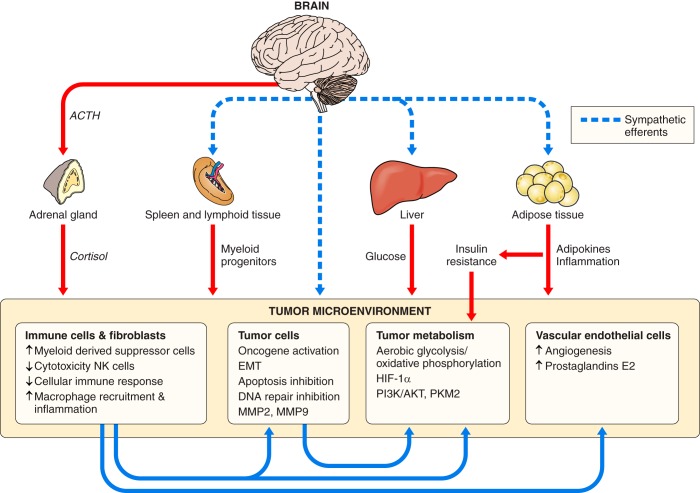
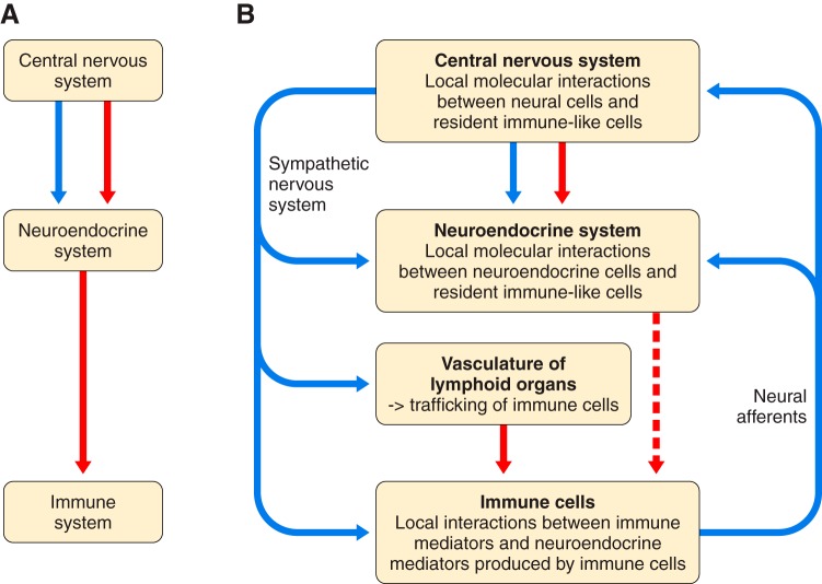
Because of the compartmentalization of disciplines that shaped the academic landscape of biology and biomedical sciences in the past, physiological systems have long been studied in isolation from each other. This has particularly been the case for the immune system. As a consequence of its ties with pathology and microbiology, immunology as a discipline has largely grown independently of physiology. Accordingly, it has taken a long time for immunologists to accept the concept that the immune system is not self-regulated but functions in close association with the nervous system. These associations are present at different levels of organization. At the local level, there is clear evidence for the production and use of immune factors by the central nervous system and for the production and use of neuroendocrine mediators by the immune system. Short-range interactions between immune cells and peripheral nerve endings innervating immune organs allow the immune system to recruit local neuronal elements for fine tuning of the immune response. Reciprocally, immune cells and mediators play a regulatory role in the nervous system and participate in the elimination and plasticity of synapses during development as well as in synaptic plasticity at adulthood. At the whole organism level, long-range interactions between immune cells and the central nervous system allow the immune system to engage the rest of the body in the fight against infection from pathogenic microorganisms and permit the nervous system to regulate immune functioning. Alterations in communication pathways between the immune system and the nervous system can account for many pathological conditions that were initially attributed to strict organ dysfunction. This applies in particular to psychiatric disorders and several immune-mediated diseases. This review will show how our understanding of this balance between long-range and short-range interactions between the immune system and the central nervous system has evolved over time, since the first demonstrations of immune influences on brain functions. The necessary complementarity of these two modes of communication will then be discussed. Finally, a few examples will illustrate how dysfunction in these communication pathways results in what was formerly considered in psychiatry and immunology to be strict organ pathologies.


Figures

FIGURE 1.
FIGURE 1.
Schematic representation of the inflammatory reflex. A: the initial model of the inflammatory reflex that was proposed originally by Tracey (231). Proinflammatory cytokines released by activated innate immune cells activate the afferent vagus nerves. This sensory input activates in a reflex-like manner neuronal cell bodies of the efferent vagus nerves. The resulting activation of the dorsal vagal complex recruits the efferent cholinergic vagus nerves that downregulate inflammation. B: the revised model of the inflammatory reflex. The parasympathetic branch of the vagus nerves activates the splenic nerves. Activation of this sympathetic nerve results in the recruitment of acetylcholine-producing T cells that downregulate inflammation.
FIGURE 2.
FIGURE 2.
Role of opioid-containing leukocytes in the regulation of inflammation-induced pain. Proinflammatory cytokines released by activated innate immune cells sensitize sensory nerve endings, resulting in an amplification and prolongation of the initial pain reaction triggered by activation of nociceptors in response to the injury. The pain response is downregulated by endogenous opioid peptides released by locally infiltrating opioid containing leukocytes in response to corticotropin releasing hormone produced by fibroblasts. [Adapted from Stein et al. (218).]
FIGURE 3.
FIGURE 3.
Immune-to-brain communication pathways. In response to pathogen-associated molecular patterns such as lipopolysaccharide sensed by Toll-like receptors and the inflammasome (not represented in the figure), activated macrophages produce and release proinflammatory cytokines in their microenvironment, including IL-1β (red circles). As presented in A, proinflammatory cytokines were initially supposed to be released in the general circulation and act at the level of circumventricular organs (CVO) where the blood-brain barrier is fenestrated. Proinflammatory cytokines were proposed to induce there the release of lipophilic prostaglandin E2 acting as secondary messengers on neurons. However, the demonstration that immunelike cells in the brain including meningeal and perivascular macrophages and microglia produce and release brain proinflammatory cytokines led to a different representation illustrated in B. The peripheral immune signal is relayed to the brain by afferent nerves. This neural message together with slowly diffusing cytokines produced at the level of circumventricular organs and prostaglandins produced by brain endothelial cells in response to circulating pathogen-associated molecular patters results in the production of brain cytokines by activated microglia. Overspill of cytokines in the general circulation can also be transported for some of them into the brain side of the blood-brain barrier (not represented in the figure).
FIGURE 4.
FIGURE 4.
Mechanisms of inflammation-induced depression. In addition to activation of the immune-to-brain communication pathways represented in FIGURE 3, peripheral proinflammatory cytokines activate the tryptophan metabolizing enzyme indoleamine 2,3-dioxygenase (IDO) that transforms tryptophan into kynurenine. Kynurenine produced at the periphery is transported into the brain where it is further metabolized into neurotoxic kynurenine metabolites including quinolinic acid by another microglia-associated enzyme, kynurenine monooxygenase (KMO). Together with glutamate released by activated microglia, quinolinic acid activates the N-methyl-d-aspartate (NMDA) receptors. Independently of this pathway, inflammatory mediators released by activated microglia and brain endothelial cells can also downregulate dopaminergic neurotransmission via oxidative stress and mitochondrial dysfunction. Activation of NMDA receptors and deficient dopaminergic neurotransmission both result in depression symptoms.
FIGURE 5.
FIGURE 5.
Mechanism of stress-induced tumor progression and dissemination. Stress facilitates the development of immunotolerance by activation of the hypothalamic pituitary adrenal axis which results in decreased cytotoxicity of natural killer cells (NK cells) and reduced cellular immune responses to the tumor antigens. At the same time, activation of the sympathetic nervous system induces profound alterations in tumor cells and in the tumor microenvironment both directly by sympathetic nerve endings innervating the organ in which the tumor develops and indirectly via the liver and adipose tissue. Red arrows represent communication via the general circulation, dashed blue arrows represent communications via nerves, and blue arrows represent communication within the tumor microenvironment. NK, natural killer; EMT, epithelial-to-mesenchymal translation of cancer cells; DNA, deoxyribonucleic acid; MMP, matrix metalloproteinase; HIF-1α, hypoxia inducible factor-1α; PI3/AKT, phosphatidylinositol 3-kinase/protein kinase B signaling pathway; PKM2, M2 isoform of pyruvate kinase. [Adapted from Cole et al. (47).]
FIGURE 6.
FIGURE 6.
Schematic representation of neuroimmune interactions. A: the initial notion that neuroimmune interactions take place via mediators released in the general circulation by the neuroendocrine system. B: summary of the knowledge presented in this review showing that long-distance communication between the central nervous system and the immune system takes place mainly via neural pathways and much less so via circulating neuroendocrine factors. Long-distance communication via neural pathways is bidirectional, from the central nervous system to the immune system and vice versa. Long-distance communication pathways modulate the functioning of local or short-range communication pathways that involve intricate interactions between resident immunelike cells (mainly microglia and astrocytes, but also endothelial cells) and neuronal cells in the nervous system, and between immune mediators and neuroendocrine mediators produced locally by immune cells in the immune system in addition to mediators released locally by nerve endings. The immune phenotype of the microenvironment of the affected organ can be modified by alterations in immune cell trafficking induced by noradrenergic or more rarely cholinergic modulation (not represented in the figure) of the vasculature of lymphoid organs. This is more likely to take place at the periphery than in the nervous system that is protected by the blood-brain barrier even if it can become leaky in some conditions. Blue arrows correspond to neural communication pathways and red arrows to humoral communication pathways.

References

    1. Ader R. Historical perspectives on psychoneuroimmunology. In: Psychoneuroimmunology, Stress, and Infection, edited by Friedman H, Klein TW, Friedman AL. Boca Raton, FL: CRC, 1995, p. 1–21.
    1. Ader R, Cohen N. Behaviorally conditioned immunosuppression. Psychosom Med 37: 333–340, 1975. doi:10.1097/00006842-197507000-00007. - DOI - PubMed
    1. Antoni MH. Psychosocial intervention effects on adaptation, disease course and biobehavioral processes in cancer. Brain Behav Immun 30, Suppl: S88–S98, 2013. doi:10.1016/j.bbi.2012.05.009. - DOI - PMC - PubMed
    1. Armaiz-Pena GN, Allen JK, Cruz A, Stone RL, Nick AM, Lin YG, Han LY, Mangala LS, Villares GJ, Vivas-Mejia P, Rodriguez-Aguayo C, Nagaraja AS, Gharpure KM, Wu Z, English RD, Soman KV, Shahzad MM, Zigler M, Deavers MT, Zien A, Soldatos TG, Jackson DB, Wiktorowicz JE, Torres-Lugo M, Young T, De Geest K, Gallick GE, Bar-Eli M, Lopez-Berestein G, Cole SW, Lopez GE, Lutgendorf SK, Sood AK. Src activation by β-adrenoreceptors is a key switch for tumour metastasis. Nat Commun 4: 1403, 2013. doi:10.1038/ncomms2413. - DOI - PMC - PubMed
    1. Arnoux I, Audinat E. Fractalkine Signaling and Microglia Functions in the Developing Brain. Neural Plast 2015: 689404, 2015. doi:10.1155/2015/689404. - DOI - PMC - PubMed

Publication types