

The Roles of Left Versus Right Anterior Temporal Lobes in Semantic Memory: A Neuropsychological Comparison of Postsurgical Temporal Lobe Epilepsy Patients
- PMID: 29351584
- PMCID: PMC6093325
- DOI: 10.1093/cercor/bhx362
The Roles of Left Versus Right Anterior Temporal Lobes in Semantic Memory: A Neuropsychological Comparison of Postsurgical Temporal Lobe Epilepsy Patients
Abstract
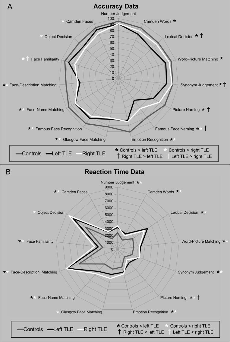
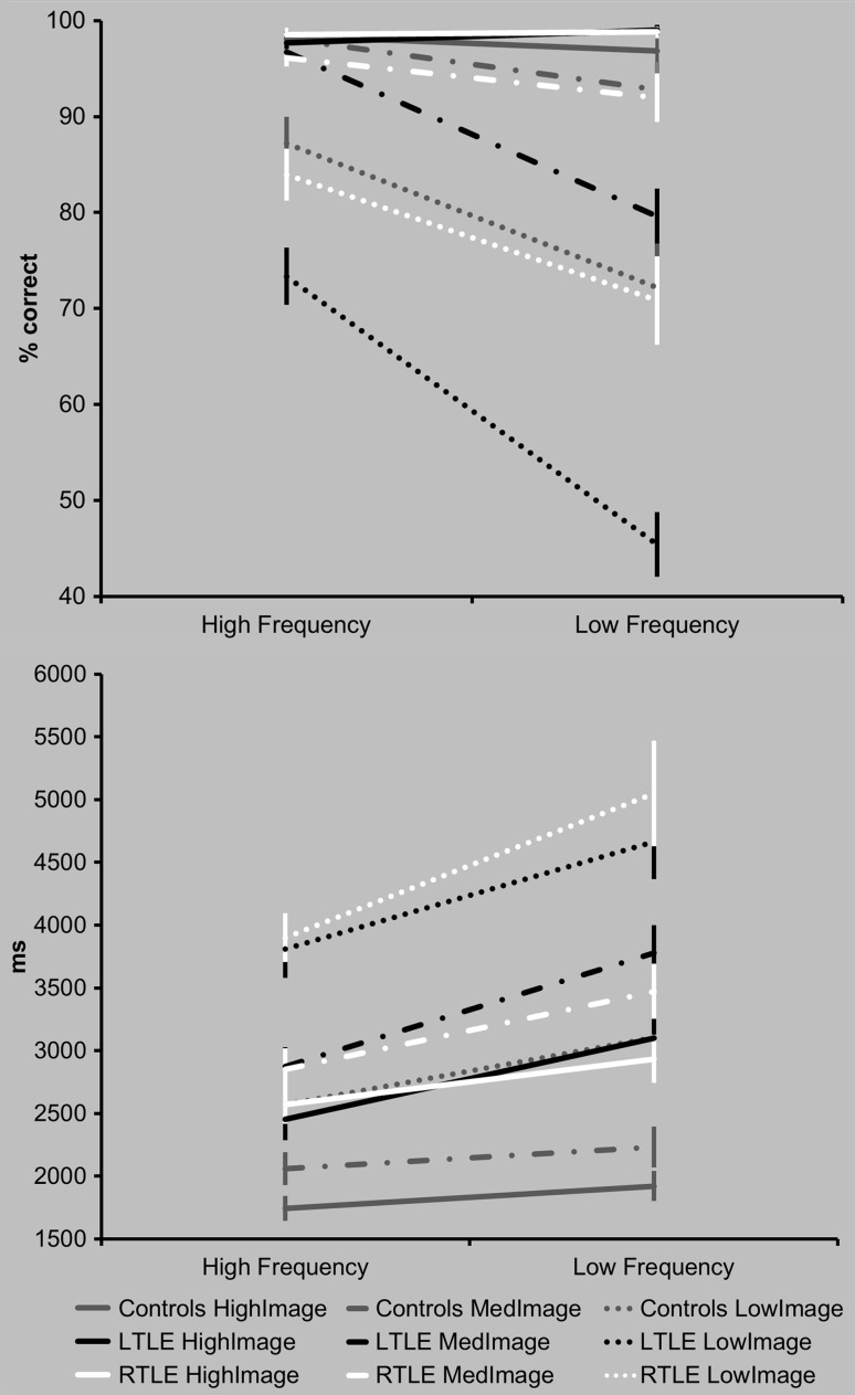
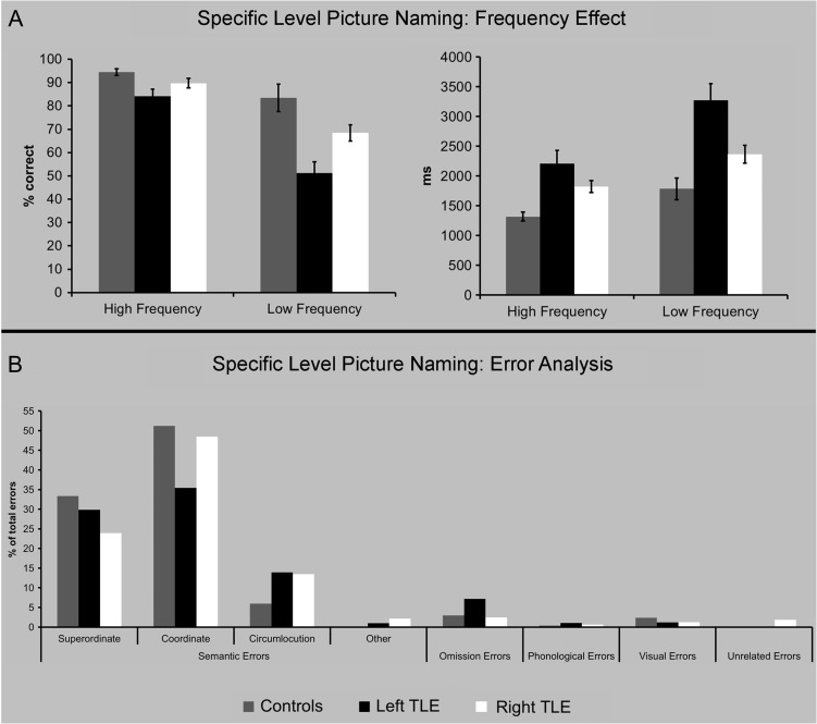
The presence and degree of specialization between the anterior temporal lobes (ATLs) is a key issue in debates about the neural architecture of semantic memory. Here, we comprehensively assessed multiple aspects of semantic cognition in a large group of postsurgical temporal lobe epilepsy (TLE) patients with left versus right anterior temporal lobectomy (n = 40). Both subgroups showed deficits in expressive and receptive verbal semantic tasks, word and object recognition, naming and recognition of famous faces and perception of faces and emotions. Graded differences in performance between the left and right groups were secondary to the overall mild semantic impairment; primarily, left resected TLE patients showed weaker performance on tasks that required naming or accessing semantic information from a written word. Right resected TLE patients were relatively more impaired at recognizing famous faces as familiar, although this effect was observed less consistently. These findings unify previous partial, inconsistent results and also align directly with fMRI and transcranial magnetic stimulation results in neurologically intact participants. Taken together, these data support a model in which the 2 ATLs act as a coupled bilateral system for the representation of semantic knowledge, and in which graded hemispheric specializations emerge as a consequence of differential connectivity to lateralized speech production and face perception regions.
Figures

References
-
- Acres K, Taylor KI, Moss HE, Stamatakis EA, Tyler LK. 2009. Complementary hemispheric asymmetries in object naming and recognition: A voxel-based correlational study. Neuropsychologia 47(8-9):1836–1843. - PubMed
-
- Adlam ALR, Patterson K, Rogers TT, Nestor PJ, Salmond CH, Acosta-Cabronero J, Hodges JR. 2006. Semantic dementia and fluent primary progressive aphasia: two sides of the same coin? Brain. 129:3066–3080. - PubMed
-
- Bi Y, Wei T, Wu C, Han Z, Jiang T, Caramazza A. 2011. The role of the left anterior temporal lobe in language processing revisited: evidence from an individual with ATL resection. Cortex. 47(5):575–587. - PubMed
-
- Binney RJ, Embleton KV, Jefferies E, Parker GJ, Lambon Ralph MA. 2010. The ventral and inferolateral aspects of the anterior temporal lobe are crucial in semantic memory: evidence from a novel direct comparison of distortion-corrected fMRI, rTMS, and semantic dementia. Cereb Cortex. 20(11):2728–2738. - PubMed
-
- Binney RJ, Henry ML, Babiak M, Pressman PS, Santos-Santos MA, Narvid J, Mandelli ML, Strain PJ, Miller BL, Rankin KP, et al. . 2016. Reading words and other people: a comparison of exception word, familiar face and affect processing in the left and right temporal variants of primary progressive aphasia. Cortex. 82:147–163. - PMC - PubMed
Publication types
MeSH terms
Grants and funding
LinkOut - more resources
Full Text Sources
Other Literature Sources
Medical
