Review
. 2018 Sep 1;29(7):667-714.
doi: 10.1089/ars.2017.7225. Epub 2018 Mar 14.

Mitochondrial Uncoupling Proteins: Subtle Regulators of Cellular Redox Signaling

Affiliations
Review

Mitochondrial Uncoupling Proteins: Subtle Regulators of Cellular Redox Signaling

Petr Ježek et al. Antioxid Redox Signal. .

Abstract

Significance: Mitochondria are the energetic, metabolic, redox, and information signaling centers of the cell. Substrate pressure, mitochondrial network dynamics, and cristae morphology state are integrated by the protonmotive force Δp or its potential component, ΔΨ, which are attenuated by proton backflux into the matrix, termed uncoupling. The mitochondrial uncoupling proteins (UCP1-5) play an eminent role in the regulation of each of the mentioned aspects, being involved in numerous physiological events including redox signaling. Recent Advances: UCP2 structure, including purine nucleotide and fatty acid (FA) binding sites, strongly support the FA cycling mechanism: UCP2 expels FA anions, whereas uncoupling is achieved by the membrane backflux of protonated FA. Nascent FAs, cleaved by phospholipases, are preferential. The resulting Δp dissipation decreases superoxide formation dependent on Δp. UCP-mediated antioxidant protection and its impairment are expected to play a major role in cell physiology and pathology. Moreover, UCP2-mediated aspartate, oxaloacetate, and malate antiport with phosphate is expected to alter metabolism of cancer cells.

Critical issues: A wide range of UCP antioxidant effects and participations in redox signaling have been reported; however, mechanisms of UCP activation are still debated. Switching off/on the UCP2 protonophoretic function might serve as redox signaling either by employing/releasing the extra capacity of cell antioxidant systems or by directly increasing/decreasing mitochondrial superoxide sources. Rapid UCP2 degradation, FA levels, elevation of purine nucleotides, decreased Mg2+, or increased pyruvate accumulation may initiate UCP-mediated redox signaling.

Future directions: Issues such as UCP2 participation in glucose sensing, neuronal (synaptic) function, and immune cell activation should be elucidated. Antioxid. Redox Signal. 29, 667-714.

Keywords: UCP2; anion transport; attenuation of superoxide formation; fatty acid cycling; mitochondrial uncoupling proteins; redox signaling.


Figures

<b>FIG. 1.</b>
FIG. 1.
Human UCP isoforms and their tissue distribution. Phylogenetic tree of human UCPs based on their primary amino acid sequence, depicted together with their major tissue/cell distribution; sequences of human UCPs were aligned using ClustalW 2.0 and displayed as rooted phylogenetic tree. Black: existence of protein verified; gray: only mRNA detected. BAT, brown adipose tissue; UCP, uncoupling protein; WAT, white adipose tissue.
<b>FIG. 2.</b>
FIG. 2.
The UCP-catalyzed protonophoretic cycle—ongoing according to the (A) FA cycling or (B) “FA shuttling.” (A) In FA cycling model, FA anion diffuses laterally within the membrane to reach a subsurface peripheral UCP binding site near the matrix (196), where it binds specifically to basic residues Arg60 and Lys271 (depicted as +) (31). The IMM potential drives the carboxylate head group through an electrostatic path composed of basic residues both inside and outside the UCP cavity (31), resulting in a transport of FA anion to the other side of the membrane (vertical arrow), that is, to the ICS-proximal lipid leaflet of ICS membranes (parts of IMM enfolded into cristae). The anion diffuses laterally (horizontal arrows) away from UCP, where it is protonated. Protonated FA diffuses rapidly back across the membrane to deliver protons electroneutrally back to the matrix by a spontaneous flip-flop mechanism, completing the cycle (182). (B) “FA shuttling” mechanism, in fact considering protein as a “carrier” where FA shuttles back and forth (wobbling) (124), actually differs so that the FA molecule cannot diffuse away from the UCP protein and stays in an unspecified way bound to the protein while exposed to the cis or trans side of the membrane either as anion or after protonation. In this case, both anionic and neutral protonated FAs are carried through the UCP protein. However, since the actual binding site was verified to face the lipid bilayer (31), this mechanism is very unlikely. FA, fatty acid; ICS, intracristal space; IMM, inner mitochondrial membrane.
<b>FIG. 3.</b>
FIG. 3.
Nascent FAs for UCP-mediated attenuation of superoxide formation are provided by the redox-activated mitochondrial phospholipase iPLA2γ. The H2O2-activated mitochondrial phospholipase iPLA2γ (PNPLA8) has been identified in mitochondria that allows a direct feedback attenuation of mitochondrial superoxide production (180, 186). Upon redox activation that typically exists at β-oxidation of FAs (+ in the arrowhead), iPLA2γ cleaves IMM phospholipids and releases nascent free FAs, which become cycling substrates enabling UCP2-mediated uncoupling. The consequent partial dissipation of Δp decreases mitochondrial superoxide formation. Moreover, FAs released by iPLA2γ serve as agonists for plasma membrane receptors such as GPR40 (186), hence FA signaling represents an amplified message. Δp, protonmotive force; GPR40, G-protein-coupled receptor-40; iPLA2γ, calcium-independent phospholipase A2γ; OMM, outer mitochondrial membrane; OXPHOS, oxidative phosphorylation.
<b>FIG. 4.</b>
FIG. 4.
Structure of UCP2according to Refs. (31) and (32). (A) UCP2 with depicted basic amino acids responsible for FA transport and purine nucleotide binding. (B) Side view. (C) Top view from the “cytosolic” side, that is, from the ICS side. H1–H6: color-coded transmembrane α-helices. The peripheral basic residues Arg60 and Lys271 are responsible for the binding of the carboxylate head group. Peripheral Arg241 together with residues inside the cavity, including Lys16, Arg88, Lys141, and Arg279 contribute to the flipping of the acidic head group through the protein cavity. In addition, the cavity-lining basic residues Arg88, Lys141 together with Arg185 participate in purine nucleotide binding (31). The structure was derived from the published NMR structure of the mitochondrial UCP2, pdb code 2LCK (32), and processed using Swiss Pdb-Viewer v. 4.1.0 (146) and the PyMOL Molecular Graphics System Version 1.8 Schrodinger, LLC.
<b>FIG. 5.</b>
FIG. 5.
Detailed structure of UCP2 purine nucleotide binding site. Visualization of UCP2 interaction with GDP within the UCP2 central cavity is depicted from the ICS side (from the top). The structure [pdb code 2LCK; (32)] was zoomed at 20 Å sphere with basic amino acid residues responsible for binding of GDP in red. Color coding of transmembrane α-helices is the same as in Figure 4. GDP, guanosine diphosphate.
<b>FIG. 6.</b>
FIG. 6.
Detailed structure of UCP2 FA binding site. Visualization of the first phase of the FA binding to UCP2 (arrow) from the matrix side (from the bottom), zoomed at 20 Å sphere (pdb code 2LCK; (32) with depicted basic amino acids responsible for correct orientation and approaching of the FA (Arg60 and Lys271) and flipping of the FA through the cavity (Arg88, Lys141, and Arg279) in red. Most of the residues act also for anion transport “channel” forming with rather a large potential field (31). Gly19 and Gly281 residues (dark blue) are influenced by conformational changes induced by purine nucleotide binding so that they might inhibit FA anion translocation (31). Color coding of transmembrane α-helices is the same as in Figure 4.
<b>FIG. 7.</b>
FIG. 7.
Localization and structure of the ucp2 gene and ucp2 promoter with important regulatory sites. The ucp2 gene locus is localized in the 11q13 region of the chromosome 11 (black arrow). The eight exons (boxes) are numbered from left to right according to the transcriptional region, including the promoter region at the beginning of the sequence. The black boxes represent the coding regions, the gray boxes represent the noncoding region of the ucp2 gene. The figure was adapted from the Ensembl genome browser for the ucp2 gene (code ENSG00000175567). The promoter region precedes the first noncoding region of the ucp2 gene and contains important binding sites for transcription factors such as Foxa1, E-box1 and 2 (helix-loop-helix protein binding sites), and TRE1 and 2 (thyroid hormone response elements). Ucp2 important polymorphisms namely −866G/A and Ala55Val are depicted as well. FOXA1, forkhead box A1.
<b>FIG. 8.</b>
FIG. 8.
Three types of redox signaling. (A) Retrograde redox signaling from the mitochondrion directed toward the cell cytosol, nucleus, plasma membrane, or other cell components is mostly executed via the H2O2 diffusion or by redox relaying enzymes (329). Besides redox-sensitive kinases and phosphatases (upper part), the prominent receivers of the redox signal are extracellular MMP, redox-sensitive channels, and upon hypoxia also PHD enzymes, which are inhibited, similarly as the FIH, both leading to HIF1-α accumulation and resulting HIF-mediated transcriptome reprogramming. (B) External redox signaling from the cell toward the mitochondrion including H2O2 activation of kinases within the ICS and hypothetical plasma membrane derived signalosomes (133, 336); likewise, redox signaling originating from norepinephrine stimulation of brown adipocytes leading to sulfenylation of Cys253 in UCP1 serves as a clear example (71), and (C) intramitochondrial redox signaling of a short range exists just within the interior of the OMM, forming tubules of mitochondrial network reticulum. A continuous compartment is represented by the matrix space, which is interrupted by the rich cristae. The cristae lumen called ICS represents numerous separate compartments, which are interconnected only via the crista outlets and the outer intermebrane space (a middle part of the sandwich of OMM and IMM). Within the matrix, typical redox regulation is exerted by acetylation of MnSOD, making it inactive, and deacetylation by sirtuin-3, activating MnSOD (390). Also, H2O2-activated PNPLA8 (iPLA2γ) (180, 186) is able to cleave FAOOH (254, 288), which are both substrates of UCP2-catalyzed H+ transport (181) and signaling molecules (162, 299, 300), leading to separate redox-sensitive signaling pathways. Within the ICS, reducing system is represented namely by CuZnSOD and thioredoxin-3 (Trx3). Within the outer intermembrane space (or hypothetically also in ICS), also MIA40/ALR SH-oxidizing protein system shifts the local environment to more prooxidant state (206). All these systems are fed by superoxide from the distinct source, the site IIIQo of complex III, under retardation of cytochrome c shuttling (e.g., upon hypoxic ALR regeneration) and upon hypoxia (376, 426). Arrows: activation; half-open line segment: inhibition. Akt, protein kinase B; FAOOH, FA hydroperoxides; FIH, factor inhibiting HIF; HIF1-α, hypoxia-inducible factor 1-α; GSH, glutathione; MAPK, mitogen-activated protein kinase; MMP, matrix metaloproteinases; MnSOD, superoxide dismutase 2; NOX, NADPH oxidase; OMM, outer mitochondrial membrane; PHD, proline hydroxylase domain; PKC, protein kinase C.
<b>FIG. 9.</b>
FIG. 9.
Sites of mitochondrial superoxide production. Overview of locations for superoxide sources (blue capital fonts) is illustrated, while their assumed relative contribution is expressed by the arrow thickness (purple arrows for superoxide, red arrows for H2O2). The sites of superoxide formation are termed according to the nomenclature introduced by Martin Brand (43), includes six sites acting at the ∼280 mV redox potential of the NADH/NAD+ isopotential pool (index F, flavin) and five sites acting at the ∼20 mV redox potential of the ubiquinol/ubiquinone (QH2/Q) isopotential pool (index Q). Among them, only the sources depending on ΔpΨ) and relying on the smoothed (unretarded) process of forward electron transport within the respiratory chain can be directly attenuated by uncoupling. Specifically, these uncoupling-attenuated sources ensure superoxide formation at the site IQ of complex I and site IIIQo of complex III. In turn, the complex I site IF increases superoxide formation at NADH >> NAD+ (at a higher substrate pressure NADH/NAD+). Typically, in pathological conditions, allowing reverse electron transfer to the complex I site IQ, this site produces the majority of superoxide; when pathology retards cytochrome c shuttling (orange elliptic arrow), the complex III site IIIQo provides major superoxide formation. The latter superoxide formation is not attenuated by uncoupling. αGPDH, α glycerolphosphate dehydrogenase; αKGDH, α ketoglutarate dehydrogenase; DH, dehydrogenase; DhODH, dihydroorotate dehydrogenase; ETF:QOR, electron-transferring-flavoprotein:ubiquinone oxidoreductase; MAO, monoaminooxidase; NAD+, nicotinamide adenine dinucleotide.
<b>FIG. 10.</b>
FIG. 10.
Influence of mild uncoupling on superoxide production. (A) Higher rates of respiration and proton pumping promoted by mild uncoupling attenuate superoxide formation—specifically at complexes I and III. (B) Regulated UCP inhibition/inactivation at simultaneous maximum ATP synthesis reaches so-called respiratory control establishing the slow electron transfer (respiration) and slow proton pumping, both promoting higher superoxide formation namely at complexes I and III. (C) Electron transfer blockage or slowdown such as resulting from retardation of cytochrome c cycling between complexes III and IV cannot be influenced by uncoupling and typically leads to a high complex III superoxide formation at site IIIQo. Alternatively, at pathological succinate accumulation leading to reverse electron transfer (“RET”), a high superoxide formation exists at the IQ site of complex I. Thicker arrows indicate higher fluxes, dashed lines represent medium fluxes, and dotted lines represent the absence of fluxes. Orientation: bottom parts represent the matrix; upper parts represent the ICS.
<b>FIG. 11.</b>
FIG. 11.
UCP2-mediated anion efflux from the matrix may substitute metabolite carriers within redox shuttles. Such efflux is driven by the ΔΨm component of Δp (similarly to the ATP3−/ADP2− antiport enabled by the ADP/ATP carrier), hence partially dissipates Δp. (A) Aspartate export (“Asp”) in a synergy with the 2-oxoglutarate carrier (“2OGC”) substituting the malate aspartate carrier within the malate/aspartate redox shuttle. (B) Malate export (“Asp”) in a synergy with the pyruvate carrier (“PyrC”) may enable so-called pyruvate/malate redox shuttle with participating cytocolic malic enzyme 1 (“ME1”) and matrix ME2. (C) Oxaloacetate export in a synergy with the 2-oxoglutarate carrier. (D) Pyruvate cycling dissipating whole Δp, enabled by the UCP2-mediated pyruvate uniport and pyruvate proton symport by the pyruvate carrier. ΔΨm, mitochondrial inner membrane potential; ALT, alanine aminotransferase; AST, aspartate aminotransferase; MDH, malate dehydrogenase; OAA, oxaloacetate.
<b>FIG. 12.</b>
FIG. 12.
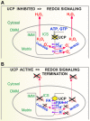
Roles for UCPs in cellular redox signaling. (A) Switching off the UCP protonophoretic function initiates novel mitochondrial retrograde or internal redox signaling by directly increasing mitochondrial superoxide production, or else promotes the ongoing cell redox signaling by decreasing the capacity of cell antioxidant systems that had to buffer the excessive mitochondrial ROS arising due to the absence of UCP2-mediated antioxidant protection. Primary superoxide is reduced to the signaling H2O2 by means of MnSOD (in the matrix) or CuZnSOD (in the ICS/intermembrane space). (B) Switching on the UCP protonophoretic function terminates the redox signaling by directly decreasing of mitochondrial superoxide production or attenuates cell redox signaling by increasing the spare capacity of cell antioxidant systems. ROS, reactive oxygen species.
<b>FIG. 13.</b>
FIG. 13.
Calcium transport is affected by UCP2 and UCP3. Participation of UCP2 (UCP3) in concert with methylation of MICU1 protein controlling MCU in complex with EMRE protein. Left panels—in the UCP absence: the protein arginine methyl transferase PRMT1 methylates the MICU1 protein at position 455, thus reducing its sensitivity to Ca2+. Right panels—in the UCP presence: the sensitivity of the Ca2+ uniport becomes unaffected by PRMT1. The scheme was derived from results in Ref. (260). EMRE, essential MCU regulator; MCU, mitochondrial calcium uniporter; MICU1, methylation of mitochondrial Ca2+ uptake 1; PRMT1, protein arginine methyl transferase 1.
<b>FIG. 14.</b>
FIG. 14.
UCP2 role in promoting fragmentation of mitochondrial network. (A) Completely continuous mitochondrial network reticulum is established when fission/fragmentation is balanced by fusion and DRP1 protein is not fully recruited toward the outer membrane. The UCP2 blockage helps to set the balance. (B) Switching on the UCP2-mediated uncoupling may set ΔΨm below a threshold required for activation of OMA1-mediated cleavage of profusion protein OPA1, and by enabling lower GTP for mitofusin 1 (“MFN1”) and mitofusin 2 GTP-ases (“MFN2”). (C, D) 3D 4Pi microscopic images of insulinoma INS1E cell mitochondrial network at continuous or fragmented morphology [measurements similar to those in Ref. (330)]. Grids: 1 μm.
<b>FIG. 15.</b>
FIG. 15.
UCP3 in cardioprotection against I/R injury. (A) Superoxide formed at instant reperfusion: high succinate accumulated in ischemic period is metabolized fast so to promote reverse electron transfer from succinate dehydrogenase (complex “II”) toward the IQ site of complex I, where a high superoxide formation occurs (73, 74, 103). (B) UCP3-mediated mild uncoupling is able to redirect electron transfer to the forward one: similarly, as depicted in Figure 3A, higher rates of respiration and proton pumping promoted by mild uncoupling attenuate superoxide formation at complex I IQ site. Derived on the basis of results in Refs. (313) and (325, 392). Thicker arrows indicate higher fluxes and dotted lines illustrate the absence of fluxes. Orientation: bottom parts represent the matrix; upper parts represent the ICS. I/R, ischemia/reperfusion.
<b>FIG. 16.</b>
FIG. 16.
UCP2 promoting tumorigenesis. Scheme of major cancer cell pathways including glycolysis, glutaminolysis, and oxidative phosphorylation, emphasizing synthesis and utilization of NADH/NAD+ and NADPH/NADP+ together with UCP2-promoted glutaminolysis. The latter occurs probably by UCP2-mediated enhancement of the aspartate efflux from the matrix, leading to suppression of glycolytic influx to the Krebs cycle by export of malate, oxaloacetae (not shown), or aspartate from the matrix. Major metabolic fluxes and their absence (fading symbols and arrows) or decrease (smaller symbols and thinner arrows) are depicted. In cancer cells, a Krebs cycle segment is largely unused that includes aconitase (“ACO”) and isocitrate dehydrogenase 3 (“IDH3”), consequently supplying less NADH for mitochondrial complex I (“CI”) of the respiratory chain (“CIII,”“CIV”), which includes also succinate dehydrogenase (“CII SDH”) of the Krebs cycle. Glutaminolytic pathway (purple arrows) supplies 2-oxoglutarate (2OG) for 2OG dehydrogenase (“2OGDH”), thus allowing pyruvate to be metabolized by mitochondria, besides for intensive lactate production. Pyruvate dehydrogenase (“PDH”) is nearly often completely inhibited by phosphorylation in cancer. Dependent on metabolic shuttles, malate (“MAL”) is either imported into the matrix or exported out. The malate export or diversion from the Krebs cycle allows malic enzyme (“ME”) reaction producing NADPH. The malate diversion efficiently leads to a lower NADH production by malate dehydrogenase (“MDH”). At sufficient Δp created by the respiratory chain proton pumping, also transhydrogenase (“TH”) synthesizes NADPH at the expense of NADH. Glutamate dehydrogenase (“GDH”) reaction, which also produces NADPH, is largely inhibited in glutaminolytic cancer cells by GTP. ACL, ATP citrate lyase; ASCT, neutral amino acid exchanger; ASP, aspartate; Cit C, mitochondrial citrate carrier; CS, citrate synthase; FH, fumarate hydratase; FASN, fatty acid synthetase; Gln C, mitochondrial glutamine carrier; GLUT, glucose transporter; MAC, mitochondrial malate aspartate carrier; MCT4, lactate transporter; OAA, oxaloacetate; OGC, oxoglutarate carrier; Pyr C, mitochondrial pyruvate carrier; S-CoA, succinyl coenzyme A; SN2, glutamine transporter. Scheme was drawn according to Refs. (415) and (378).
<b>FIG. 17.</b>
FIG. 17.
UCP2 roles in macrophage activation. UCP2 is highly expressed in resting macrophage. After LPS stimulation, glucose oxidation increases on the expense of glutaminolysis, due to MAPK pathway/mediated downregulation of UCP2 expression. This is communicated by p38 and JNK information signaling pathways leading to suppression of UCP2 expression. Low glutamine levels stop promoting UCP2 mRNA translation. Resulting higher mitochondrial H2O2 release due to blockage of the UCP2-mediated antioxidant action further stimulate the ERK and NFκB pathways providing the more strengthening downregulation of UCP2 expression. The scheme was modified from Ref. (119). TLR, toll-like receptor. Arrows, ongoing regulations or fluxes; dotted arrows, slow or inhibited regulations or fluxes; dotted boxes.

Similar articles

Cited by

References

    1. Abrahams JP, Leslie AGW, Lutter R, and Walker JE. Structure at 2.8 Â resolution of F1-ATPase from bovine heart mitochondria. Nature 370: 621–628, 1994 - PubMed
    1. Adams AE, Carroll AM, Fallon PG, and Porter RK. Mitochondrial uncoupling protein 1 expression in thymocytes. Biochim Biophys Acta 1777: 772–776, 2008 - PubMed
    1. Adams AE, Hanrahan O, Nolan DN, Voorheis HP, Fallon P, and Porter RK. Images of mitochondrial UCP 1 in mouse thymocytes using confocal microscopy. Biochim Biophys Acta 1777: 115–117, 2008 - PubMed
    1. Adams AE, Kelly OM, and Porter RK. Absence of mitochondrial uncoupling protein 1 affects apoptosis in thymocytes, thymocyte/T-cell profile and peripheral T-cell number. Biochim Biophys Acta 1797: 807–816, 2010 - PubMed
    1. Affourtit C. and Brand MD. Uncoupling protein-2 contributes significantly to high mitochondrial proton leak in INS-1E insulinoma cells and attenuates glucose-stimulated insulin secretion. Biochem J 409: 199–204, 2008 - PubMed

Publication types

Substances