. 2018 Jan 19;18(1):20.
doi: 10.1186/s12906-018-2088-x.

Combination of Pelargonium sidoides and Coptis chinensis root inhibits nuclear factor kappa B-mediated inflammatory response in vitro and in vivo

Affiliations

Combination of Pelargonium sidoides and Coptis chinensis root inhibits nuclear factor kappa B-mediated inflammatory response in vitro and in vivo

Sang Mi Park et al. BMC Complement Altern Med. .

Abstract

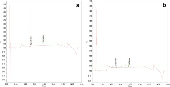
Background: Pelargonium sidoides (PS) and Coptis chinensis root (CR) have traditionally been used to treat various diseases, including respiratory and gastrointestinal infections, dysmenorrhea, and hepatic disorders. The present study was conducted to evaluate the anti-inflammatory effects of a combination of PS and CR in vitro and in vivo.

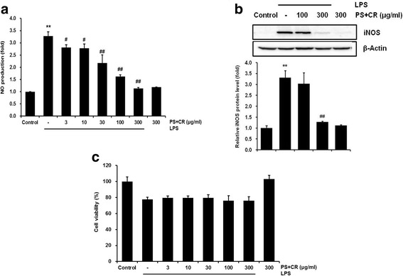
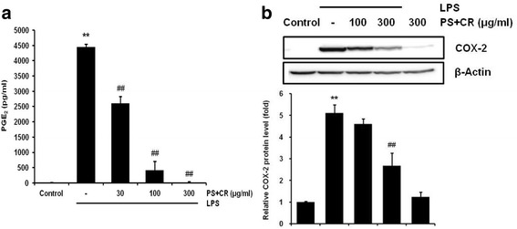
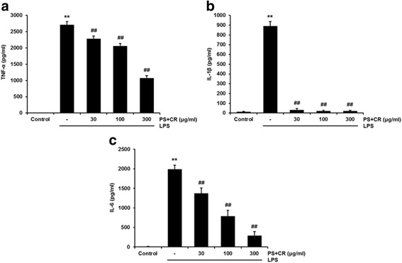
Methods: The in vitro effects of PS + CR on the induction of inflammation-related proteins were evaluated in lipopolysaccharide (LPS)-stimulated RAW 264.7 cells. The levels of nitric oxide (NO) and of inflammatory cytokines and prostaglandin E2 (PGE2) were measured using the Griess reagent and enzyme-linked immunosorbent assay (ELISA) methods, respectively. The expression of inflammation-related proteins was confirmed by Western blot. Additionally, the effects of PS + CR on paw edema volume, skin thickness, and numbers of infiltrated inflammatory cells, mast cells, COX-2-, iNOS-, and TNF-α-immunoreactive cells in dorsum and ventrum pedis skin were evaluated in a rat model of carrageenan (CA)-induced paw edema.

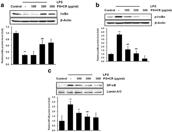
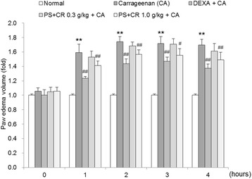
Results: PS + CR significantly reduced production of NO, PGE2 and three pro-inflammatory cytokines (tumor necrosis factor-α (TNF-α), interleukin (IL)-1β, and IL-6) and also decreased levels of inducible nitric oxide synthase (iNOS) and cyclooxygenase-2 (COX-2). Treatment with PS + CR significantly reduced the protein expression levels of LPS-stimulated nuclear factor kappa B (NF-κB) and phosphorylated inhibitor of NF-κB (p-I-κBα). Additionally, PS + CR significantly inhibited the increases in paw swelling, skin thickness, infiltrated inflammatory cells, mast cell degranulation, COX-2-, iNOS-, and TNF-α-immunoreactive cells in the rat model of CA-induced acute edematous paw.

Conclusions: These results demonstrate that PS + CR exhibits anti-inflammatory properties through decreasing the production of pro-inflammatory mediators (NO, PGE2, TNF-α, IL-1β, and IL-6), suppressing NF-κB signaling in LPS-induced RAW 264.7 cells. Additionally, the results of the CA-induced rat paw edema assay revealed an anti-edema effect of PS + CR. Furthermore, it is suggested that PS + CR also inhibits acute edematous inflammation by suppressing mast cell degranulation and inflammatory mediators (COX-2, iNOS, and TNF-α). Thus, PS + CR may be a potential candidate for the treatment of various inflammatory diseases, and it may also contribute to a better understanding of the molecular mechanisms underlying inflammatory response regulation.

Keywords: Coptis chinensis root; Histopathology; Inflammation; Nuclear factor kappa B; Paw edema; Pelargonium sidoides.


Conflict of interest statement

Ethics approval

All animal experiments were approved by the Animal Ethical Care Committee of Daegu Haany University (Approval number: DHU2016–089) and conducted in accordance with the guidelines for the care and use of laboratory animals at Daegu Haany University.

Consent for publication

Not applicable.

Competing interests

The authors declare that they have no competing interests.

Publisher’s Note

Springer Nature remains neutral with regard to jurisdictional claims in published maps and institutional affiliations.

Figures

Fig. 1
Fig. 1
Analysis of compounds in a mixture of Pelargonium sidoides and Coptis chinensis root (PS + CR) by ultra-performance liquid chromatography (UPLC). UPLC chromatograms of standard compounds; epicatechin: 230 nm, berberine: 345 nm. a standard compounds. b PS + CR extracts
Fig. 2
Fig. 2
Inhibition of nitric oxide (NO) production and inducible nitric oxide synthase (iNOS) expression by PS + CR. RAW 264.7 cells were treated with 3–300 μg/ml PS + CR for 1 h prior to the addition of lipopolysaccharide (LPS) (1 μg/ml) and further incubated for 24 h. a Effects of PS + CR on NO production. Secreted nitrite concentrations in the culture medium were assayed using the Griess reagent method. b Effects of PS + CR on iNOS protein expression. Equal amounts of total protein (50 μg) were separated by SDS-PAGE. β-Actin was used as a loading control, and the bar chart shows the quantitative evaluation of iNOS bands by densitometry. (c) Effects of PS + CR plus LPS on cell viability. Cell viability was measured by 3-(4,5-dimethylthiazol)-2,5-diphenyltetrazolium bromide (MTT) assay after 24 h of incubation. Values represent the mean ± S.D. of three independent experiments (significant compared to the control, **p < 0.01; significant compared to LPS alone, #p < 0.05 or ##p < 0.01)
Fig. 3
Fig. 3
Inhibition of the LPS-induced prostaglandin E2 (PGE2) secretion and cyclooxygenase-2 (COX-2) expression by PS + CR. RAW 264.7 cells were treated with 30–300 μg/ml of PS + CR for 1 h prior to the addition of LPS (1 μg/ml) and further incubated for 24 h. a Effects of PS + CR on the levels of PGE2. PGE2 concentrations in the culture medium were assayed using the PGE2 ELISA kit as detailed in the Methods section. b Effects of PS + CR on COX-2 protein expression. Equal amounts of total protein (50 μg) were separated by SDS-PAGE. β-Actin was used as a loading control, and the bar chart shows the quantitative evaluation of COX-2 bands by densitometry. Values represent the mean ± S.D. of three independent experiments (significant compared to the control, **p < 0.01; significant compared to LPS alone, ##p < 0.01)
Fig. 4
Fig. 4
Inhibition of the LPS-induced secretion of pro-inflammatory cytokines by PS + CR. RAW 264.7 cells were treated with 30–300 μg/ml of PS + CR for 1 h prior to the addition of LPS (1 μg/ml) and further incubated for 24 h. Levels of TNF-α (a) IL-1β (b) and IL-6 (c). As detailed in the Methods section, the concentrations of cytokines in the culture medium were assayed using a kit. Values represent the mean ± S.D. of three independent experiments (significant compared to the control, **p < 0.01; significant compared to LPS alone, ##p < 0.01)
Fig. 5
Fig. 5
Inhibition of LPS-induced nuclear factor-kappa B (NF-κB) protein expression by PS + CR. (a, b) Levels of I-κBα and phospho-I-κBα. RAW 264.7 cells were treated with 100 and 300 μg/ml of PS + CR for 1 h prior to the addition of lipopolysaccharide (LPS) (1 μg/ml) and further incubated for 30 min. Cytosol fraction proteins were separated by SDS-PAGE. β-Actin was used as a loading control, and the bar chart shows the quantitative evaluation of I-κBα and phospho-I-κBα bands by densitometry. (c) The levels of NF-κB. RAW 264.7 cells were treated with 100 and 300 μg/ml of PS + CR for 1 h prior to the addition of LPS (1 μg/ml) and further incubated for 1 h. Nuclear fraction proteins were separated by SDS-PAGE. Lamin A/C was used as a loading control, and the bar chart shows the quantitative evaluation of NF-κB bands by densitometry. Values represent the mean ± S.D. of three independent experiments (significant compared to the control; **p < 0.01, significant compared to LPS alone, #p < 0.05 or ##p < 0.01)
Fig. 6
Fig. 6
Inhibition of carrageenan (CA)-induced paw edema by PS + CR. PS + CR was orally administered to rats at 0.3 or 1.0 g/kg/day for four days prior to the induction of paw edema. Paw edema was induced by subcutaneous injection of a 1% solution of CA dissolved in saline (0.1 ml per animal) into the right hind paw of each rat. The swelling volume of the paw was measured up to 4 h after the CA injection at intervals of 1 h using a plethysmometer. Dexamethasone (DEXA) (1 mg/kg, p.o.) was used as a positive control. Values represent the mean ± S.D. of five animals (significant compared to the normal, **p < 0.01; significant compared to CA alone, #p < 0.05 or ##p < 0.01)
Fig. 7
Fig. 7
Inhibition of increased paw skin thickness and infiltrating inflammatory cells by PS + CR. Histological profiles and infiltrated inflammatory cells in the dorsum pedis skin (a) and ventrum pedis skin (b). CA treatment markedly increased skin thickness (arrow) and inflammatory cell infiltration due to edematous changes. Scale bars = 60 μm
Fig. 8
Fig. 8
Representative immunohistochemical profiles of COX-2, iNOS, and TNF-α in paw skin. Marked increases of COX-2, iNOS, and TNF-α-positive cells were detected in the epithelium and dermis of the dorsum (a) and ventrum pedis skin tissues (b) in CA-treated rats compared with normal rats. Scale bars = 60 μm

References

    1. Zhang X, Mosser DM. Macrophage activation by endogenous danger signals. J Pathol. 2008;214:161–178. doi: 10.1002/path.2284. - DOI - PMC - PubMed
    1. Jang KJ, Choi SH, Yu GJ, Hong SH, Chung YH, Kim CH, Yoon HM, Kim GY, Kim BW, Choi YH. Anti-inflammatory potential of total saponins derived from the roots of Panax Ginseng in lipopolysaccharide-activated RAW 264.7 macrophages. Exp Ther Med. 2016;11:1109–1115. doi: 10.3892/etm.2015.2965. - DOI - PMC - PubMed
    1. Kim SY, Park SM, Hwangbo M, Lee JR, Byun SH, Ku SK, Cho IJ, Kim SC, Jee SY, Park SJ. Cheongsangbangpung-tang ameliorated the acute inflammatory response via the inhibition of NF-kappaB activation and MAPK phosphorylation. BMC Complement Altern Med. 2017;17:46. doi: 10.1186/s12906-016-1501-6. - DOI - PMC - PubMed
    1. Fitzpatrick FA. Inflammation, carcinogenesis and cancer. Int Immunopharmacol. 2001;1(9–10):1651–1667. doi: 10.1016/S1567-5769(01)00102-3. - DOI - PubMed
    1. Kapadia M, Sakic B. Autoimmune and inflammatory mechanisms of CNS damage. Prog Neurobiol. 2011;95(3):301–333. doi: 10.1016/j.pneurobio.2011.08.008. - DOI - PubMed

LinkOut - more resources