. 2018 Apr 1;78(7):1713-1725.
doi: 10.1158/0008-5472.CAN-17-1423. Epub 2018 Jan 19.

Targeting the SphK1/S1P/S1PR1 Axis That Links Obesity, Chronic Inflammation, and Breast Cancer Metastasis

Affiliations

Targeting the SphK1/S1P/S1PR1 Axis That Links Obesity, Chronic Inflammation, and Breast Cancer Metastasis

Masayuki Nagahashi et al. Cancer Res. .

Abstract

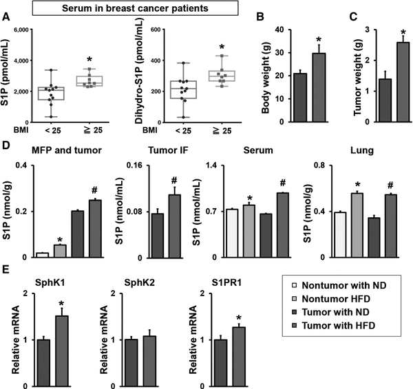
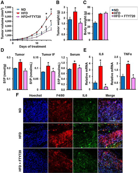
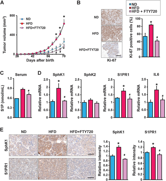
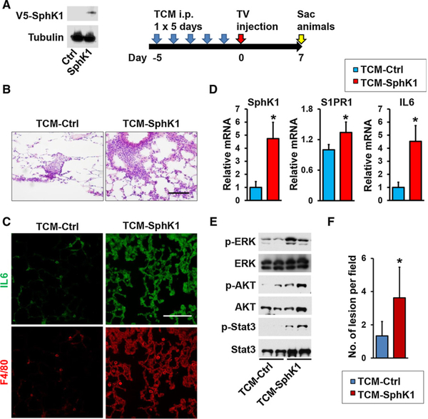
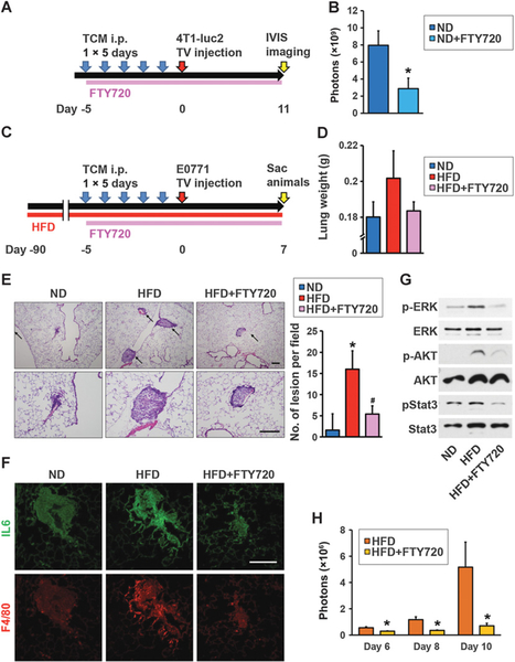
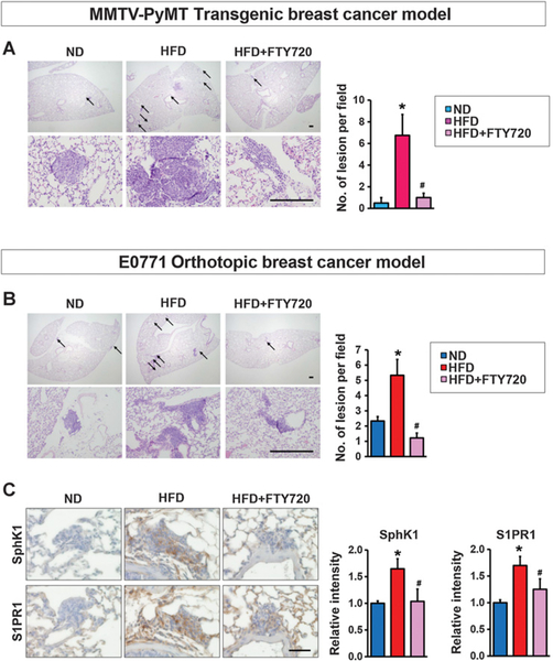
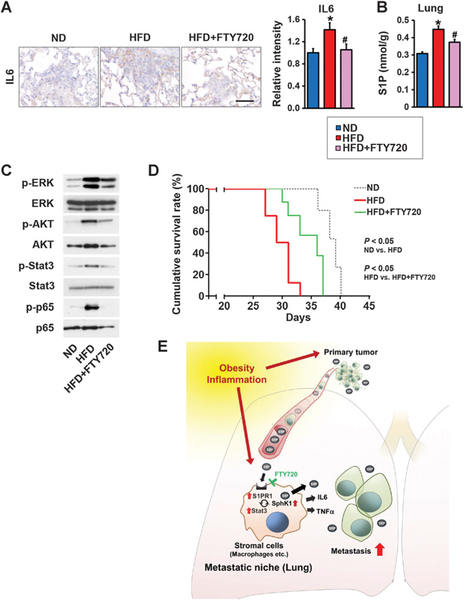
Although obesity with associated inflammation is now recognized as a risk factor for breast cancer and distant metastases, the functional basis for these connections remain poorly understood. Here, we show that in breast cancer patients and in animal breast cancer models, obesity is a sufficient cause for increased expression of the bioactive sphingolipid mediator sphingosine-1-phosphate (S1P), which mediates cancer pathogenesis. A high-fat diet was sufficient to upregulate expression of sphingosine kinase 1 (SphK1), the enzyme that produces S1P, along with its receptor S1PR1 in syngeneic and spontaneous breast tumors. Targeting the SphK1/S1P/S1PR1 axis with FTY720/fingolimod attenuated key proinflammatory cytokines, macrophage infiltration, and tumor progression induced by obesity. S1P produced in the lung premetastatic niche by tumor-induced SphK1 increased macrophage recruitment into the lung and induced IL6 and signaling pathways important for lung metastatic colonization. Conversely, FTY720 suppressed IL6, macrophage infiltration, and S1P-mediated signaling pathways in the lung induced by a high-fat diet, and it dramatically reduced formation of metastatic foci. In tumor-bearing mice, FTY720 similarly reduced obesity-related inflammation, S1P signaling, and pulmonary metastasis, thereby prolonging survival. Taken together, our results establish a critical role for circulating S1P produced by tumors and the SphK1/S1P/S1PR1 axis in obesity-related inflammation, formation of lung metastatic niches, and breast cancer metastasis, with potential implications for prevention and treatment.Significance: These findings offer a preclinical proof of concept that signaling by a sphingolipid may be an effective target to prevent obesity-related breast cancer metastasis. Cancer Res; 78(7); 1713-25. ©2018 AACR.


Conflict of interest statement

Disclosure of Potential Conflicts of Interest

No potential conflicts of interest were disclosed.

Figures

Figure 1.
Figure 1.
Obesity increased circulating levels of S1P in humans and mice with breast cancer. A, S1P and dihydro-S1P levels in serum from preoperative breast cancer patients with BMI < 25 kg/m2 (n = 11), or BMI 25 kg/m2 (n = 8) were measured by LC-ESI-MS/MS. The S1P and dihydro-S1P levels are shown in the box plot. The central rectangle spans the first quartile to the third quartile. A segment inside the rectangle shows the median and “whiskers” above and below the box shows the value of the minimum and maximum. All data points are also shown as dots. *, P < 0.05. B–E, The SphK1/S1P/S1PR1 axis in HFD promoted breast cancer progression. B, Prior to implantation of E0771 cells into the chest mammary fat pad under direct vision, C57Bl/6 mice were fed ND or HFD for 12 weeks and body weight was measured. C, Tumors were harvested 30 days after the implantation, and the tumor weight was measured. Data are means ± SEM. *, P < 0.05. D,, Levels of S1P in mammary fat pad (MFP) and breast tumors, tumor IF, serum without or with breast tumors, and lung without or with breast tumors from mice fed with ND or HFD were measured by LC-ESI-MS/MS. E,, Expression of Sphk1, Sphk2, and S1pr1 in breast tumors was determined by qPCR and normalized to Gapdh mRNA. Data are expressed as means ± SEM. *, P < 0.05.
Figure 2.
Figure 2.
The SphK1/S1P/S1PR1 axis connects obesity, chronic inflammation, and breast cancer progression. A–E, Mice were treated as above and when tumors reached 5 mm in diameter, HFD-fed mice were treated by gavage daily with PBS or FTY720 (1 mg/kg). A, Tumor volumes were measured on the indicated days. B–E, After treatment for 18 days, tumors were harvested and tumor and body weights determined (B and C). D, Levels of S1P in breast tumors, tumor IF, and serum were measured by LC-ESI-MS/MS. E, Tnfα and IL6 mRNA levels in tumors were determined by qPCR and normalized to Gapdh mRNA. Data are expressed as means ± SEM. *, P < 0.05 versus ND; #, P < 0.05 versus HFD. F, Immunofluorescence analysis of tumors stained for IL6 (green), F4/80 (red), and Hoechst (blue). Scale bar, 100 μm.
Figure 3.
Figure 3.
Targeting the SphK1/S1P/S1PR1 axis with FTY720 mitigates HFD-induced inflammation and tumorigenesis in MMTV-PyMT transgenic mice. A, Beginning at 3 weeks of age, MMTV-PyMT transgenic mice were fed ND or HFD and treated daily by gavage with PBS or FTY720 (1 mg/kg). A, Spontaneous tumor sizes were determined on the indicated days. B, Tumor sections were stained with Ki67 antibody and percent Ki67–positive cells determined. Scale bar, 50 μm. C, Levels of S1P in serum were measured by LC-ESI-MS/MS. D, Expression of Sphk1, Sphk2, S1pr1, and IL6 in breast tumors was determined by qPCR and normalized to Gapdh mRNA. E, Breast tumors were immunostained with anti-SphK1 or anti-S1PR1. Scale bar, 50 μm. Relative intensity of the immunostaining was quantified. Data are expressed as means ± SEM. *, P < 0.05 versus ND; #, P < 0.05 versus HFD.
Figure 4.
Figure 4.
S1P in tumor conditioned medium increased macrophage recruitment and induced factors and signaling pathways important for lung premetastatic niches. A, Equal amounts lysates of E0771 cells with SphK1 overexpression or control (Ctrl) analyzed by immunoblotting with the indicated antibodies (left). Schematic overview of regimen for examination of premetastatic niche formation in the lung utilizing TCM (right). B–E, Mice were treated with TCM from Sphk1-overexpressing E0771 cells (TCM-Sphk1) or control E0771 cells (TCM-Ctrl) for 5 days, followed by systemic tumor challenge. Lungs were harvested 7 days later. B, H&E staining of lung sections. Scale bar, 100 μm. C, Immunofluorescence analysis of lung sections stained for IL6 (green), F4/80 (red), and Hoechst (blue). Scale bar, 100 μm. D, Expression of Sphk1, S1pr1, and IL6 mRNA in lungs determined by qPCR and normalized to levels of Gapdh mRNA. Data are means ± SEM. *, P < 0.05. E, Equal amounts of lung lysates analyzed by immunoblotting with the indicated antibodies. F, Quantitation of metastatic lesions in the mouse model with TV injection of 4T1-luc2. Data are means ± SD. *, P < 0.05 versus TCM-Ctrl.
Figure 5.
Figure 5.
FTY720 suppressed HFD-induced inflammation and lung-seeding ability of breast cancer cells. A, Schematic overview of treatment regimen for lung colonization. Mice were fed ND and treated with TCM from Sphk1-overexpressing 4T1-luc2 cells (TCM-Sphk1) for 5 days and also daily treated orally with PBS or with FTY720 (1 mg/kg) as indicated. At day 6, 4T1 cells were injected intravenously and tumor burden was quantified by in vivo bioluminescence on the indicated days. B, Tumor burden of mice with tail vein injection of 4T1-luc2 cells treated without or with FTY720 quantified by in vivo bioluminescence on day 11. Data are means ± SEM. *, P < 0.05 versus ND. C, Schematic overview of treatment regimen for lung colonization. Mice were fed ND or HFD for 12 weeks and treated with TCM from Sphk1-overexpressing E0771 cells (TCM-Sphk1) for 5 days and also daily treated orally with PBS or with FTY720 (1 mg/kg) as indicated. At day 6, E0771 cells were injected intravenously and 7 days later, lungs were harvested. D, Lung weights. E, H&E staining of lung sections. Top and bottom panels show lower (×40) and higher (×100) magnifications, respectively. Arrows, cancer cell–seeding lesions. Scale bar, 200 μm. Quantitation of cancer cell–seeding lesions. Data are means ± SEM. *, P < 0.05 versus ND; #, P < 0.05 versus HFD. F, Immunofluorescence analysis of lung sections stained for IL6 (green) and F4/80 (red). Scale bar, 100 μm. G, Equal amounts of lung lysates analyzed by immunoblotting, with the indicated antibodies. H, Tumor burden of HFD-fed mice with tail vein injection of 4T1-luc2 cells treated without or with FTY720 quantified by in vivo bioluminescence on the indicated days. Data are means ± SEM. *, P < 0.05 versus HFD, respectively.
Figure 6.
Figure 6.
FTY720 suppresses lung metastasis in HFD-fed MMTV-PyMT transgenic and E0771 syngeneic orthotopic breast cancer mice. A, MMTV-PyMT transgenic mice were fed with ND or HFD and treated daily by gavage with PBS or FTY720 (1 mg/kg) as indicated. Lungs were harvested when mice were 10 weeks old. C, H&E staining of lung sections. Top and bottom panels show lower (×20) and higher (×200) magnifications, respectively. Arrows, metastatic lesions. Scale bar, 100 μm. Quantitation of metastatic lesions. Data are means ± SEM. *, P < 0.05 versus ND; #, P < 0.05 versus HFD. B and C, C57Bl/6 mice were fed with ND or HFD for 12 weeks, before E0771 cells were implanted into the chest mammary fat pad under direct vision. When tumor sizes reached 5 mm in diameter, mice fed HFD were randomized into two groups and treated by gavage with PBS or FTY720 (1 mg/kg/day). Lungs were examined 18 days later. B, H&E staining of lung sections. Top and bottom panels show lower (×20) and higher (×200) magnifications, respectively. Arrows, metastatic lesions. Scale bar, 100 μm. Quantitation of metastatic lesions. Data are means ± SEM. *, P < 0.05 versus ND; #, P < 0.05 versus HFD. C, Lung sections were immunostained with anti-SphK1 or anti-S1PR1. Scale bar, 50 μm. Relative intensity of immunostaining was quantified. Data are means ± SEM. *, P < 0.05 versus PBS; #, P < 0.05 versus HFD.
Figure 7.
Figure 7.
FTY720 mitigates obesity-related lung inflammation and S1P signaling, and prolongs survival of breast cancer-bearing mice. A–D, C57Bl/6 mice were fed ND or HFD for 12 weeks. Tumor-bearing mice fed HFD were randomized into two groups 2 days after implantation and then treated by gavage with PBS or FTY720 (1 mg/kg/day) and 18 days later, lungs were examined. A, Lung sections were immunostained with anti-IL6. Scale bar, 50 μm. Relative intensity of the immunostaining was quantified. B, Levels of S1P in lungs were measured by LC-ESI-MS/MS. Data are means ± SEM. *, P < 0.05 versus. ND; #, P < 0.05 versus HFD. C, Equal amounts of lung lysates were analyzed by Western blotting with the indicated antibodies. D, Kaplan–Meier cumulative survival curves for mice fed with ND, HFD, or HFD plus FTY720 treatment. Data from days after E0771 implantation are shown. E, Scheme illustrating the role of SphK1/S1P/S1PR1 axis in the link between obesity, inflammation, and breast cancer progression and lung metastasis and targeting this axis with FTY720 for treatment. See text for more details.

References

    1. Flegal KM, Carroll MD, Ogden CL, Curtin LR. Prevalence and trends in obesity among US adults, 1999–2008. JAMA 2010;303: 235–41. - PubMed
    1. Calle EE, Rodriguez C, Walker-Thurmond K, Thun MJ. Overweight, obesity, and mortality from cancer in a prospectively studied cohort of U.S. adults. N Engl J Med 2003;348:1625–38. - PubMed
    1. Niraula S, Ocana A, Ennis M, Goodwin PJ. Body size and breast cancer prognosis in relation to hormone receptor and menopausal status: a meta-analysis. Breast Cancer Res Treat 2012;134:769–81. - PubMed
    1. Neuhouser ML, Aragaki AK, Prentice RL, Manson JE, Chlebowski R, Carty CL, et al. Overweight, obesity, and postmenopausal invasive breast cancer risk: a secondary analysis of the women’s health initiative randomized clinical trials. JAMA Oncol 2015;1:611–21. - PMC - PubMed
    1. Demark-Wahnefried W, Platz EA, Ligibel JA, Blair CK, Courneya KS, Meyerhardt JA, et al. The role of obesity in cancer survival and recurrence. Cancer Epidemiol Biomarkers Prev 2012;21:1244–59. - PMC - PubMed

Publication types

MeSH terms