. 2018 Feb;7(2):305-324.
doi: 10.1530/EC-17-0373. Epub 2018 Jan 19.

EDC IMPACT: Molecular effects of developmental FM 550 exposure in Wistar rat placenta and fetal forebrain

Affiliations

EDC IMPACT: Molecular effects of developmental FM 550 exposure in Wistar rat placenta and fetal forebrain

Kylie D Rock et al. Endocr Connect. 2018 Feb.

Abstract

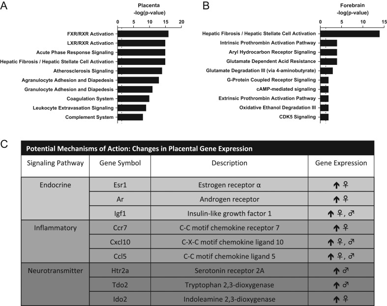
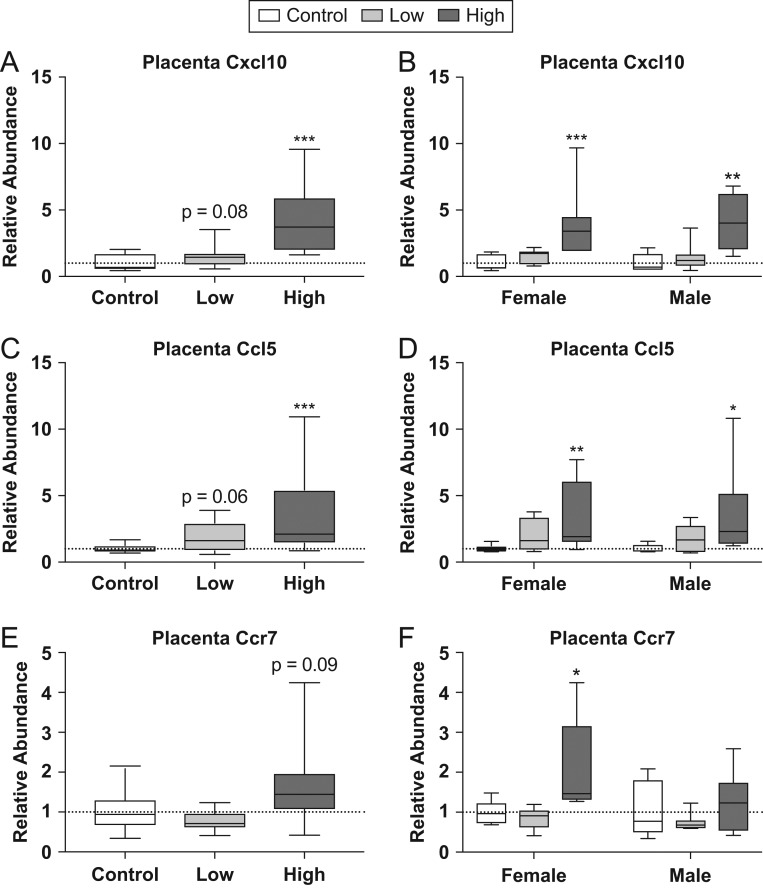
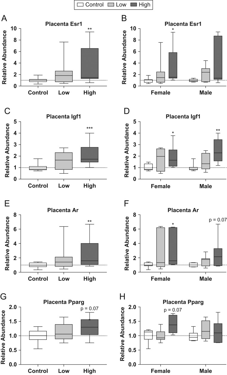
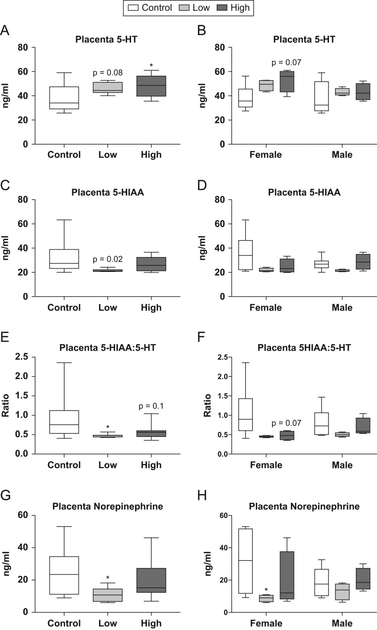
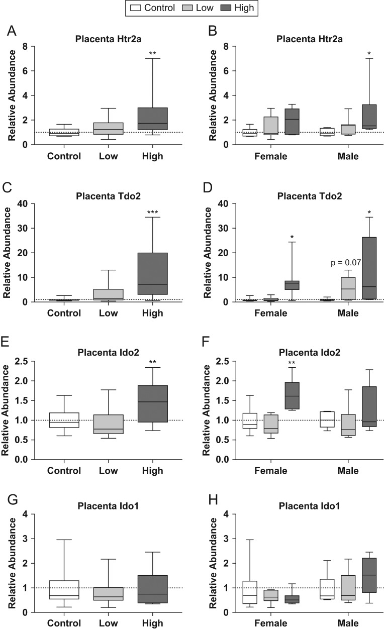
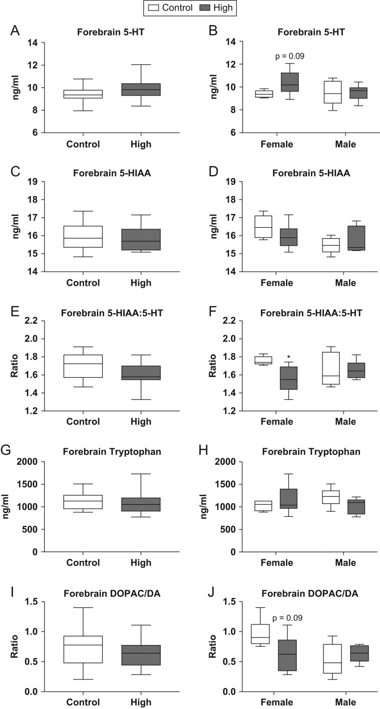
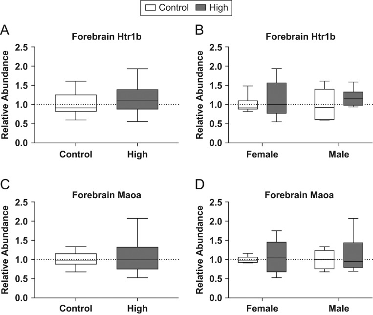
Firemaster 550 (FM 550) is a flame retardant (FR) mixture that has become one of the most commonly used FRs in foam-based furniture and baby products. Human exposure to this commercial mixture, composed of brominated and organophosphate components, is widespread. We have repeatedly shown that developmental exposure can lead to sex-specific behavioral effects in rats. Accruing evidence of endocrine disruption and potential neurotoxicity has raised concerns regarding the neurodevelopmental effects of FM 550 exposure, but the specific mechanisms of action remains unclear. Additionally, we observed significant, and in some cases sex-specific, accumulation of FM 550 in placental tissue following gestational exposure. Because the placenta is an important source of hormones and neurotransmitters for the developing brain, it may be a critical target of toxicity to consider in the context of developmental neurotoxicity. Using a mixture of targeted and exploratory approaches, the goal of the present study was to identify possible mechanisms of action in the developing forebrain and placenta. Wistar rat dams were orally exposed to FM 550 (0, 300 or 1000 µg/day) for 10 days during gestation and placenta and fetal forebrain tissue collected for analysis. In placenta, evidence of endocrine, inflammatory and neurotransmitter signaling pathway disruption was identified. Notably, 5-HT turnover was reduced in placental tissue and fetal forebrains indicating that 5-HT signaling between the placenta and the embryonic brain may be disrupted. These findings demonstrate that environmental contaminants, like FM 550, have the potential to impact the developing brain by disrupting normal placental functions.

Keywords: EDC; behavior; brain; endocrine disruptors; flame retardants; serotonin; sexually dimorphic.


Figures

Figure 1
Figure 1
Top 10 canonical pathways identified by IPA analysis, in placenta (A) and fetal forebrain (B) and changes in placental gene expression verified by qRT-PCR.
Figure 2
Figure 2
Effects of prenatal FM 550 exposure on the relative abundance of genes related to inflammatory signaling in placental tissue. Significant upregulation of Cxcl10 and Ccl5 was observed at the highest dose when males and females were group for analysis (A and C). Similarly, significant upregulation was observed at the highest dose for Cxcl10 and Ccl5 in males and females when analyzed by sex (B and D). No significant effect of exposure was observed for Ccr7 when combined for analysis (E), however, when analyzed by sex, high-dose females showed a significant increase in expression (F). For each dose n = 7/sex. Graphs depict median with whiskers from minimum to maximum (*P ≤ 0.05, **P ≤ 0.01, ***P ≤ 0.001).
Figure 3
Figure 3
Effects of prenatal FM 550 exposure on the relative abundance of genes related to endocrine signaling in placental tissue. Significant upregulation of Esr1, Igf1 and Ar was observed at the highest dose when males and females were grouped for analysis (A, C, and E). Similarly, significant upregulation of Igf1 and Ar was in males and females exposed to the highest dose, while upregulation was found for Esr1 in high-dose females only (B, D, and E). No significant effect of exposure was observed for either Pparg in placental tissue (G and H). For each dose n = 7/sex. Graphs depict median with whiskers from minimum to maximum (*P ≤ 0.05, **P ≤ 0.01, ***P ≤ 0.001).
Figure 4
Figure 4
Effects of prenatal FM 550 exposure on neurotransmitter and neurotransmitter metabolite levels in whole placenta. A dose–response relationship was observed in placental 5-HT levels when sexes were grouped for analysis (A). No significant effect of either dose was observed for 5-HT levels when analyzed by sex; however, females showed a similar pattern compared to the grouped analysis (B). A significant reduction in 5-HIAA was observed at the low dose when sexes were grouped for analysis (C), and a consistent trend was observed for both sexes when analyzed by sex. Additionally, the ratio of 5-HIAA:5-HT was significantly reduced at the lowest dose in the grouped analysis (E) with a similar trend observed in the female analysis (F). Finally, a significant reduction in norepinephrine was observed at the lowest dose in both the grouped and female specific analysis (G and H). For the control group n = 6/sex while the low and the high dose n = 4/sex. Graphs depict median with whiskers from minimum to maximum (*P ≤ 0.05).
Figure 5
Figure 5
Effects of prenatal FM 550 exposure on the relative abundance of genes related to 5-HT signaling and metabolism in placental tissue. Significant upregulation of Htr2a, Tdo2 and Ido2 was observed at the highest dose in the grouped analysis (A, C, and E). When analyzed in a sex-specific manner, significant upregulation was observed at the highest dose in males for Htr2a (B), both sexes for Tdo2 (D), and in females only for Ido2 (D). No significant effect exposure was observed for Ido1 in placental tissue (G and H). For each dose n = 7/sex. Graphs depict median with whiskers from minimum to maximum (*P ≤ 0.05, **P ≤ 0.01, ***P ≤ 0.001).
Figure 6
Figure 6
Effects of prenatal FM 550 exposure on neurotransmitter and neurotransmitter metabolite levels in fetal forebrain. 5-HT levels were not significantly altered in fetal forebrain (A) although there was a trend for higher levels in females (B). No significant effect of exposure was observed for 5-HIAA (C); however, a significant difference between male and female controls was observed (D). A significant reduction in the ratio of 5-HIAA:5-HT was observed at the highest dose but only in females (F). No significant effect of exposure was found for tryptophan or DOPAC:DA (G, H, I and J). For each dose group n = 6/sex. Graphs depict median with whiskers from minimum to maximum (*P ≤ 0.05).
Figure 7
Figure 7
Effects of prenatal FM 550 exposure on the relative abundance of genes related to 5-HT signaling and metabolism in fetal forebrain. No significant effect of exposure was observed for Htr1b or Maoa in the fetal forebrain (A, B, C and D). For each dose n = 6/sex. Graphs depict median with whiskers from minimum to maximum.

References

    1. Stapleton HM, Klosterhaus S, Eagle S, Fuh J, Meeker JD, Blum A, Webster TF. Detection of organophosphate flame retardants in furniture foam and U.S. house dust. Environmental Science and Technology 2009. 43 7490–7495. (10.1021/es9014019) - DOI - PMC - PubMed
    1. Grandjean P, Landrigan PJ. Neurobehavioural effects of developmental toxicity. Lancet Neurology 2014. 13 330–338. (10.1016/S1474-4422(13)70278-3) - DOI - PMC - PubMed
    1. Kalkbrenner AE, Schmidt RJ, Penlesky AC. Environmental chemical exposures and autism spectrum disorders: a review of the epidemiological evidence. Current Problems in Pediatric and Adolescent Health Care 2014. 44 277–318. (10.1016/j.cppeds.2014.06.001) - DOI - PMC - PubMed
    1. Landrigan PJ, Lambertini L, Birnbaum LS. A research strategy to discover the environmental causes of autism and neurodevelopmental disabilities. Environmental Health Perspectives 2012. 120 a258–a260. (10.1289/ehp.1104285) - DOI - PMC - PubMed
    1. Bonnin A, Levitt P. Fetal, maternal, and placental sources of serotonin and new implications for developmental programming of the brain. Neuroscience 2011. 197 1–7. (10.1016/j.neuroscience.2011.10.005) - DOI - PMC - PubMed