

Right Ventricular Myofilament Functional Differences in Humans With Systemic Sclerosis-Associated Versus Idiopathic Pulmonary Arterial Hypertension
- PMID: 29352073
- PMCID: PMC5976528
- DOI: 10.1161/CIRCULATIONAHA.117.033147
Right Ventricular Myofilament Functional Differences in Humans With Systemic Sclerosis-Associated Versus Idiopathic Pulmonary Arterial Hypertension
Abstract
Background: Patients with systemic sclerosis (SSc)-associated pulmonary arterial hypertension (PAH) have a far worse prognosis than those with idiopathic PAH (IPAH). In the intact heart, SSc-PAH exhibits depressed rest and reserve right ventricular (RV) contractility compared with IPAH. We tested whether this disparity involves underlying differences in myofilament function.
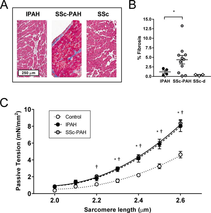
Methods: Cardiac myocytes were isolated from RV septal endomyocardial biopsies from patients with SSc-PAH, IPAH, or SSc with exertional dyspnea but no resting PAH (SSc-d); control RV septal tissue was obtained from nondiseased donor hearts (6-7 per group). Isolated myocyte passive length-tension and developed tension-calcium relationships were determined and correlated with in vivo RV function and reserve. RV septal fibrosis was also examined.
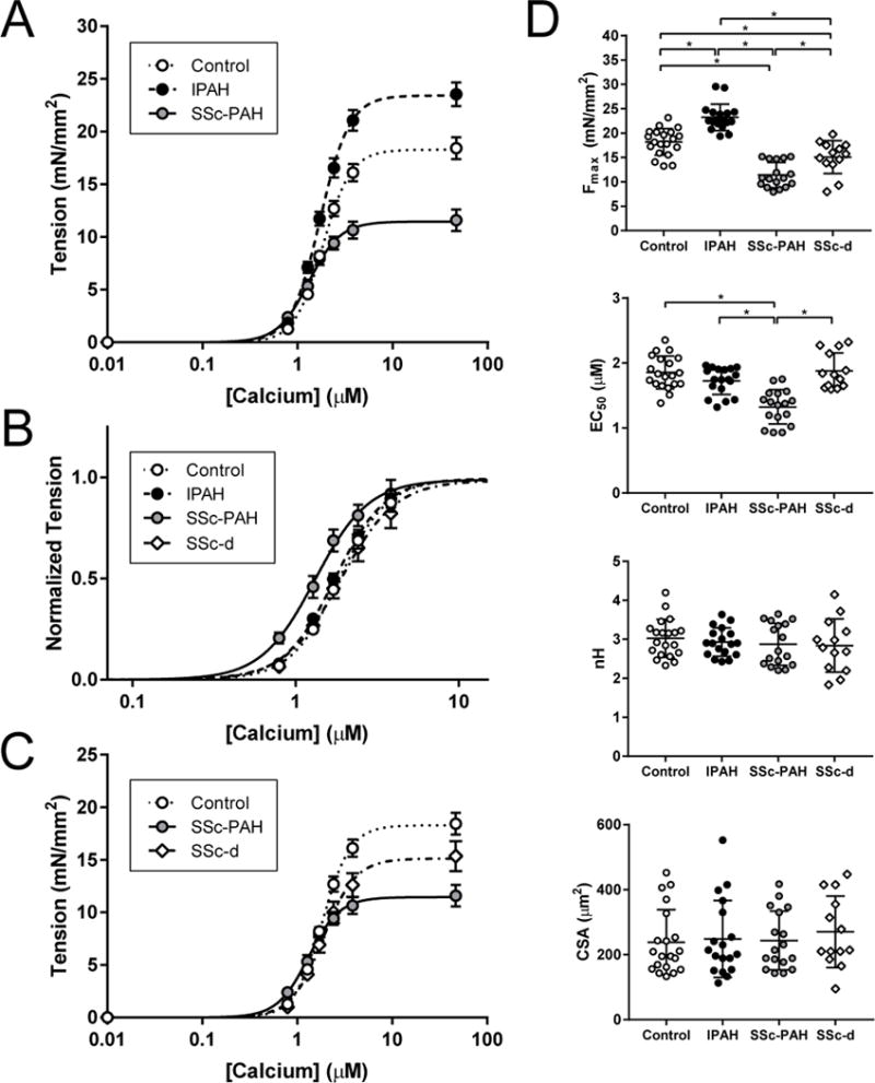
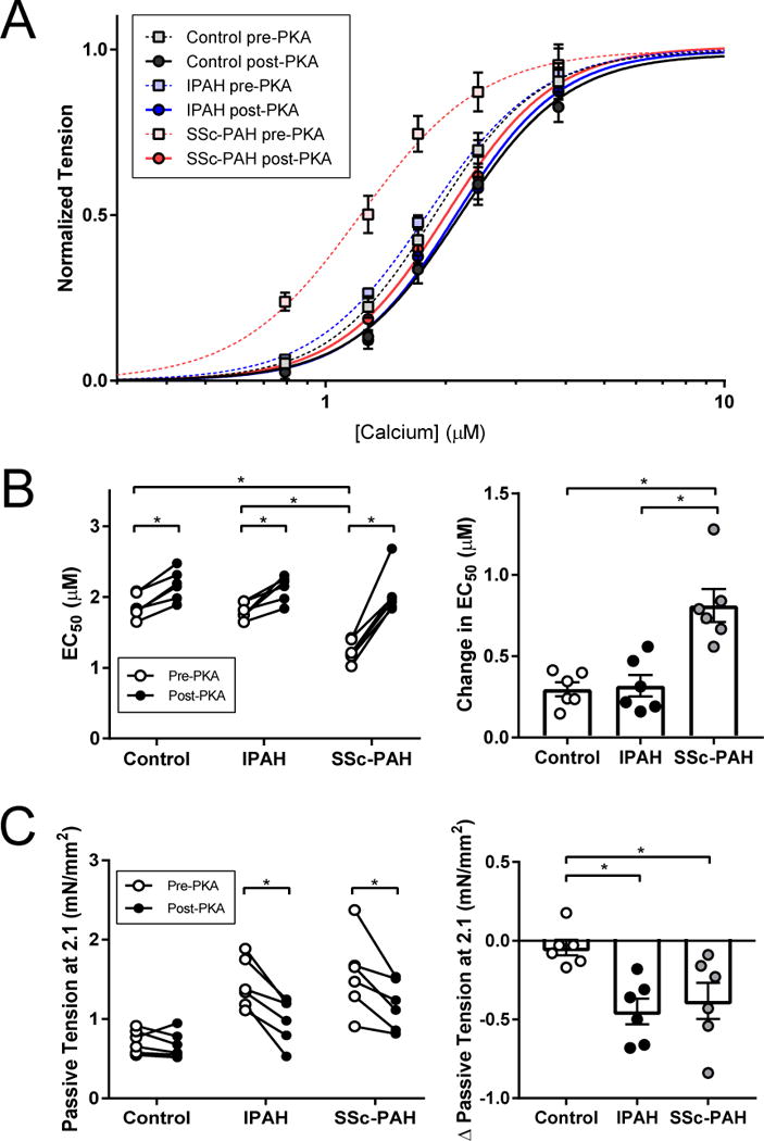
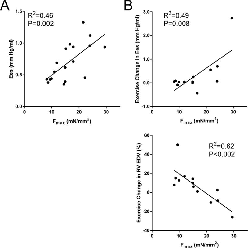
Results: Myocyte passive stiffness from length-tension relations was similarly increased in IPAH and SSc-PAH compared with control, although SSc-PAH biopsies had more interstitial fibrosis. More striking disparities were found between active force-calcium relations. Compared with controls, maximal calcium-activated force (Fmax) was 28% higher in IPAH but 37% lower in SSc-PAH. Fmax in SSc-d was intermediate between control and SSc-PAH. The calcium concentration required for half-maximal force (EC50) was similar between control, IPAH, and SSc-d but lower in SSc-PAH. This disparity disappeared in myocytes incubated with the active catalytic subunit of protein kinase A. Myocyte Fmax directly correlated with in vivo RV contractility assessed by end-systolic elastance (R2 =0.46, P=0.002) and change in end-systolic elastance with exercise (R2 =0.49, P=0.008) and was inversely related with exercise-induced chamber dilation (R2 =0.63, P<0.002), which also was a marker of depressed contractile reserve.
Conclusions: A primary defect in human SSc-PAH resides in depressed sarcomere function, whereas this is enhanced in IPAH. These disparities correlate with in vivo RV contractility and contractile reserve and are consistent with worse clinical outcomes in SSc-PAH. The existence of sarcomere disease before the development of resting PAH in patients with SSc-d suggests that earlier identification and intervention may prove useful.
Keywords: heart ventricles; hypertension, pulmonary; myofibrils; protein kinases; scleroderma, systemic.
© 2018 American Heart Association, Inc.
Figures
References
-
- Vonk Noordegraaf A, Haddad F, Chin KM, Forfia PR, Kawut SM, Lumens J, Naeije R, Newman J, Oudiz RJ, Provencher S, Torbicki A, Voelkel NF, Hassoun PM. Right heart adaptation to pulmonary arterial hypertension: physiology and pathobiology. J Am Coll Cardiol. 2013;62:D22–D33. doi: 10.1016/j.jacc.2013.10.027. - DOI - PubMed
-
- Ruiz-Cano MJ, Escribano P, Alonso R, Delgado J, Carreira P, Velazquez T, Sanchez MAG, Sáenz de la Calzada C. Comparison of baseline characteristics and survival between patients with idiopathic and connective tissue disease-related pulmonary arterial hypertension. J Heart Lung Transplant. 2009;28:621–627. doi: 10.1016/j.healun.2009.02.016. - DOI - PubMed
-
- Argula RG, Karwa A, Lauer A, Gregg D, Silver RM, Feghali-Bostwick C, Schanpp LM, Egbert K, Usher BW, Ramakrishnan V, Hassoun PM, Strange C. Differences in Right Ventricular Functional Changes during Treatment between Systemic Sclerosis-associated Pulmonary Arterial Hypertension and Idiopathic Pulmonary Arterial Hypertension. Ann Am Thorac Soc. 2017;14:682–689. doi: 10.1513/AnnalsATS.201608-655OC. - DOI - PMC - PubMed
Publication types
MeSH terms
Substances
Grants and funding
LinkOut - more resources
Full Text Sources
Other Literature Sources
Medical
