. 2018 Mar 9;293(10):3651-3662.
doi: 10.1074/jbc.RA117.001681. Epub 2018 Jan 19.

Mycolyltransferase from Mycobacterium tuberculosis in covalent complex with tetrahydrolipstatin provides insights into antigen 85 catalysis

Affiliations

Mycolyltransferase from Mycobacterium tuberculosis in covalent complex with tetrahydrolipstatin provides insights into antigen 85 catalysis

Christopher M Goins et al. J Biol Chem. .

Abstract

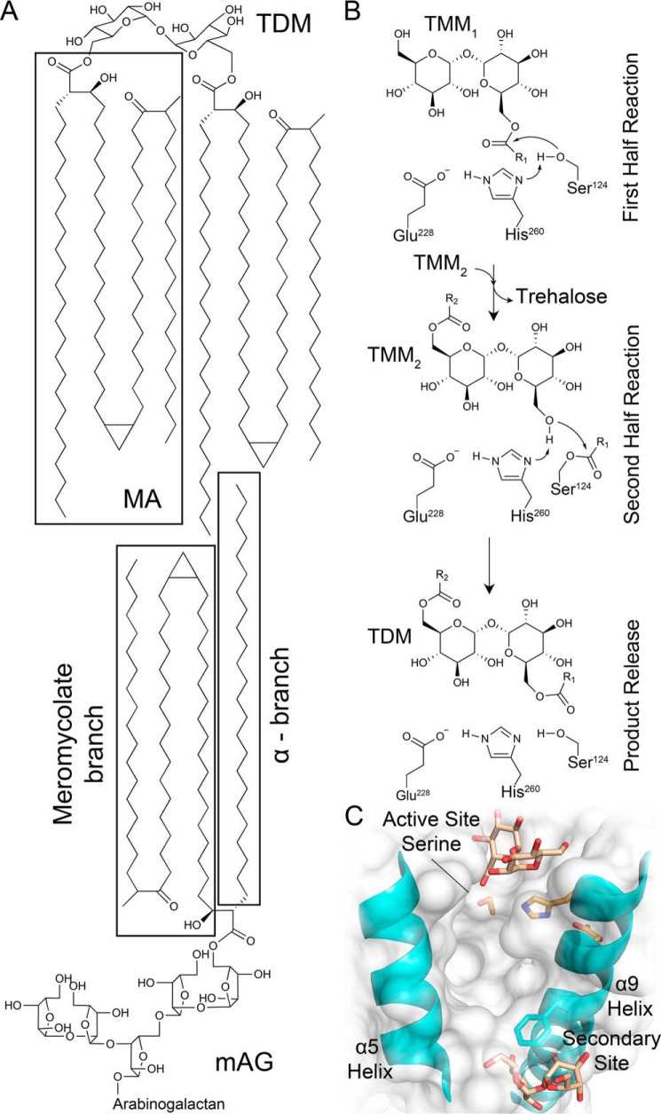
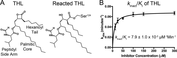
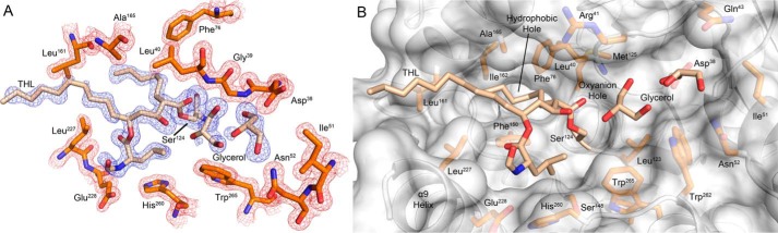
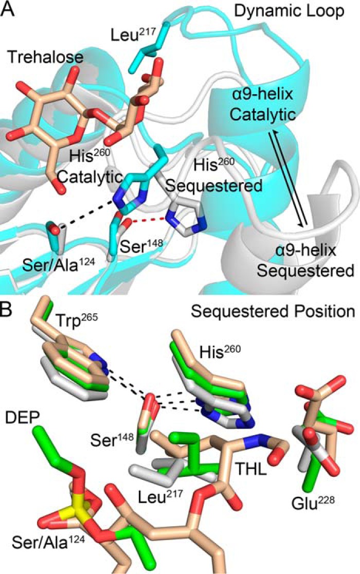
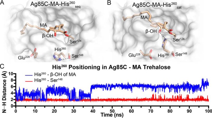
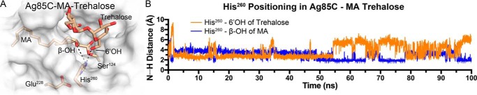
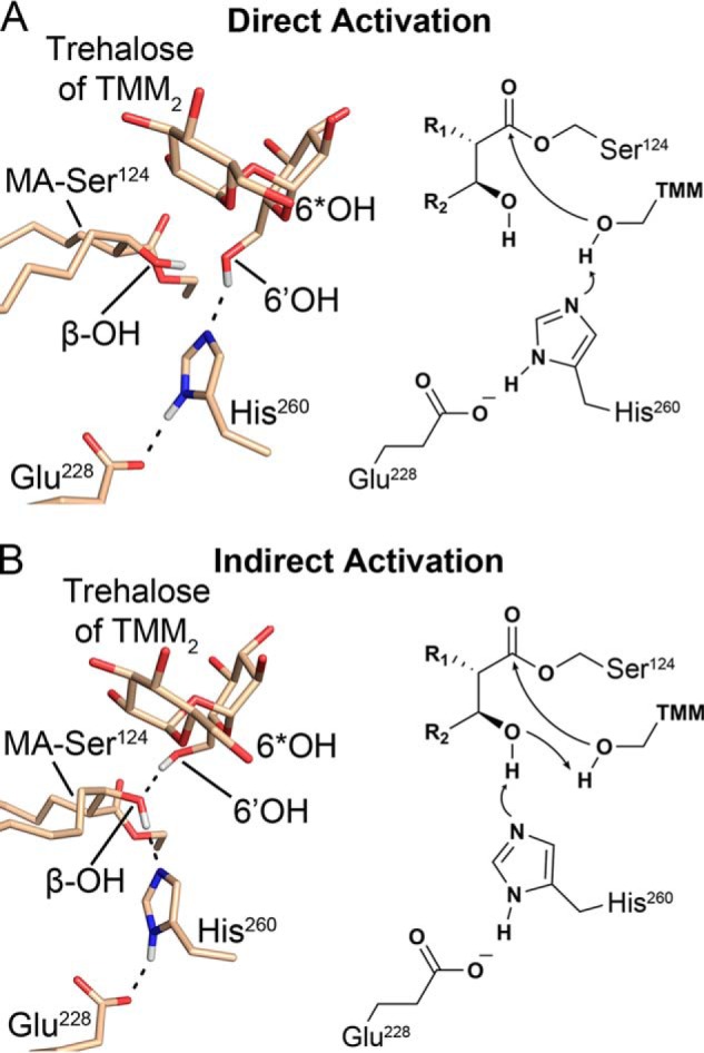
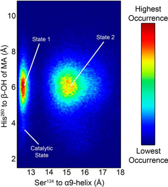
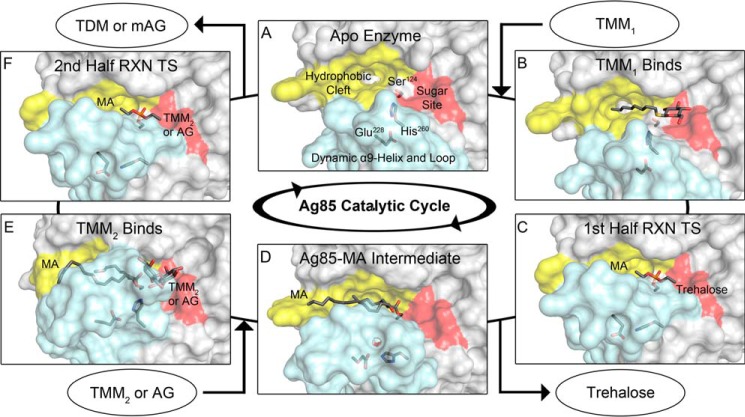
Mycobacterium tuberculosis antigen 85 (Ag85) enzymes catalyze the transfer of mycolic acid (MA) from trehalose monomycolate to produce the mycolyl arabinogalactan (mAG) or trehalose dimycolate (TDM). These lipids define the protective mycomembrane of mycobacteria. The current model of substrate binding within the active sites of Ag85s for the production of TDM is not sterically and geometrically feasible; additionally, this model does not account for the production of mAG. Furthermore, this model does not address how Ag85s limit the hydrolysis of the acyl-enzyme intermediate while catalyzing acyl transfer. To inform an updated model, we obtained an Ag85 acyl-enzyme intermediate structure that resembles the mycolated form. Here, we present a 1.45-Å X-ray crystal structure of M. tuberculosis Ag85C covalently modified by tetrahydrolipstatin (THL), an esterase inhibitor that suppresses M. tuberculosis growth and mimics structural attributes of MAs. The mode of covalent inhibition differs from that observed in the reversible inhibition of the human fatty-acid synthase by THL. Similarities between the Ag85-THL structure and previously determined Ag85C structures suggest that the enzyme undergoes structural changes upon acylation, and positioning of the peptidyl arm of THL limits hydrolysis of the acyl-enzyme adduct. Molecular dynamics simulations of the modeled mycolated-enzyme form corroborate the structural analysis. From these findings, we propose an alternative arrangement of substrates that rectifies issues with the previous model and suggest a direct role for the β-hydroxy of MA in the second half-reaction of Ag85 catalysis. This information affords the visualization of a complete mycolyltransferase catalytic cycle.

Keywords: acyltransferase; antigen 85; glycolipid; hydrolase; lipid esterase; mycolyltransferase; serine esterase; structural biology; tetrahydrolipstatin; α/β-hydrolase.


Conflict of interest statement

The authors declare that they have no conflicts of interest with the contents of this article

Figures

Figure 1.
Figure 1.
M. tuberculosis Ag85 reaction products, mechanism, and trehalose-binding sites. A, Ag85s transfer MA onto the 6′-hydroxy of TMM and the 5-hydroxy of AG to form TDM and mAG. The depicted MA has a cis-keto meromycolate branch (4, 10). B, ping–pong reaction mechanism for Ag85C formation of TDM (R1 and R2 = MA chains) adapted from Ronning et al. (7). C, identified trehalose-binding sites adjacent to the Ag85B active site and a secondary site (Protein Data Bank code 1F0P) (9).
Figure 2.
Figure 2.
Covalent inhibition of Ag85C by THL. A, chemical structure of THL and resulting structure upon covalent attack by Ag85C. B, kinact/KI plot of THL inhibition. Error bars represent S.D. of triplicate reactions.
Figure 3.
Figure 3.
Ag85C-THL structure. A, FoFc likelihood-weighted omit map (blue) contoured to 3.0 σ for Ser124 modified by THL and glycerol molecules in the active site and 2FoFc map (red) contoured to 1.5 σ for surrounding active site residues. B, surface rendering of Ag85C with Ser124 modified with THL and two glycerol molecules occupying the trehalose-binding portion of the active site.
Figure 4.
Figure 4.
Observed structural changes between the active and acyl-enzyme intermediate forms. A, catalytic triad disruption results in relaxation of the α9-helix with His260 adopting a sequestered position (white, Ag85C-S124A, Protein Data Bank code 4QEK; cyan, trehalose-bound Ag85B, Protein Data Bank code 1F0P) (9, 12). B, consistent displacement of His260 results in hydrogen bond formation with Ser148. The aliphatic side arm of THL mimics the hydrophobic interactions of the side chain of Leu217 in the diethyl phosphate–modified Ag85C and S124A mutant structures (wheat, Ag85C-THL, Protein Data Bank code 5VNS; green, Ag85C-diethyl phosphate, Protein Data Bank code 1DQY; white, Ag85C-S124A, Protein Data Bank code 4QEK) (7, 12).
Figure 5.
Figure 5.
MD models of the mycolated Ag85C intermediate. A, Ag85C-MA-His260seq. B, Ag85C-MA-His260cat. C, hydrogen bonding of His260 with Ser148 in the sequestered position was stable during MD simulations. When His260 was placed in the catalytic position without an acceptor molecule present the side chain freely samples conformations, making limited hydrogen-bonding interactions with the β-hydroxy of MA.
Figure 6.
Figure 6.
MD model of the mycolated Ag85C intermediate with acceptor molecule present. A, Ag85C-MA-trehalose MD model. B, in the catalytic position when an acceptor molecule is present, His260 exclusively sampled both the 6′-hydroxy of trehalose and the β-hydroxy of MA.
Figure 7.
Figure 7.
Nucleophilic activation pathways for the second half-reaction (6*OH denotes the mycolated hydroxy of TMM). A, direct activation of the 6′-hydroxy of TMM for the second half-reaction by His260, stabilized by the β-hydroxy of MA. B, indirect activation scheme proceeding through a proton transfer from the β-hydroxy of MA to His260 followed by the proton transfer from the 6′-hydroxy to deprotonated β-hydroxy of MA.
Figure 8.
Figure 8.
Heat map of Ag85C-MA-His260cat conformations sampled during the REMD simulation. The starting catalytic position was not frequently sampled when the α9-helix remained kinked toward the active site in the catalytic position; instead His260 randomly sampled phase space, similar to what was observed in the initial MD simulation (state 1). In state 2, the α9-helix relaxes, similar to what is observed in the sequestered form. Although His260 is positioned toward Ser148, a stable hydrogen bond to Ser148 is never observed.
Figure 9.
Figure 9.
Proposed structure-based catalytic cycle of Ag85s. The light blue surface corresponds to the α9-helix and dynamic loop, yellow corresponds to the MA-binding site, and red corresponds to the sugar-binding site. A, apoenzyme exhibits a large TMM1-binding site (Protein Data Bank code 1DQZ) (7). B, Ag85C octyl thioglucoside structure mimics the initial TMM1 binding event (Protein Data Bank code 1VA5) (8). C, tetrahedral transition state (TS) of the first half-reaction (RXN) represented by the Ag85C-diethyl phosphate structure with His260 sequestered and the α9-helix relaxed (Protein Data Bank code 1DQY) (7). A free trehalose molecule leaves, completing the first half-reaction. D, model of the Ag85C-MA intermediate based on the Ag85C-THL structure. E, the second half-reaction proceeds through the binding of TMM2 or AG to the sugar-binding site. The α9-helix and His260 are restored to the catalytic position as a result of acceptor binding. The model of Ag85C-MA-trehalose is shown. F, transition state for the second half-reaction, again modeled by Ag85C-diethyl phosphate, leading to the formation of TDM or mAG and subsequent product release (Protein Data Bank code 1DQY) (7).

References

    1. Pai M., Behr M. A., Dowdy D., Dheda K., Divangahi M., Boehme C. C., Ginsberg A., Swaminathan S., Spigelman M., Getahun H., Menzies D., and Raviglione M. (2016) Tuberculosis. Nat. Rev. Dis. Primers 2, 16076 10.1038/nrdp.2016.76 - DOI - PubMed
    1. Jackson M., McNeil M. R., and Brennan P. J. (2013) Progress in targeting cell envelope biogenesis in Mycobacterium tuberculosis. Future Microbiol. 8, 855–875 10.2217/fmb.13.52 - DOI - PMC - PubMed
    1. Kaur D., Guerin M. E., Skovierová H., Brennan P. J., and Jackson M. (2009) Biogenesis of the cell wall and other glycoconjugates of Mycobacterium tuberculosis. Adv. Appl. Microbiol. 69, 23–78 10.1016/S0065-2164(09)69002-X - DOI - PMC - PubMed
    1. Marrakchi H., Lanéelle M. A., and Daffé M. (2014) Mycolic acids: structures, biosynthesis, and beyond. Chem. Biol. 21, 67–85 10.1016/j.chembiol.2013.11.011 - DOI - PubMed
    1. Belisle J. T., Vissa V. D., Sievert T., Takayama K., Brennan P. J., and Besra G. S. (1997) Role of the major antigen of Mycobacterium tuberculosis in cell wall biogenesis. Science 276, 1420–1422 10.1126/science.276.5317.1420 - DOI - PubMed

Publication types

MeSH terms

LinkOut - more resources