. 2018 Mar 16;293(11):3965-3980.
doi: 10.1074/jbc.M117.816769. Epub 2018 Jan 19.

DeSUMOylation of MKK7 kinase by the SUMO2/3 protease SENP3 potentiates lipopolysaccharide-induced inflammatory signaling in macrophages

Affiliations

DeSUMOylation of MKK7 kinase by the SUMO2/3 protease SENP3 potentiates lipopolysaccharide-induced inflammatory signaling in macrophages

Yimin Lao et al. J Biol Chem. .

Abstract

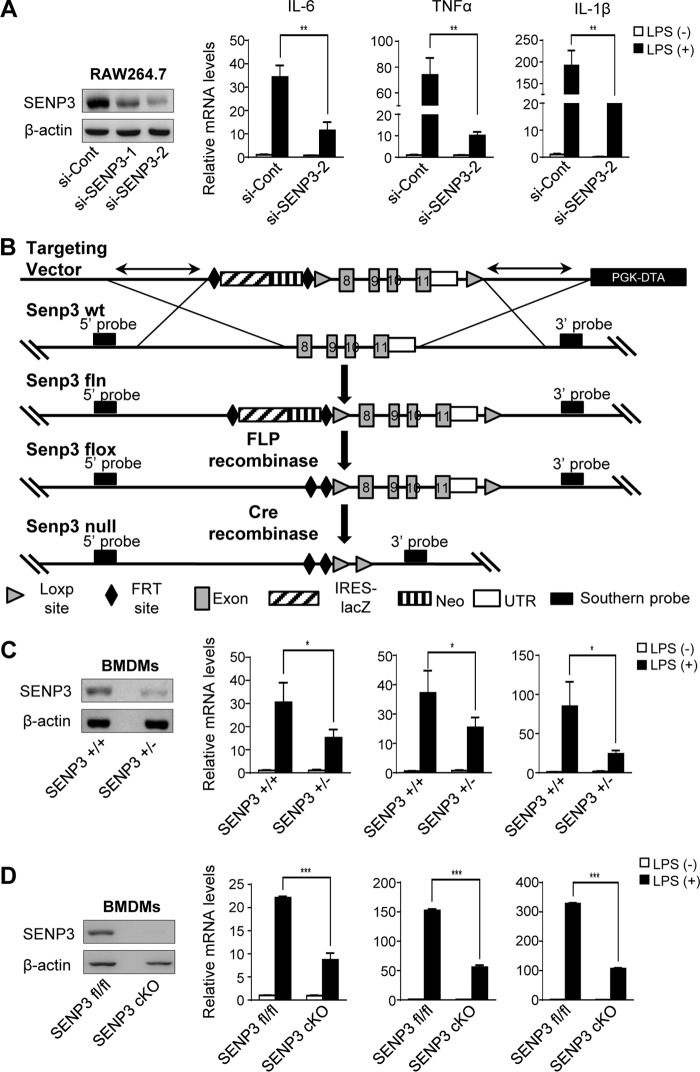
Protein SUMOylation has been reported to play a role in innate immune response, but the enzymes, substrates, and consequences of the specific inflammatory signaling events are largely unknown. Reactive oxygen species (ROS) are abundantly produced during macrophage activation and required for Toll-like receptor 4 (TLR4)-mediated inflammatory signaling. Previously, we demonstrated that SENP3 is a redox-sensitive SUMO2/3 protease. To explore any links between reversible SUMOylation and ROS-related inflammatory signaling in macrophage activation, we generated mice with Senp3 conditional knock-out in myeloid cells. In bacterial lipopolysaccharide (LPS)-induced in vitro and in vivo inflammation models, we found that SENP3 deficiency markedly compromises the activation of TLR4 inflammatory signaling and the production of proinflammatory cytokines in macrophages exposed to LPS. Moreover, Senp3 conditional knock-out mice were significantly less susceptible to septic shock. Of note, SENP3 deficiency was associated with impairment in JNK phosphorylation. We found that MKK7, which selectively phosphorylates JNK, is a SENP3 substrate and that SENP3-mediated deSUMOylation of MKK7 may favor its binding to JNK. Importantly, ROS-dependent SENP3 accumulation and MKK7 deSUMOylation rapidly occurred after LPS stimulation. In conclusion, our findings indicate that SENP3 potentiates LPS-induced TLR4 signaling via deSUMOylation of MKK7 leading to enhancement in JNK phosphorylation and the downstream events. Therefore this work provides novel mechanistic insights into redox regulation of innate immune responses.

Keywords: ROS; SENP3; c-Jun N-terminal kinase (JNK); inflammation; innate immunity; macrophage; sumoylation.


Conflict of interest statement

The authors declare that they have no conflicts of interest with the contents of this article

Figures

Figure 1.
Figure 1.
SENP3 deficiency decreases LPS-induced cytokine production in macrophages. A, RAW 264.7 cells transfected with nonspecific siRNA (si-Cont) or SENP3 siRNA (si-SENP3) were incubated with LPS (100 ng/ml) for 6 h. mRNA levels of proinflammatory cytokines IL-6, TNFα, and IL-1β were assessed by qRT-PCR. The knockdown efficiency of two siRNAs of SENP3 was determined by IB. B, a strategy of Senp3flox/flox Lyz2-cre mouse generation was shown. A mouse model expressing a myeloid cell-specific deletion of SENP3 was generated using transgenic mice bearing loxp sites flanking exon 8 to exon 11 of the Senp3 gene (SENP3flox) and mice expressing a Cre recombinase transgene from the Lysozyme M locus (Lys M-Cre also known as Lyz2-Cre). C, BMDMs isolated from Senp3+/+ and Senp3+/− mice were incubated with LPS (100 ng/ml) for 6 h. mRNA levels of proinflammatory cytokines IL-6, TNFα, and IL-1β were assessed by qRT-PCR. The SENP3 level was determined by IB. D, BMDMs isolated from Senp3flox/flox (Senp3fl/fl) and Senp3flox/flox Lyz2-cre (Senp3 cKO) mice were incubated with LPS (100 ng/ml) for 6 h. mRNA levels of proinflammatory cytokines IL-6, TNFα, and IL-1β were assessed by qRT-PCR. SENP3 level was determined by IB. Graphs show the mean ± S.D. and data (A, C, and D) shown are representative of three independent experiments. *, p < 0.05; **, p < 0.01; ***, p < 0.001.
Figure 2.
Figure 2.
SENP3 deficiency selectively attenuates MAPK signaling and JNK phosphorylation in macrophages. A, RAW 264.7 cells transfected with si-Cont or si-SENP3 were stimulated with LPS (100 ng/ml) for the indicated time. IκBα degradation was assessed by IB. B, NF-κB-luciferase (left panel) or AP-1-luciferase (right panel) and Renilla were transfected into RAW 264.7 cells together with the indicated siRNA. 48 h after transfection, cells were stimulated with LPS (100 ng/ml) for 6 h followed by luciferase reporter assays. Graphs show the mean ± S.D. and data shown are representative of three independent experiments. ns, no statistical difference; *, p < 0.05. C, RAW 264.7 cells transfected with si-Cont or si-SENP3 were stimulated with LPS (100 ng/ml) for the indicated time (left panel). The cells were overexpressed with RGS-His-SENP3 (RH-SENP3 for short) (right panel). Phosphorylated JNK (p-JNK), p38 (p-p38), and ERK (p-ERK) were assessed by IB. D, Senp3fl/fl and Senp3 cKO BMDMs stimulated with LPS (100 ng/ml) for the indicated time. p-JNK, p-p38, and p-ERK were assessed by IB. E, RAW 264.7 cells were stimulated with LPS (100 ng/ml) for 30 min. Immunofluorescence of p-JNK was performed and representative pictures were shown in left panel. Quantification of p-JNK fluorescence intensity was shown in the right panel. p-JNK, green; 4′,6-diamidino-2-phenylindole (DAPI), blue. Scale bar, 10 μm. Graphs are shown as mean ± S.D. (n = 40).
Figure 3.
Figure 3.
SENP3 catalyzes deSUMOylation of MKK7 at Lys-18 site. A, the interaction of SENP3 with MKK7 was determined by co-IP. HEK293T cells were transfected with FLAG–SENP3 and -MKK7 plasmids for 48 h. Co-IP was performed using FLAG-M2 beads for immunoprecipitation and using anti-MKK7 antibodies for IB (upper panel). HEK293T cells were transfected with GFP–SENP3 and FLAG–MKK7 plasmids for 48 h. Co-IP was performed using FLAG-M2 beads for immunoprecipitation and using anti-GFP antibodies for IB (bottom panel). B, TLR4 293 cells were administrated with LPS (100 ng/ml) for 30 min. The interaction of endogenous SENP3 and MKK7 was determined by co-IP. C, SUMO3 conjugates of MKK7 were determined by denaturing co-IP or Ni-bead pull-down. FLAG–MKK7 were transfected into HEK293T cells along with RH–SUMO3, UBC9, and GFP–SENP3 or GFP–SENP3 mutant (C532A) as indicated for 48 h. Cell lysates were immunoprecipitated with FLAG-M2 beads, and then analyzed by IB with the indicated antibodies (upper panel). FLAG–MKK7 were transfected into HEK293T cells along with RH-SUMO3, UBC9, and GFP–SENP3 as indicated for 48 h. Cells were lysed and RH–SUMO3 was pulled down using Ni-NTA beads and then analyzed by IB as indicated (bottom panel). D, SUMO3 conjugates of MKK7 were determined by denaturing co-IP or Ni-bead pull-down. HEK293T cells were transfected with FLAG-tagged MKK7 WT or mutated K18R and K400R, RH–SUMO3 and UBC9 as indicated for 48 h. Cell lysates were immunoprecipitated with FLAG-M2 beads, and then analyzed by IB with the indicated antibodies (upper panel). Cells were lysed and RH–SUMO3 was pulled down using Ni-NTA beads and then analyzed by IB as indicated (bottom panel). Arrowheads indicated SUMO3-conjugated MKK7 in (C and D). *, nonspecific bands.
Figure 4.
Figure 4.
SENP3-mediated deSUMOylation of MKK7 potentiates JNK activation. A, TLR4 293 cells transfected with si-Cont or si-SENP3 for 48 h were incubated with LPS (100 ng/ml) for the indicated time, and p-JNK was assessed by IB. B, TLR4 293 cells with si-SENP3 were transfected with FLAG-tagged MKK7 WT or SUMO-less mutant K18R. The cells were incubated with LPS (100 ng/ml) for the indicated time and then p-JNK was determined by IB. C, TLR4 293 cells with si-SENP3 were co-transfected with RH–SUMO3, UBC9, and FLAG-tagged MKK7 WT or SUMO-less mutant K18R. The cells were incubated with LPS (100 ng/ml) for the indicated time and then p-JNK was determined by IB. D, TLR4 293 cells transfected with FLAG-tagged MKK7 WT or SUMO fusion plasmid for 48 h were stimulated with LPS (100 ng/ml) for the indicated time. p-JNK was analyzed by IB. E, TLR4 293 cells transfected with FLAG-tagged MKK7 WT or SUMO3 fusion for 48 h were stimulated with LPS (100 ng/ml) for 30 min. The interaction of MKK7 with p-JNK was determined by co-IP. F, TLR4 293 cells were transfected with FLAG-tagged MKK7 WT or SUMO3 fusion for 48 h. Immunoprecipitation kinase assay was performed. Phosphorylated JNK (p-JNK) and c-Jun (p-c-Jun) were harvested from anti-c-Jun-precipitated cell lysates. The levels of p-JNK and p-c-Jun were analyzed by IB and quantified in three independent experiments; graphs are shown as mean ± S.D. G, GST–MKK7 and GST–MKK7–SUMO3 were expressed in bacteria and purified. GST pull-down assays were used to test the binding capability between GST–MKK7 or GST–MKK7–SUMO3 and JNK1. JNK1 (1 μg) was tested for binding to GST–Sepharose with GST–MKK7 or GST–MKK7–SUMO3. JNK1 bound with MKK7 was determined by IB.
Figure 5.
Figure 5.
SENP3 rapidly accumulates and deSUMOylates MKK7 after LPS stimulation. A, RAW 264.7 cells were stimulated with LPS (100 ng/ml) in the presence or absence of 5 mm NAC for the indicated time. NAC was pretreated for 2 h. SENP3 accumulation was monitored by IB. B, BMDMs were stimulated with LPS (100 ng/ml) in the presence or absence of 5 mm NAC for the indicated time. SENP3 accumulation was monitored by IB. C, BMDMs were exposed to LPS (100 ng/ml) for the indicated time. The ROS level was determined by DCFH-DA staining and flow-cytometric analysis. NAC was pretreated for 2 h. Graphs show the mean ± S.D. of three independent experiments. ***, p < 0.001. D, RAW 264.7 cells were exposed to LPS (100 ng/ml) for the indicated time. SENP3 levels in cytoplasmic and nuclear fractions were evaluated by IB. Tubulin and poly(ADP-ribose) polymerase (PARP) were taken as the internal controls for cytoplasm and nucleus, respectively. To show SENP3 levels comparable between the cytoplasmic and nuclear fractions based on protein concentration measurements, the sample volume ratio of cytoplasmic fractions versus nuclear fractions was about 10:1. E, TLR4 293 cells were transfected with RH–SUMO3 for 48 h followed by stimulation with LPS (100 ng/ml) for the indicated time. Endogenous MKK7 that conjugated with RH–SUMO3 was detected by co-IP. F, Senp3fl/fl and Senp3 cKO BMDMs were stimulated with LPS (100 ng/ml) for the indicated time. Endogenous MKK7 that conjugated with endogenous SUMO3 in the cells was detected by co-IP. Arrowheads indicated SUMO3-conjugated MKK7 in E and F; *, nonspecific bands.
Figure 6.
Figure 6.
Senp3 cKO mice had less severe inflammatory responses and higher survival rates in LPS-induced endotoxin shock. A–C, Senp3fl/fl and Senp3 cKO mice (n = 6–8 per group) were intraperitoneally injected with LPS (30 mg/kg) for 6 h. A, IL-6 and TNFα in mice serum were assessed by ELISA. B, TNFα and IL-6 expression in livers, lungs, and spleens (n = 6–8 per group) were monitored by qRT-PCR. Relative mRNA level of LPS injection group is to control group. C, IL-6 and TNFα in mice livers were assessed by ELISA. D, Senp3fl/fl and Senp3 cKO mice (n = 10 per group) were injected with LPS (30 mg/kg, intraperitoneally) and then monitored for survival for up to 48 h. Graphs show the mean ± S.D. and data (A–C) are representative of three independent experiments. *, p < 0.05; **, p < 0.01; ***, p < 0.001.
Figure 7.
Figure 7.
A schematic model of SENP3 function in LPS-induced TLR4 signaling. Under the resting condition, more MKK7 are conjugated by SUMO2/3 in macrophages. This prevents MKK7 from binding to JNK, thus attenuating the JNK phosphorylation and activation. LPS induces ROS production and consequently SENP3 accumulation in addition to activation of TLR4. SENP3 catalyzes the de-SUMOylation of MKK7 and potentiates JNK-mediated expression of inflammatory cytokines.

Similar articles

Cited by

References

    1. Liu J., Qian C., and Cao X. (2016) Post-translational modification control of innate immunity. Immunity 45, 15–30 10.1016/j.immuni.2016.06.020 - DOI - PubMed
    1. Kawai T., and Akira S. (2010) The role of pattern-recognition receptors in innate immunity: update on Toll-like receptors. Nature immunology 11, 373–384 10.1038/ni.1863 - DOI - PubMed
    1. Medzhitov R. (2007) Recognition of microorganisms and activation of the immune response. Nature 449, 819–826 10.1038/nature06246 - DOI - PubMed
    1. Cook D. N., Pisetsky D. S., and Schwartz D. A. (2004) Toll-like receptors in the pathogenesis of human disease. Nat. Immunol. 5, 975–979 10.1038/ni1116 - DOI - PubMed
    1. Gay N. J., Symmons M. F., Gangloff M., and Bryant C. E. (2014) Assembly and localization of Toll-like receptor signalling complexes. Nat. Rev. Immunol. 14, 546–558 10.1038/nri3713 - DOI - PubMed

Publication types

MeSH terms