. 2018 Jan 19;9(2):45.
doi: 10.1038/s41419-017-0064-x.

PI3K/mTOR inhibition promotes the regression of experimental vascular malformations driven by PIK3CA-activating mutations

Affiliations

PI3K/mTOR inhibition promotes the regression of experimental vascular malformations driven by PIK3CA-activating mutations

Laura di Blasio et al. Cell Death Dis. .

Abstract

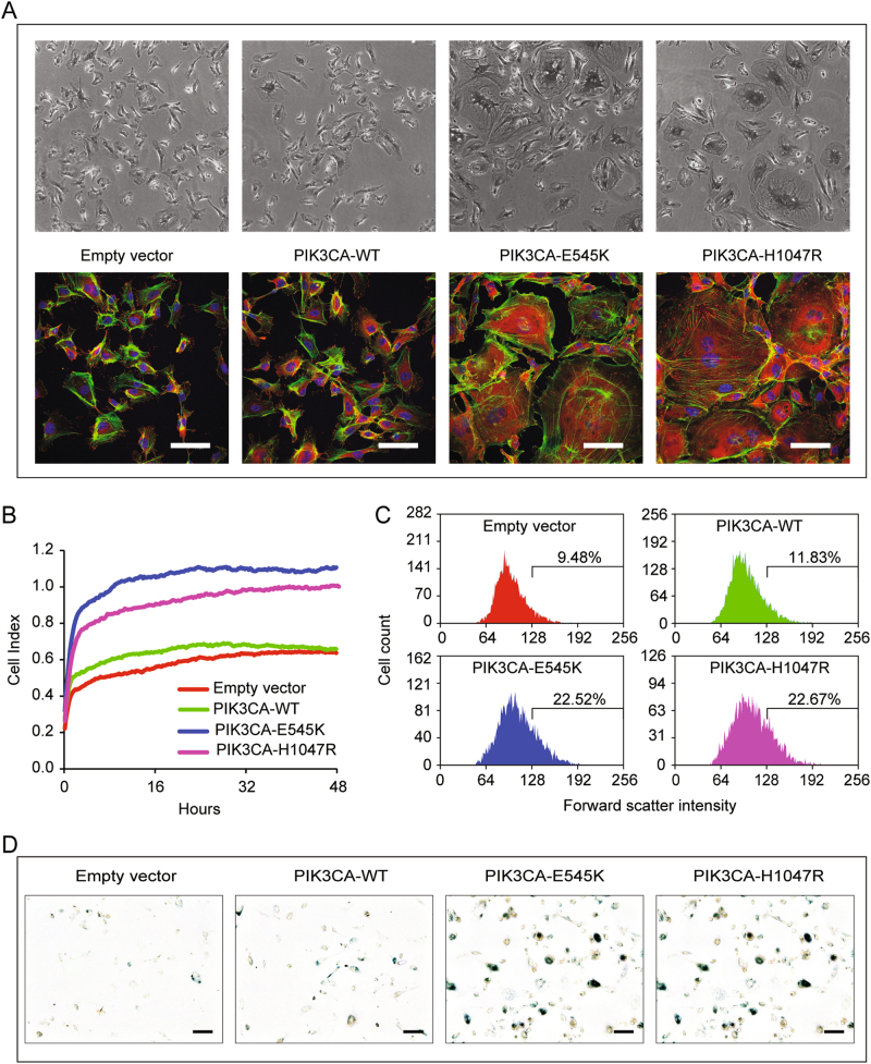
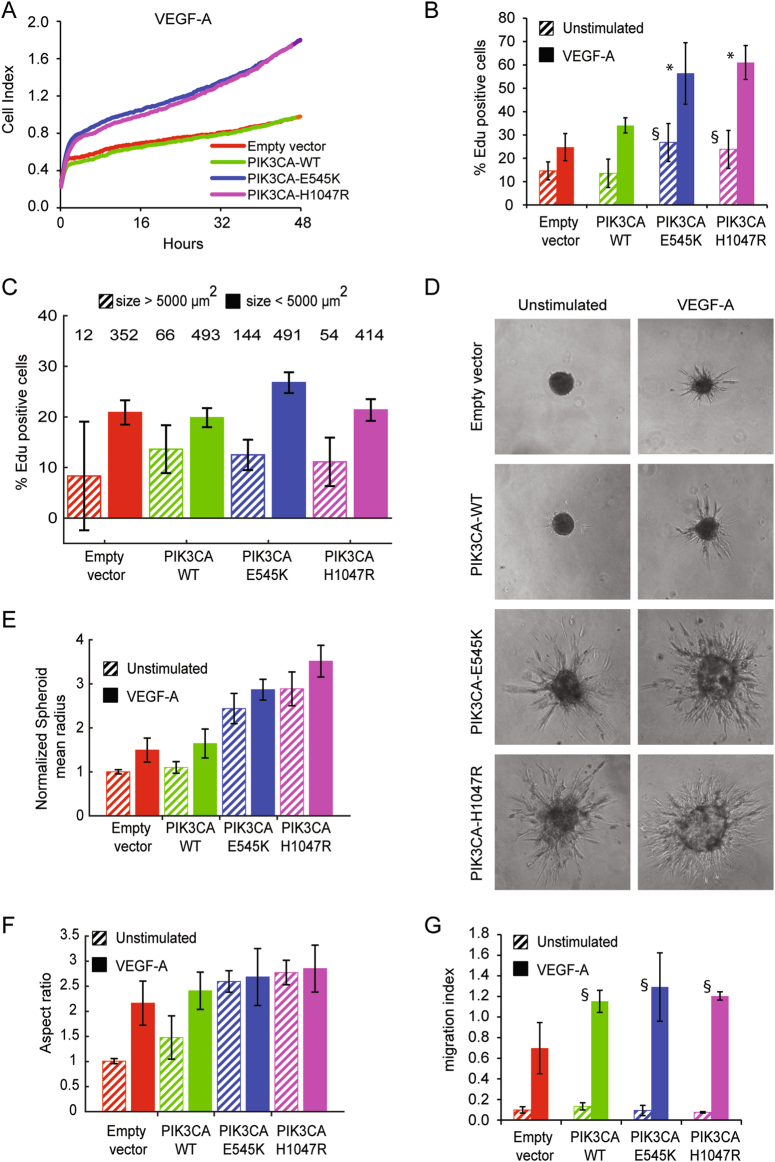
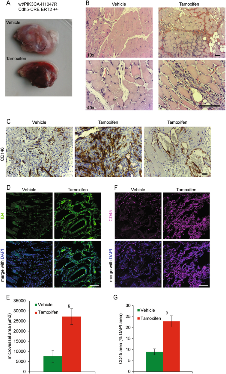
Somatic activating mutations within the PIK3CA gene have been recently detected in sporadic lymphatic and venous malformations, and in vascular malformations (VM) associated to overgrowth syndromes, such as CLOVES and Klippel-Trenaunay syndrome. Although VM are often limited to specific tissue areas and can be well treated, in extended or recurrent lesions novel therapeutic approaches are needed. We generated a mouse model of VM by local expression of PIK3CA-activating mutation in endothelial cells. PIK3CA-driven lesions are characterized by large areas of hemorrhage, hyperplastic vessels, infiltrates of inflammatory cells, and elevated endothelial cell density. Such vascular lesions are ameliorated by administration of dual PI3K/mTOR inhibitor, BEZ235, and mTOR inhibitor, Everolimus. Unexpectedly, the expression of PIK3CA-activating mutations in human endothelial cells results in both increased proliferation rates and senescence. Moreover, active forms of PIK3CA strongly promote the angiogenic sprouting. Treatment with PI3K/mTOR inhibitors restores normal endothelial cell proliferation rate and reduces the amount of senescent cells, whereas treatment with Akt inhibitor is less effective. Our findings reveal that PIK3CA mutations have a key role in the pathogenesis of VM and PIK3CA-driven experimental lesions can be effectively treated by PI3K/mTOR inhibitors.


Conflict of interest statement

Ethics approval and consent to participate

All experiments with animals were conducted according to the local regulations and with permission of local animal welfare officers.

Competing interests

The authors declare that they have no competing interests.

Figures

Fig. 1
Fig. 1. Mice expressing Pik3caH1047R in developing and adult vascular EC are not viable
a Transgenic mice that express latent Pik3caH1047R mutant allele (H1047R) were crossed with mice expressing Cre recombinase under endothelial promoter (Tie2Cre). The Mendelian expected ratio for endothelial Pik3caH1047R allele was the 50% of newborn mice, but only mice carrying wild-type alleles were identified. b We recovered live embryos with PIK3CA mutations until mouse embryonic day 9.5. These embryos showed growth delay (top) and evident vascular defects (bottom, in red endomucin staining). c Transgenic mice that express latent Pik3caH1047R mutant allele (H1047R) were crossed with mice expressing Tamoxifen-inducible Cre recombinase under VE-Cadherin promoter (Cdh5-CreERT2). Mice treated with single administration of Tamoxifen did not survive >2 weeks after Cre induction
Fig. 2
Fig. 2. PIK3CA-activating mutations induce morphological alterations and senescence in EC
a Human primary endothelial cells were transduced with retroviral vectors carrying activating mutations of PIK3CA (PIK3CA-H1047R and PIK3CA-E545K), wild-type PIK3CA (WT), or with empty vector. EC-expressing PIK3CA mutants showed morphological alterations. Large cells with abnormal stress fibers and intracellular vesicles are observable (green: Phalloidin, red: vinculin, blue: DAPI). Scale bar 50 µm. b Cell surface area is measured in real-time by impedance system with xCELLigence technology in absence of growth factors. EC-H1047R and EC-E545K adhere on substrate and occupy more surface than EC-WT or normal cells. Data were plotted as the mean cell index from three wells at each time points; P-values were calculated at 24 h, *P < 0.005, vs. control wild-type PIK3CA endothelial cells. c Transduced EC are analyzed by flow cytometer for forward scatter intensity. The expression of PIK3CA mutants increase the percentage of cells with higher volume as indicated. The reported experiments are representative of three independent experiments. d β-galactosidase staining show senescent cells. Image manipulation has been done to highlight positive cells, as described in the Methods section. Original pictures are in supplemental Fig. 1c
Fig. 3
Fig. 3. PIK3CA-activating mutations increase proliferation rate and sprouting formation
a Cell surface area is measured in real-time by impedance system with xCELLigence technology in presence of VEGF-A. EC- H1047R and EC-E545K grew faster than EC-WT or normal cells. Slopes for each sample are reported in supplemental Fig. 1e. b DNA replication rate was quantified by Click-iT EdU Alexa Fluor 647 Imaging kit. EC-H1047R and EC-E545K showed higher DNA replication rates. The percentage of EdU-positive nuclei is indicated in the graph. Data were plotted as the mean from three independent experiments; *P < 0.005 and §P < 0.001, vs. control wild-type PIK3CA endothelial cells. c After Click-iT EdU assay, VEGF-A-stimulated EdU-positive cells are divided into two categories: normal ( < 5000 µm2) and large (>5000 µm2); the percentage of positive large cells among the total number of large cells, and correspondingly the percentage of positive normal cells among the total number of normal cells was calculated. For each category, total number of cells in the category is indicated on the top of the graph area. d Spheroids of the indicated EC were embedded in a collagen gel and stimulated or not with VEGF-A to generate capillary-like sprouts. Spheroids of EC-expressing active forms of PIK3CA produced sprouts even in absence of VEGF-A. e Quantification of spheroids growth. Equivalent radii of the spheroids were normalized with control average radius (unstimulated empty vector EC). f To quantify the sprouting, an aspect ratio measure was used, defined as the ratio between the equivalent ratios obtained by perimeter and area of the spheroids. g Chemotaxis assay was performed by means of Boyden Chamber; EC were induced to migrate by VEGF-A. Data were plotted as the mean from three independent experiments; §P < 0.001, vs. endothelial cells transduced with empty vector
Fig. 4
Fig. 4. Localized expression of Pik3caH1047R in mice induces vascular malformations
a, b Vehicle or 4-OH Tamoxifen were locally injected in posterior limbs of Pik3caH1047R/Cdh5-CreERT2 mice. After 1 week, animals were sacrificed and muscles were dissected and analyzed by H&E staining. c Vessels of the same samples were analyzed by immunohistochemistry with anti-CD146 antibody. d, f Frozen tissues of vehicle or 4-OH Tamoxifen-injected mice were analyzed by immunofluorescence with anti-IB4 to stain vessels (green in d, scale bar 100 µm) and anti-CD45 to stain recruited inflammatory cells (magenta in f, scale bar 100 µm). e, g Quantification of microvessel area (IB4-positive area in the total field area) and CD45-positive area in samples from vehicle or 4-OH Tamoxifen-injected mice; §P < 0.005, vs. vehicle treated
Fig. 5
Fig. 5. The dual PI3K/mTOR inhibitor BEZ235 rescues the cell phenotype induced by PIK3CA-activating mutations
a EC were serum starved and then stimulated with VEGF-A in presence or absence of the indicated inhibitors (BEZ235, Everolimus, MK2206); corresponding lysates were then separated by SDS-PAGE and analyzed with the indicated antibodies. b DNA replication rate was measured by Click-iT EdU Alexa Fluor 647 Imaging kit after treatment of EC with the indicated inhibitors. The percentage of EdU-positive nuclei is indicated in the graph. Data were plotted as the mean from three independent experiments; *P < 0.001 and §P < 0.05, vs. the same VEGF-stimulated and untreated cells. c EC treated with the indicated inhibitors were analyzed by flow cytometer. The percentage of cells with linear Forward Scatter > 150 (large cells) is indicated in the graph. Data were plotted as the mean from three independent experiments; *P < 0.01 and §P < 0.05, vs. the same VEGF-stimulated and untreated cells
Fig. 6
Fig. 6. Vascular PIK3CA-driven lesions are ameliorated by BEZ235 administration
a, b Localized Pik3caH1047R expression was induced in mice by 4-OH Tamoxifen injection in the posterior leg. One week later, we started the treatment with BEZ235 or Everolimus. Explanted muscles were examined and sections of the same tissues were analyzed by H&E. c Vessels of the same samples were analyzed by immunohistochemistry with anti-CD146 antibody. d Frozen tissues of vehicle or 4-OH Tamoxifen-injected mice, treated or not with BEZ235 or Everolimus, were analyzed by immunofluorescence with anti-CD31 to stain vessels (in green, merged with DAPI in blue). e Microvessel area (CD31-positive area in the total field area) was quantified; §P < 0.05, vs. vehicle treated, *P < 0.05 vs. Tamoxifen treated

Similar articles

Cited by

References

    1. Samuels Y, Ericson K. Oncogenic PI3K and its role in cancer. Curr. Opin. Oncol. 2006;18:77–82. doi: 10.1097/01.cco.0000198021.99347.b9. - DOI - PubMed
    1. Samuels Y, et al. High frequency of mutations of the PIK3CA gene in human cancers. Science. 2004;304:554. doi: 10.1126/science.1096502. - DOI - PubMed
    1. Keppler-Noreuil KM, et al. PIK3CA-related overgrowth spectrum (PROS): diagnostic and testing eligibility criteria, differential diagnosis, and evaluation. Am. J. Med. Genet. A. 2015;167A:287–295. doi: 10.1002/ajmg.a.36836. - DOI - PMC - PubMed
    1. Lindhurst MJ, et al. Mosaic overgrowth with fibroadipose hyperplasia is caused by somatic activating mutations in PIK3CA. Nat. Genet. 2012;44:928–933. doi: 10.1038/ng.2332. - DOI - PMC - PubMed
    1. Kurek KC, et al. Somatic mosaic activating mutations in PIK3CA cause CLOVES syndrome. Am. J. Hum. Genet. 2012;90:1108–1115. doi: 10.1016/j.ajhg.2012.05.006. - DOI - PMC - PubMed

Publication types

MeSH terms

Substances