. 2018 Jan 19;8(1):1218.
doi: 10.1038/s41598-018-19526-4.

Blocking Zika virus vertical transmission

Affiliations

Blocking Zika virus vertical transmission

Pinar Mesci et al. Sci Rep. .

Erratum in

  • Author Correction: Blocking Zika virus vertical transmission.
    Mesci P, Macia A, Moore SM, Shiryaev SA, Pinto A, Huang CT, Tejwani L, Fernandes IR, Suarez NA, Kolar MJ, Montefusco S, Rosenberg SC, Herai RH, Cugola FR, Russo FB, Sheets N, Saghatelian A, Shresta S, Momper JD, Siqueira-Neto JL, Corbett KD, Beltrão-Braga PCB, Terskikh AV, Muotri AR. Mesci P, et al. Sci Rep. 2018 Jun 5;8(1):8794. doi: 10.1038/s41598-018-26959-4. Sci Rep. 2018. PMID: 29867187 Free PMC article.

Abstract

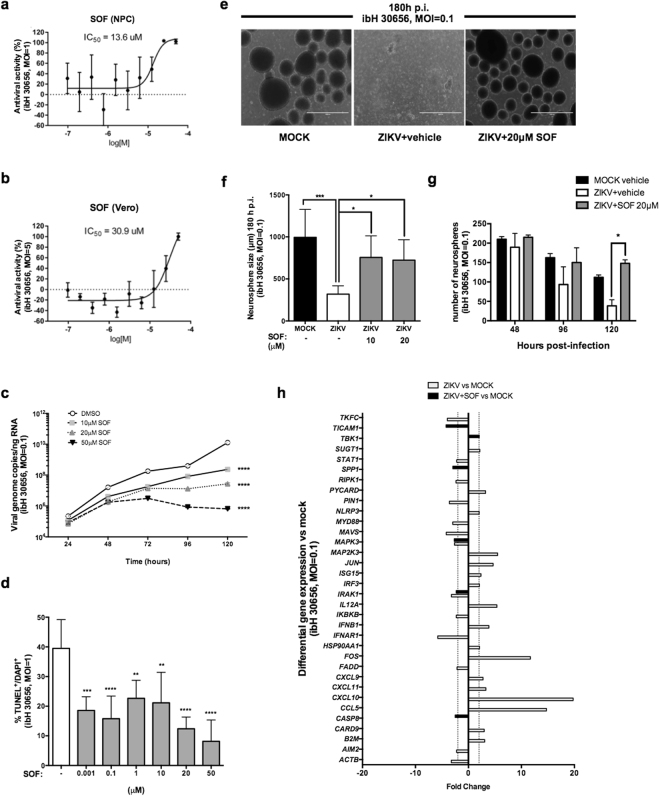
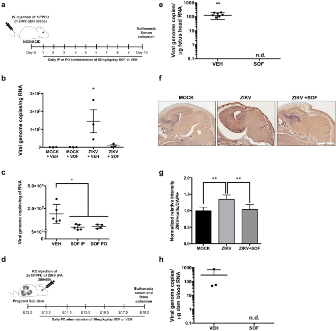
The outbreak of the Zika virus (ZIKV) has been associated with increased incidence of congenital malformations. Although recent efforts have focused on vaccine development, treatments for infected individuals are needed urgently. Sofosbuvir (SOF), an FDA-approved nucleotide analog inhibitor of the Hepatitis C (HCV) RNA-dependent RNA polymerase (RdRp) was recently shown to be protective against ZIKV both in vitro and in vivo. Here, we show that SOF protected human neural progenitor cells (NPC) and 3D neurospheres from ZIKV infection-mediated cell death and importantly restored the antiviral immune response in NPCs. In vivo, SOF treatment post-infection (p.i.) decreased viral burden in an immunodeficient mouse model. Finally, we show for the first time that acute SOF treatment of pregnant dams p.i. was well-tolerated and prevented vertical transmission of the virus to the fetus. Taken together, our data confirmed SOF-mediated sparing of human neural cell types from ZIKV-mediated cell death in vitro and reduced viral burden in vivo in animal models of chronic infection and vertical transmission, strengthening the growing body of evidence for SOF anti-ZIKV activity.


Conflict of interest statement

The authors declare that they have no competing interests.

Figures

Figure 1
Figure 1
The RdRp domain is conserved between HCV and ZIKV. (a) Phylogenetic dendrogram showing HCV and ZIKV belonging to Hepacivirus and Flavivirus genera, respectively, within the Flaviviridae family. Phylogenetic tree of RNA polymerase domain of HCV and ZIKV strains was based on Maximum-likelihood approach. This tree is intended to highlight the degree of similarity (branch length) of the RNA polymerase from different viruses. The longer the branch is in the horizontal dimension, the larger the amount of change. The units of branch length are amino acid substitutions per site. (b) Schematic of HCV and ZIKV protein structures. Both viruses have their RNA-dependent polymerases (RdRp) within the NS5B and NS5 domains, respectively. (c) Capsid protein; prM: precursor of Membrane protein; E: Envelope protein; p7: Viroporin, NS: Non-structural proteins. (c) RNA polymerase amino acid sequence conservation and DPP domain between different members of Flaviviradae family including HCV, West Nile virus, Japanese encephalitis, Dengue virus and ZIKV. Highlighted sequences correspond to amino acid sequence conservation between strains. Light gray corresponds to amino acid conservation in 4 strains, and dark gray correspond to amino acid conservation in 5 strains (all represented strains). The blue box indicated by the arrow corresponds to the GDD amino acid domain that is conserved in all 5 represented virus strains (highlighted as dark gray).
Figure 2
Figure 2
SOF blocks ZIKV replication in vitro. (a,b) Dose-response curves of SOF against ZIKV in NPCs and in Vero cells, respectively. SOF was tested in 10 points dose-response with two-fold dilution starting at 50 μM, in triplicates. The graphs show the antiviral activity (%) based on reduction of the cytopathic effect by quantification of the cytopathic effects of ZIKV ± SEM on NPCs (a) and Vero cells (b,c). Multi-step growth curve of vehicle or SOF-treated ZIKV-infected (ibH 30656, MOI = 0.1) NPCs at different concentrations over time as measured by viral genome copies of RNA using qRT-PCR. Data represents means ± SD (n = 3 replicates); two-way ANOVA, followed by Tukey’s multiple comparison tests. ****P < 0.0001 compared to DMSO (vehicle) treated NPCs at 120 hours p.i. Note that SD cannot be seen in the log scale. (d) The percentages of apoptotic cells were calculated, averaged, and graphed accordingly (ibH 30656, MOI = 0.1, 96 hours p.i.) in the presence of SOF at different doses (1 nM, 100 nM, 1 μM, 10 μM, 20 μM and 50 μM). One-way ANOVA tests with Tukey multiple comparisons were performed to compare to ZIKV-infected different groups. The presented values are means of TUNEL+/DAPI+ percentage ± SD (n = 6 images per condition), **P < 0.01, ***P < 0.001, ****P < 0.0001 TUNEL+/DAPI+ percentage decreases with different doses of SOF. (e) Representative images of mock-infected human neurospheres (left panel), ZIKV-infected vehicle-treated neurospheres (middle panel) and ZIKV-infected SOF-treated (20 μM) (right panel) neurospheres image (ibH30656, MOI = 0.1, 180 hours p.i.), scale bar: 500 μm. (f) Quantification of neurosphere diameter (μm) at 180 hours p.i. One-way ANOVA tests with Tukey multiple comparison were performed to compare different groups. The bars represent the number of spheres counted averaged and plotted for each condition ± SD (n = 6 images captured per condition), *P < 0.05, ***P < 0.001 (n ≥ 100 neurospheres counted per condition). (g) Neurosphere amount was counted over time after infection with ZIKV (ibH30656, MOI = 0.1 at 48, 96 and 120 hours p.i.) treated with vehicle or with SOF (20 μM). Bars represent the number of neurospheres ± SD at a given time (48, 96 and 120 hours p.i.). Two-way ANOVA tests with Tukey multiple comparison were performed to compare different groups, *P < 0.05 (n = 2 independent experiments). (h) Human antiviral response expression signature in human NPCs in monolayer at 120 hours p.i.: mock, ZIKV-infected (ibH30656, MOI 0.1) vehicle or SOF-treated (50 μM) (threshold = two-fold). Bars represent fold changes detected by qPCR from 96 different genes tested, between ZIKV-infected compared to mock-infected NPCs (in white) and between ZIKV + SOF-treated compared to mock-infected NPCs (in black). Note that there are fewer dysregulated genes between ZIKV-infected compared to mock NPCs upon SOF treatment (less black bars) than the ZIKV-infected untreated ones.
Figure 3
Figure 3
SOF blocks ZIKV replication in vivo. (a) Schematic of the experimental design for mice infection: 8 weeks-old NOD/SCID female mice were intra-venously injected with 108 PFU of ZIKV (ibH 30656). One day p.i. mice were randomized to receive either vehicle or SOF at 50 mg/kg/day IP or PO. (b) RNA viral load was measured in the serum of infected mice by qRT-PCR. One-way ANOVA tests with Tukey multiple comparison were performed to compare to MOCK-vehicle (VEH) group, *P < 0.05 (n = 3 technical replicates, n = 3 mice per group). Each dot represents the average of n = 3 technical replicates per mouse ± SEM. (c) RNA viral load was measured in the serum of infected mice by qRT-PCR. Each dot represents a mouse: vehicle n = 5 mice, SOF IP n = 5 mice, SOF PO n = 5 and monitored for 10 days before euthanasia and blood collection. One-way ANOVA tests with Tukey multiple comparison were performed to compare to vehicle group, *P < 0.05. Bars represent ± SD. (d) Schematic of the experimental design for infection of SJL pregnant mice. SJL dams were ZIKV-infected (PA 259459) with 2 × 105 PFU at E12.5. At E13.5, they were randomly assigned to receive vehicle or SOF (50 mg/kg/day) PO. At E18.5, mice were euthanized for blood and fetus collection. (e) RNA viral load was measured in the fetus heads mice by qRT-PCR. Each dot represents a fetus head (vehicle-treated n = 8 pooled from 3 independent litters, 1 did not amplify and SOF-treated, n = 6 fetus heads from 3 independent litters). Student’s t-test was performed to compare the two groups; Bars represent the average viral load ± SD (**P < 0.01), n. d. = not detected. (f) Immunohistochemistry against Flavivirus Group Antigen (brown) and counterstained in Mayer’s hematoxylin (blue) in brain cross-sections of E18.5 fetuses from mock-infected, ZIKV-infected vehicle-treated, or ZIKV-infected, SOF-treated dams. Scale bars 4 mm. (g) All data are represented as mean ± SD (n = 6 per condition: 3 embryos, 2 slides each embryo per condition) and were analyzed with one-way ANOVA (Turkey’s Multiple Comparison post test). **P < 0.01. (h) RNA viral load was measured in the dam serum by qRT-PCR. Each dot represents one mouse ± SD (vehicle-treated n = 3 and SOF-treated, n = 3). n. d. = not detected. Student’s t-test was performed to compare the two groups (P = 0.2902).

References

    1. Campos GS, Bandeira AC, Sardi SI. Zika virus outbreak, Bahia, Brazil. Emerg. Infect. Dis. 2015;21:1885–1886. doi: 10.3201/eid2110.150847. - DOI - PMC - PubMed
    1. Mlakar J, et al. Zika Virus Associated with Microcephaly. N. Engl. J. Med. 2016;374:951–8. doi: 10.1056/NEJMoa1600651. - DOI - PubMed
    1. van der Linden V, et al. Description of 13 Infants Born During October 2015–January 2016 With Congenital Zika Virus Infection Without Microcephaly at Birth — Brazil. MMWR. Morb. Mortal. Wkly. Rep. 2016;65:1343–1348. doi: 10.15585/mmwr.mm6547e2. - DOI - PubMed
    1. Xu M, et al. Identification of small-molecule inhibitors of Zika virus infection and induced neural cell death via a drug repurposing screen. Nat. Med. 2016;22:1101–1107. doi: 10.1038/nm.4184. - DOI - PMC - PubMed
    1. Retallack H, et al. Zika virus cell tropism in the developing human brain and inhibition by azithromycin. Proc. Natl. Acad. Sci. USA. 2016;113:201618029. doi: 10.1073/pnas.1618029113. - DOI - PMC - PubMed

Publication types