

2,5-Hexanedione induces dopaminergic neurodegeneration through integrin αMβ2/NADPH oxidase axis-mediated microglial activation
- PMID: 29352205
- PMCID: PMC5833449
- DOI: 10.1038/s41419-017-0091-7
2,5-Hexanedione induces dopaminergic neurodegeneration through integrin αMβ2/NADPH oxidase axis-mediated microglial activation
Erratum in
-
Correction: 2,5-Hexanedione induces dopaminergic neurodegeneration through integrin αMβ2/NADPH oxidase axis-mediated microglial activation.Cell Death Dis. 2022 Dec 13;13(12):1036. doi: 10.1038/s41419-022-05493-2. Cell Death Dis. 2022. PMID: 36513641 Free PMC article. No abstract available.
Abstract
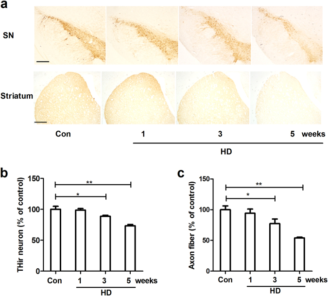
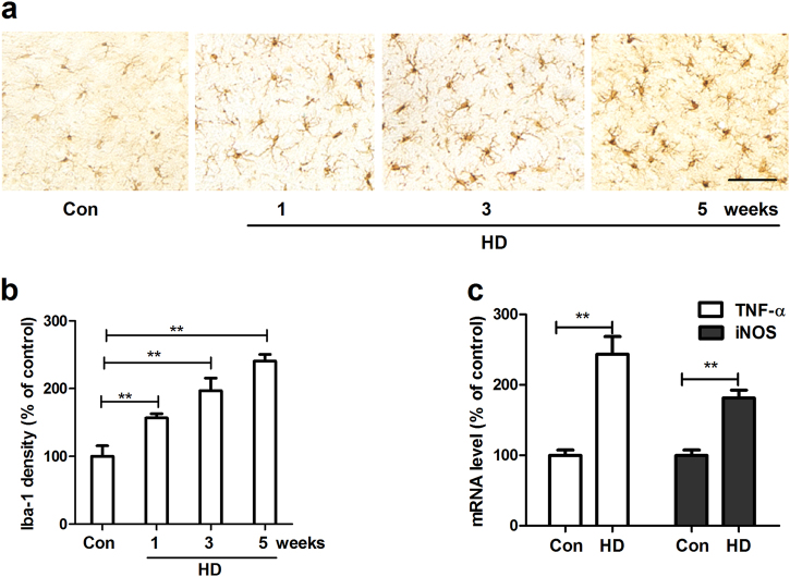
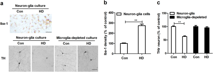
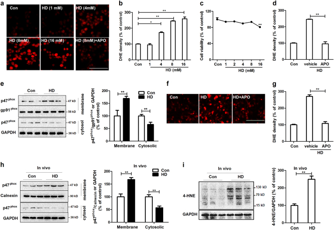
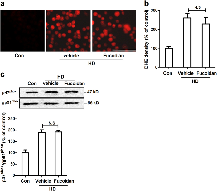
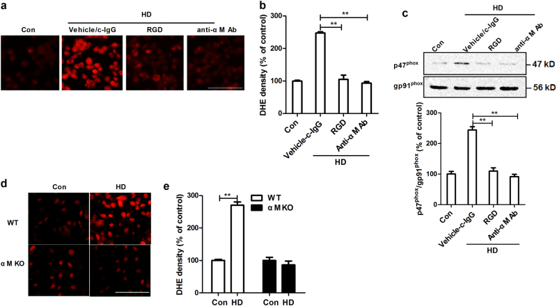
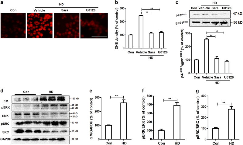
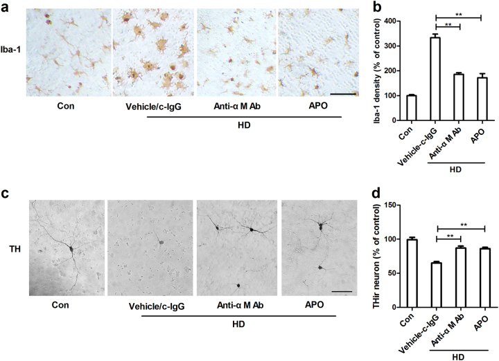
Recent study demonstrated that chronic exposure to solvents increases the risk of Parkinson's disease (PD), the second most common neurodegenerative disorder characterized by progressive dopaminergic neurodegeneration in the substantia nigra (SN). n-Hexane, a widely used organic solvent, displays central-peripheral neurotoxicity, which is mainly mediated by its active metabolite, 2,5-hexanedione (HD). However, whether HD exposure contributes to PD remains unclear. In this study, we found that rats exposed to HD displayed progressive dopaminergic neurodegeneration in the nigrostriatal system. Microglial activation was also detected in HD-treated rats, which occurred prior to degeneration of dopaminergic neurons. Moreover, depletion of microglia markedly reduced HD-induced dopaminergic neurotoxicity. Mechanistic study revealed an essential role of microglial integrin αMβ2-NADPH oxidase (NOX2) axis in HD-elicited neurotoxicity. HD activated NOX2 by inducing membrane translocation of NOX2 cytosolic subunit, p47phox. Integrin αMβ2 was critical for HD-induced NOX2 activation since inhibition or genetic deletion of αMβ2 attenuated NOX2-generated superoxide and p47phox membrane translocation in response to HD. Src and Erk, two downstream signals of αMβ2, were recognized to bridge HD/αMβ2-mediated NOX2 activation. Finally, pharmacological inhibition of αMβ2-NOX2 axis attenuated HD-induced microglial activation and dopaminergic neurodegeneration. Our findings revealed that HD exposure damaged nigrostriatal dopaminergic system through αMβ2-NOX2 axis-mediated microglial activation, providing, for the first time, experimental evidence for n-hexane exposure contributing to the etiology of PD.
Conflict of interest statement
The authors declare that they have no competing interests.
Figures
Similar articles
-
Role and mechanism of microglial activation in iron-induced selective and progressive dopaminergic neurodegeneration.Mol Neurobiol. 2014 Jun;49(3):1153-65. doi: 10.1007/s12035-013-8586-4. Epub 2013 Nov 26. Mol Neurobiol. 2014. PMID: 24277523 Free PMC article.
-
Complement receptor 3 mediates NADPH oxidase activation and dopaminergic neurodegeneration through a Src-Erk-dependent pathway.Redox Biol. 2018 Apr;14:250-260. doi: 10.1016/j.redox.2017.09.017. Epub 2017 Sep 27. Redox Biol. 2018. PMID: 28978491 Free PMC article.
-
Activation of neuronal NADPH oxidase NOX2 promotes inflammatory neurodegeneration.Free Radic Biol Med. 2023 May 1;200:47-58. doi: 10.1016/j.freeradbiomed.2023.03.001. Epub 2023 Mar 2. Free Radic Biol Med. 2023. PMID: 36870375 Free PMC article.
-
Targeting microglia-mediated neurotoxicity: the potential of NOX2 inhibitors.Cell Mol Life Sci. 2012 Jul;69(14):2409-27. doi: 10.1007/s00018-012-1015-4. Epub 2012 May 13. Cell Mol Life Sci. 2012. PMID: 22581365 Free PMC article. Review.
-
Critical role of the Mac1/NOX2 pathway in mediating reactive microgliosis-generated chronic neuroinflammation and progressive neurodegeneration.Curr Opin Pharmacol. 2016 Feb;26:54-60. doi: 10.1016/j.coph.2015.10.001. Epub 2015 Oct 26. Curr Opin Pharmacol. 2016. PMID: 26498406 Free PMC article. Review.
Cited by
-
The Promiscuous Profile of Complement Receptor 3 in Ligand Binding, Immune Modulation, and Pathophysiology.Front Immunol. 2021 Apr 29;12:662164. doi: 10.3389/fimmu.2021.662164. eCollection 2021. Front Immunol. 2021. PMID: 33995387 Free PMC article. Review.
-
NOX2 Activation in COVID-19: Possible Implications for Neurodegenerative Diseases.Medicina (Kaunas). 2021 Jun 11;57(6):604. doi: 10.3390/medicina57060604. Medicina (Kaunas). 2021. PMID: 34208136 Free PMC article. Review.
-
Proapoptotic effects of 2,5‑hexanedione on pheochromocytoma cells via oxidative injury.Mol Med Rep. 2019 Oct;20(4):3249-3255. doi: 10.3892/mmr.2019.10546. Epub 2019 Aug 1. Mol Med Rep. 2019. PMID: 31432125 Free PMC article.
-
Cold-inducible protein RBM3 mediates hypothermic neuroprotection against neurotoxin rotenone via inhibition on MAPK signalling.J Cell Mol Med. 2019 Oct;23(10):7010-7020. doi: 10.1111/jcmm.14588. Epub 2019 Aug 22. J Cell Mol Med. 2019. PMID: 31436914 Free PMC article.
-
Microglial Activation Mediates Noradrenergic Locus Coeruleus Neurodegeneration via Complement Receptor 3 in a Rotenone-Induced Parkinson's Disease Mouse Model.J Inflamm Res. 2021 Apr 9;14:1341-1356. doi: 10.2147/JIR.S299927. eCollection 2021. J Inflamm Res. 2021. PMID: 33859489 Free PMC article.
References
-
- Tanner CM. Is the cause of Parkinson’s disease environmental or hereditary? Evidence from twin studies. Adv. Neurol. 2003;91:133. - PubMed
Publication types
MeSH terms
Substances
LinkOut - more resources
Full Text Sources
Other Literature Sources
Medical
Research Materials
Miscellaneous
