. 2018 Jan 19;8(1):1195.
doi: 10.1038/s41598-018-19480-1.

Handheld magnetic probe with permanent magnet and Hall sensor for identifying sentinel lymph nodes in breast cancer patients

Affiliations

Handheld magnetic probe with permanent magnet and Hall sensor for identifying sentinel lymph nodes in breast cancer patients

Masaki Sekino et al. Sci Rep. .

Abstract

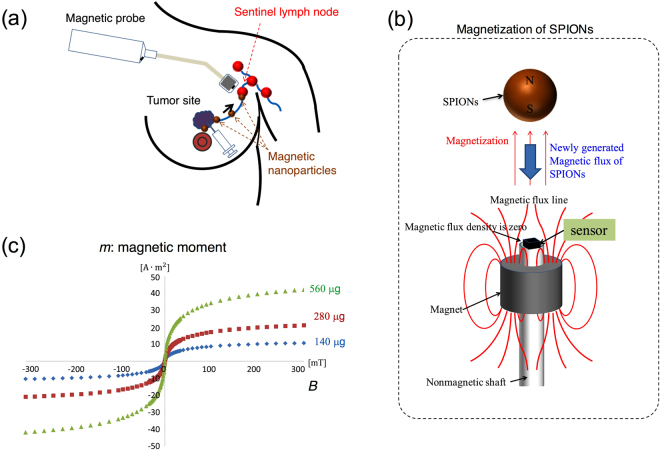
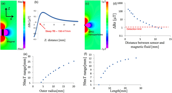
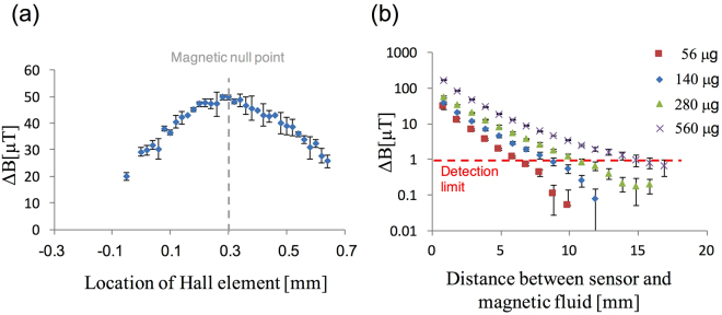
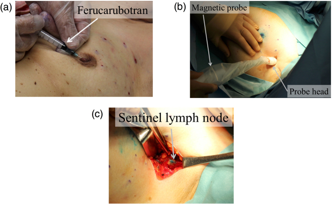
The newly developed radioisotope-free technique based on magnetic nanoparticle detection using a magnetic probe is a promising method for sentinel lymph node biopsy. In this study, a novel handheld magnetic probe with a permanent magnet and magnetic sensor is developed to detect the sentinel lymph nodes in breast cancer patients. An outstanding feature of the probe is the precise positioning of the sensor at the magnetic null point of the magnet, leading to highly sensitive measurements unaffected by the strong ambient magnetic fields of the magnet. Numerical and experimental results show that the longitudinal detection length is approximately 10 mm, for 140 μg of iron. Clinical tests were performed, for the first time, using magnetic and blue dye tracers-without radioisotopes-in breast cancer patients to demonstrate the performance of the probe. The nodes were identified through transcutaneous and ex-vivo measurements, and the iron accumulation in the nodes was quantitatively revealed. These results show that the handheld magnetic probe is useful in sentinel lymph node biopsy and that magnetic techniques are widely being accepted as future standard methods in medical institutions lacking nuclear medicine facilities.


Conflict of interest statement

The authors declare that they have no competing interests.

Figures

Figure 1
Figure 1
(a) Principle of the technique based on magnetic detection of SLNs using a handheld magnetic probe and magnetic nanoparticles for breast cancer patients. Injected magnetic nanoparticles accumulate in the SLNs via the axillary lymphatic system and are detected by a magnetometer. (b) Schematic of the magnetic field lines produced by the ring-shaped permanent magnet. There are two magnetic null points on the surface of the magnet along the axis. The magnetic sensor is located at a magnetic null point. (c) Superparamagnetic characteristics of Resovist (Ferucarbotran); the magnetic moment measured by the SQUID apparatus as a function of the applied magnetic field from −300 to 300 mT.
Figure 2
Figure 2
Numerical results: (a) Two-dimensional distribution of the longitudinal magnetic field BZ of the permanent magnet. (b) BZ of the magnet on the Z-axis (X = 0 mm). ∇B (dBZ/dz) is approximately 158 mT/mm around the magnetic null point (Z = 0.3 mm). (c) Two-dimensional distribution of BZ of 140 μg of iron (5 μL of Resovist) of fully magnetized SPIO located at (X, Z) = (0, 10.4) mm. (d) Magnetic field strength ΔB at the sensor position as a function of the distance from the magnetic sensor to the SPIONs location on the Z-axis. Red dotted line represents the sensor detection limit of 1 μT. Relationships between (e) the outer radius and (f) the length of the magnet, and the distance Z50 of the point where the BZ of the magnet is 50 mT from the magnet surface.
Figure 3
Figure 3
(a) Signals detected with 140 μg of iron (5 μL of Resovist), as functions of the Hall element position: the gray dotted line shows that the magnetic null point is at approximately 0.3 mm. (b) Measured signals for four different volumes, 56, 140, 280, and 560 μg of iron. The red dotted line shows the detection limit of the sensor.
Figure 4
Figure 4
Procedure of the clinical test for the magnetic technique involving SPIO and blue dye tracers, using the handheld magnetic probe with a permanent magnet and Hall sensor for SLNB; (a) injection of SPIO tracers around the subareolar region, (b) transcutaneous detection of the SLNs before incision, and (c) SLNs detection and excision in the axilla after incision. In the injection phase (a), the blue dye tracer is injected at the same location after the injection of the SPIO tracer.
Figure 5
Figure 5
(a) Blue color of Patent blue and brown color of Resovist (Ferucarbotran) in the excised node. (b) Magnetic moments of a half volume of the excised node and 140 μg of SPIONs measured by the SQUID apparatus. (c) Histopathology of the excised node. The distribution of iron staining (Perl’s Prussian blue) is generally in the cortex of the node. Metastasis was observed in one excised node.

Similar articles

Cited by

References

    1. Krag DN, Weaver DL, Alex JC, Fairbank JT. Surgical resection and radiolocalization of the sentinel lymph node in breast cancer using a gamma probe. Surg Oncol. 1993;2:335–339. doi: 10.1016/0960-7404(93)90064-6. - DOI - PubMed
    1. Giuliano AE, Kirgan DM, Guenther JM, Morton DL. Lymphatic mapping and sentinel lymphadenectomy for breast cancer. Ann. Surg. 1994;220:391–401. doi: 10.1097/00000658-199409000-00015. - DOI - PMC - PubMed
    1. Wiechmann L, et al. Presenting features of breast cancer differ by molecular subtype. Ann. Surg. Oncol. 2009;16:2705–2710. doi: 10.1245/s10434-009-0606-2. - DOI - PubMed
    1. Britten AJ. A method to evaluate intra-operative gamma probes for sentinel lymph node localization. Eur. J. Nucl. Med. 1999;26:76–83. doi: 10.1007/s002590050362. - DOI - PubMed
    1. Wydra D, Matuszewski R, Romanowicz G, Bandurski T. Evaluation of surgical gamma probes for sentinel node localization in cervical and vulvar cancer. Nucl. Med. Rev. Cent. East. Eur. 2005;8(2):105–110. - PubMed

Publication types